Treatment
- Manual Release, hydration, and stretching of the flexor retinaculum
- Proprioceptive exercises of the ankle to improve feedback information to the brain and thus increasing stability
- Strengthening of the intrinsic muscles of the foot and ankle including the tibialis posterior and flexor digitorum...
- Ensure adequate glide and slide of the tibial nerve with nerve...
What is the flexor retinaculum of the foot?
The flexor retinaculum of the foot is a strong fibrous band that covers the tendons of the muscles that flex the foot such as walking on the toes like a ballerina.
What are the symptoms of a tear of the flexor retinaculum?
This creates a suboptimal function of flexor hallucis longus, the primary toe flexor tendon. Symptoms of an isolated tear of the flexor retinaculum include pain, numbness, and tingling on the arch of the foot that’s provoked by walking, standing, and running.
What happens to the flexor retinaculum extra-articular when injured?
During ankle strains and strain of the inner ankle, the flexor retinaculum extra-articular can become injured. A tear can occur which once healed can create a scar.
What is the flexor retinaculum made of?
A flexor retinaculum consists of a fibrous band of fascia, which is a sheet of dense connective tissue that covers or binds other body structures. The flexor retinaculum of the foot, also known as the laciniate ligament, covers the tendons of the flexor muscles of the ankle.

Can you tear your flexor retinaculum?
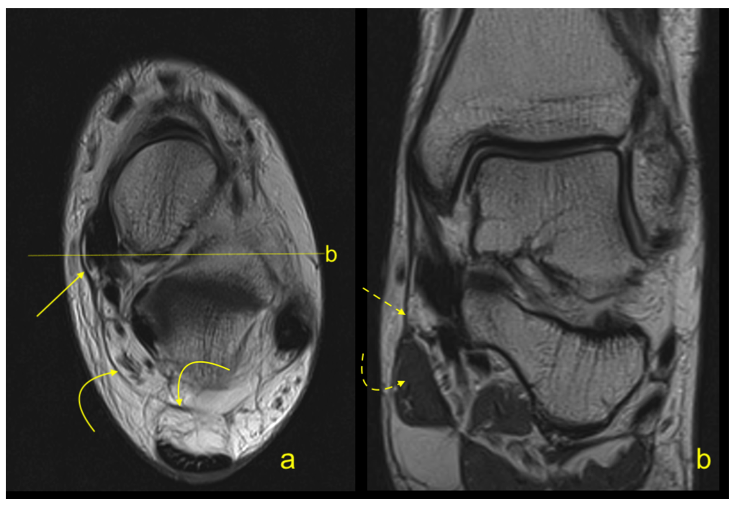
Medial flexor retinaculum injuries are not uncommon, but medial flexor retinaculum periosteal avulsion injuries are rare. This patient sustained a medial flexor retinaculum tear readily characterized at computed tomography by an associated proximal retinacular avulsion fracture from the posteromedial tibia.
What causes flexor retinaculum pain?
This can occur from swollen varicose veins, a tumor (noncancerous) on the tibial nerve, and swelling caused by other conditions, such as diabetes. As pressure increases in the tarsal tunnel, the nerve is the most sensitive to the pressure and is squeezed against the flexor retinaculum.
How do you treat retinaculum?
TREATMENT. Restrictions of the retinacula can be treated quite effectively with Manual Therapy and a series of corrective exercises. Treating with manual therapy involves breaking restrictions between the retinaculum and the tendon.
What happens if you tear your retinaculum?
A rupture of the extensor retinaculum could result in a loss of dorsiflexion power, prominent tendons in the anterior aspect of the ankle and local inflammation due to lack of appropriate tracking or sliding disturbance of the tendons.
Can retinaculum repair itself?
Background: Superior peroneal retinaculum tears are often mistaken for lateral ankle instability. These tears often do not heal readily by themselves and must be identified so that proper treatment can begin.
Is retinaculum a ligament?
The flexor retinaculum is a fibrous connective tissue band that forms the anterior roof of the carpal tunnel. Many experts consider the flexor retinaculum synonymous with the transverse carpal ligament and the annular ligament; for this discussion, they will be considered the same structure.
How long does it take retinaculum to heal?
Recovery from surgery requires a moderately long period, usually in the order of 2-6 weeks of immobilization, in order to allow the retinaculum and any bony procedures to heal. This is followed by four to six weeks of fairly graduated and intensive rehabilitation.
What is a retinaculum tear?
In order for the tendon to move out of place, the peroneal retinaculum has to be torn. This is a ligament that hold the tendons in place behind the lateral malleolus (lower end the fibula).
How long does tendonitis take to heal in the foot?
It usually takes two to three months to recover from foot or ankle tendonitis, but it can take much longer without the proper treatment so early diagnosis and treatment is essential. If tendonitis is not treated early or resolved early, severe foot deformities may develop including worsening of flat feet and arthritis.
Can you walk with a torn tendon in your foot?
Can You Walk with a Torn Foot Tendon? The quick answer is yes, typically you can walk with a torn ligament or tendon in the foot. Walking may be painful but you can typically still walk.
Where is the flexor retinaculum in the foot?
The flexor retinaculum of the foot extends from the medial malleolus above, to the calcaneus below. This converts a series of bony grooves into canals for the passage of the tendons of the flexor muscles and the posterior tibial vessels and tibial nerve into the sole of the foot, known as the tarsal tunnel.
Can a tendon move out of place?
Subluxation is more likely in people with certain genetic anatomical differences, but tendons can also snap out of place as a result of an injury. Tendon ruptures can also occur. These injuries may be due to a combination of immediate trauma and chronic trauma.
What muscle attaches to the flexor retinaculum?
The tendons of the palmaris longus and flexor carpi ulnaris are partly attached to the surface of the retinaculum; below, the short muscles of the thumb and little finger originate from the flexor retinaculum.
How do you treat tendonitis on top of foot?
Rest the affected foot for two to three days. Use it as little as possible to give the tendons a break. While you are resting your foot, put ice on it for 20 minutes every two or three hours. Wrap an elastic bandage around the injured area to reduce inflammation, or use a brace.
What muscles go through the flexor retinaculum?
pronator teres muscle.flexor carpi radialis muscle.palmaris longus muscle. palmar aponeurosis.flexor carpi ulnaris muscle.
Why does my superior extensor retinaculum hurt?
Running with a sore superior extensor retinaculum might cause injury. Runners continually place strain on the joints and tissues of the lower body, and an intense or poorly designed training schedule, or improper running technique, might damage the tissues in your feet or legs.
What is the flexor retinaculum?
Flexor retinaculum is a strong fibrous band which bridges the anterior concavity of the carpal bones thus converts it into a tunnel, the carpal tunnel.
Which carpal bone gives attachments to both flexor and extensor retinacula?
These four bony points are all palpable in the living hand and it should be noted that pisiform is the only carpal bone that gives attachments to both flexor and extensor retinacula. On either side the retinaculum has a slip.
Which side of the retina has a slip?
On either side the retinaculum has a slip.
What is the best approach to tendinopathy?
The best approach to all tendinopathys is reduce or modify the activity and then institute a loading program.
Can you treat a swollen foot with insoles?
You can't treat it with insoles.
What is the flexor retinaculum?
A flexor retinaculum consists of a fibrous band of fascia, which is a sheet of dense connective tissue that covers or binds other body structures. The flexor retinaculum of the foot, also known as the laciniate ligament, covers the tendons of the flexor muscles of the ankle. The specific tendons covered are the tibialis posterior, ...
Where does the flexor retinaculum pass through?
They pass through the flexor retinaculum immediately posterior to (behind) the medial malleolus, which is the network of nerve tissue and muscle that surrounds the ankle joint.
Which tendons help to flex the foot?
The specific tendons covered are the tibialis posterior, flexor digitorum longus, and flexor hallucis longus, which all help to flex the foot so that the toes point downward. The function of the flexor retinaculum of the foot is to prevent subluxation, or partial dislocation, of these tendons.