
What does the incus do in the ear?
The incus or anvil is a bone in the middle ear. The anvil-shaped small bone is one of three ossicles in the middle ear. The incus receives vibrations from the malleus, to which it is connected laterally, and transmits these to the stapes medially. The incus is so-called because of its resemblance to an anvil (Latin: Incus). Left incus.
What is the structure of the incus?
Structure. The incus is the second of the ossicles, three bones in the middle ear which act to transmit sound. It is shaped like an anvil, and has a long and short crus extending from the body, which articulates with the malleus.:862 The short crus attaches to the posterior ligament of the incus.
Why is the incus called the anvil?
The incus is so-called because of its resemblance to an anvil ( Latin: Incus ). The incus is the second of the ossicles, three bones in the middle ear which act to transmit sound. It is shaped like an anvil, and has a long and short crus extending from the body, which articulates with the malleus.
What is the function of the malleus and incus?
The malleus, resting on the membrane, conveys vibrations to the incus. This in turn conveys vibrations to the stapes. [2] "Incus" means "anvil" in Latin.
See more
What is the incus?
At the end of the long crus is the lenticular process, a hooked-shaped part of the incus that forms a joint with the head of the stapes. The short crus attaches to the back wall of the middle ear cavity, which houses the ossicles. The center of the incus is also known as the body.
What are the three bones in the middle ear?
There are three bones located in the middle ear: the incus, the malleus and the stapes. Collectively, all three bones comprise the ossicles.
What is the shape of the incus?
It is shaped like an anvil, which is why ‘the anvil’ is a widely used alternative name for the bone. The bone has a few basic regions. One of its surfaces, called the head, forms a joint with the malleus ossicle. The incus also has two extensions known as the long and short crus.
Where is the short crus located?
The short crus attaches to the back wall of the middle ear cavity, which houses the ossicles. The center of the incus is also known as the body. Last medically reviewed on January 22, 2018.
Where does sound travel in the brain?
These vibrations then travel into the cochlea, where sound is translated into nervous system signals that are sent to the brain. The incus lays at the center of the ossicles, connecting the malleus to the stapes. It is shaped like an anvil, which is why ‘the anvil’ is a widely used alternative name for the bone.
What is the incus in the middle ear?
Auditory tube, laid open by a cut in its long axis. The incus or anvil is a bone in the middle ear. The anvil -shaped small bone is one of three ossicles in the middle ear. The incus receives vibrations from the malleus, to which it is connected laterally, and transmits these to the stapes medially.
Why is the incus called the incus?
The incus is so-called because of its resemblance to an anvil ( Latin: Incus ).
What is the second ossicle?
The incus is the second of the ossicles, three bones in the middle ear which act to transmit sound. It is shaped like an anvil, and has a long and short crus extending from the body, which articulates with the malleus. The short crus attaches to the posterior ligament of the incus.
What is the short crus?
The short crus attaches to the posterior ligament of the incus. The long crus articulates with the stirrup at the lenticular process. The superior ligament of the incus attaches at the body of the incus to the roof of the tympanic cavity .
Who discovered the incus?
"Incus" means "anvil" in Latin. Several sources attribute the discovery of the incus to the anatomist and philosopher Alessandro Achillini. The first brief written description of the incus was by Berengario da Carpi in his Commentaria super anatomia Mundini (1521). Andreas Vesalius, in his De humani corporis fabrica, was the first to compare the second element of the ossicles to an anvil, thereby giving it the name incus. The final part of the long limb was once described as a "fourth ossicle" by Pieter Paaw in 1615.
Which part of the ear receives vibrations?
Main article: Hearing. Vibrations in the middle ear are received via the tympanic membrane. The malleus, resting on the membrane, conveys vibrations to the incus. This in turn conveys vibrations to the stapes.
What are the three bones that make up the ossicles?
The ossicles are actually tiny bones — the smallest in the human body. The three bones are named after their shapes: the malleus (hammer), incus (anvil) and stapes (stirrup). The ossicles further amplify the sound.
What are the red hair cells in a rodent's cochlea?
In green are four rows of hair cells that respond to sound vibrations, and in red are auditory nerve fibers that convey sound information from the hair cells to the brain. Researchers at Johns Hopkins are studying the molecular mechanisms that guide the formation of hair cells. Studies such as these might be a step towards less invasive treatments for deafness in which molecular cues can be used to biologically regenerate hair cells in the cochlea.
What is the outer ear?
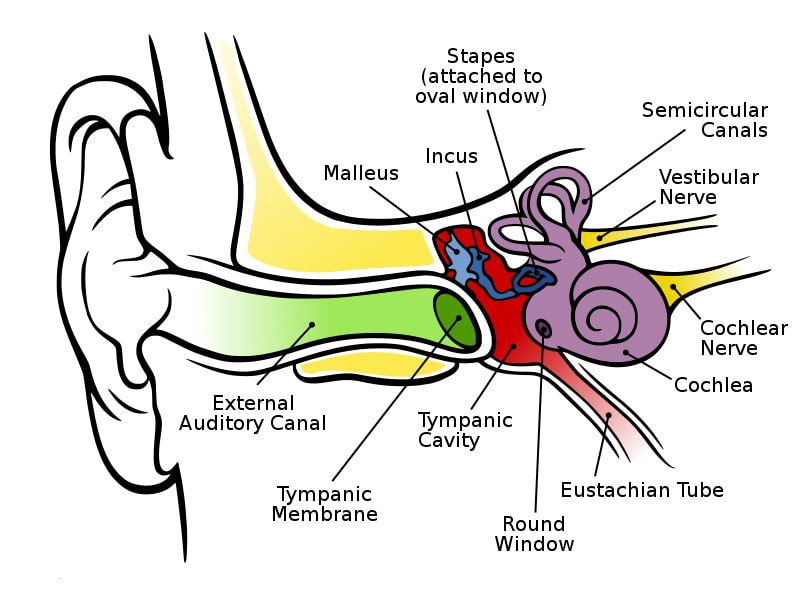
The Outer Ear. The auricle ( pinna) is the visible portion of the outer ear. It collects sound waves and channels them into the ear canal (external auditory meatus), where the sound is amplified. The sound waves then travel toward a flexible, oval membrane at the end of the ear canal called the eardrum, or tympanic membrane.
How many nerve endings are set into motion?
As the fluid moves, 25,000 nerve endings are set into motion. These nerve endings transform the vibrations into electrical impulses that then travel along the eighth cranial nerve (auditory nerve) to the brain. The brain then interprets these signals, and this is how we hear.
Which bone is responsible for equalizing the pressure between the air outside the ear and that within the middle ear?
The ossicles further amplify the sound. The tiny stapes bone attaches to the oval window that connects the middle ear to the inner ear. The Eustachian tube , which opens into the middle ear, is responsible for equalizing the pressure between the air outside the ear and that within the middle ear.
What causes the cochlea to ripple?
Once the vibrations cause the fluid inside the cochlea to ripple, a traveling wave forms along the basilar membrane. Hair cells—sensory cells sitting on top of the basilar membrane—ride the wave. Hair cells near the wide end of the snail-shaped cochlea detect higher-pitched sounds, such as an infant crying.
What is the membrane of the cochlea called?
An elastic partition runs from the beginning to the end of the cochlea, splitting it into an upper and lower part. This partition is called the basilar membrane because it serves as the base, or ground floor, on which key hearing structures sit.
What are the three bones that vibrate in the middle ear?
The eardrum vibrates from the incoming sound waves and sends these vibrations to three tiny bones in the middle ear. These bones are called the malleus, incus, and stapes . The bones in the middle ear amplify, or increase, the sound vibrations and send them to the cochlea, a snail-shaped structure filled with fluid, in the inner ear.
How does hearing work?
Hearing depends on a series of complex steps that change sound waves in the air into electrical signals. Our auditory nerve then carries these signals to the brain. Also available: Journey of Sound to the Brain, an animated video.
What happens when hair cells bend?
As the hair cells move up and down, microscopic hair-like projections (known as stereocilia) that perch on top of the hair cells bump against an overlying structure and bend. Bending causes pore-like channels, which are at the tips of the stereocilia, to open up.
Which nerve carries electrical signals to the brain?
The auditory nerve carries this electrical signal to the brain, which turns it into a sound that we recognize and understand.
What are the three chambers of the cochlea?
The cochlea is filled with fluid (perilymph and endolymph) and is divided into three chambers called the scala vestibuli, scala media, and the scala tympani . Two of these fluid-filled chambers sense pressure changes (caused by sound) while the third chamber contains the organ of Corti, the cochlear duct and the basilar membrane.
What is the fluid in the semicircular canals?
The semicircular canals are filled with a fluid called endolymph and function to provide the body with a proper sense of balance. Directly adjacent to the semicircular canals, prior to the beginning of the snail-shaped tube that forms the cochlea is the round window. 5 . The Anatomy of the Ear.
How early does the inner ear form?
Anatomical Variations. Embryonically, the inner ear begins to form as early on as 4 weeks gestation. The cochlea itself is typically formed by 18 weeks gestation. The gene SOX2 is largely responsible for the formation of the cochlea and mutations in SOX2 are associated with sensorineural hearing loss. 3 .
Where are the hair cells located?
2 . Also located within the cochlea are tiny hair cells. They are specifically found within the organ of Corti and are essential for proper hearing. 3 . At birth we have about 12,000 hair cells.
What is the purpose of cochlear implants?
A cochlear implant is an electronic device that can improve hearing in individuals who experience deafness or profound hearing loss as a result of damage to the cochlea. It has several parts including a microphone a speech processor, a transmitter and receiver, and an electrode array.
What is pulsatile tinnitus?
Pulsatile tinnitus is when you can hear what sounds like your own heartbeat in your ears. Tinnitus is strongly associated with exposure to loud noises, sensorineural hearing loss and is also thought to be the result of damage to the hair cells in the cochlea. 10 .
Where do stapes strike?
The stapes strikes the oval window and vibrations are further conducted through the perilymph (fluid) located inside of the cochlea. Sound vibrations continue on through the scala vestibuli and scala tympani eventually displacing the round window. 3

Overview
The incus (plural incudes) or anvil is a bone in the middle ear. The anvil-shaped small bone is one of three ossicles in the middle ear. The incus receives vibrations from the malleus, to which it is connected laterally, and transmits these to the stapes medially. The incus is so-called because of its resemblance to an anvil (Latin: Incus).
Structure
The incus is the second of the ossicles, three bones in the middle ear which act to transmit sound. It is shaped like an anvil, and has a long and short crus extending from the body, which articulates with the malleus. The short crus attaches to the posterior ligament of the incus. The long crus articulates with the stirrup at the lenticular process.
The superior ligament of the incus attaches at the body of the incus to the roof of the tympanic ca…
Function
Vibrations in the middle ear are received via the tympanic membrane. The malleus, resting on the membrane, conveys vibrations to the incus. This in turn conveys vibrations to the stapes.
History
"Incus" means "anvil" in Latin. Several sources attribute the discovery of the incus to the anatomist and philosopher Alessandro Achillini. The first brief written description of the incus was by Berengario da Carpi in his Commentaria super anatomia Mundini (1521). Andreas Vesalius, in his De humani corporis fabrica, was the first to compare the second element of the ossicles to an anvil, thereby giving it the name incus. The final part of the long limb was once described as a "f…
Additional images
• Ossicles
• Tympanic cavity. Facial canal. Internal carotid artery.
• Auditory ossicles. Tympanic cavity. Deep dissection.
• Aditory ossicles. Incus and malleus. Deep dissection.
See also
• Hearing – Sensory perception of sound by living organisms
• Ear – Organ of hearing and balance
• Ossicles – Three bones in each middle ear that are among the smallest bones in the human body
External links
• The Anatomy Wiz Incus