Clavicle / Ribs
- The clavicle and ribs act as landmarks when assessing the adequacy of inspiration taken by the patient
- The anterior end of approximately 5-7 ribs should be visible above the point at which the mid-clavicular line intersects the diaphragm
- Less than 5 ribs indicates incomplete inspiration
- More than 7 ribs suggests lung hyper-expansion
How many ribs should be visible on a chest xray?
Clavicle / Ribs The clavicle and ribs act as landmarks when assessing the adequacy of inspiration taken by the patient The anterior end of approximately 5-7 ribs should be visible above the point at which the mid-clavicular line intersects the diaphragm Less than 5 ribs indicates incomplete inspiration
How many ribs should be visible above the mid-clavicular line?
The anterior end of approximately 5-7 ribs should be visible above the point at which the mid-clavicular line intersects the diaphragm Less than 5 ribs indicates incomplete inspiration More than 7 ribs suggests lung hyper-expansion On this normal X-ray the anterior end of the 7th rib (asterisk) intersects the diaphragm at the mid-clavicular line
What is the role of X-rays in the workup of ribs?
When X-rays of the ribs, the state of the bone mechanism is visualized, and the spine can be partially seen. The degree of ionizing radiation is not considered dangerous to human health, so X-rays can be considered a good alternative to ultrasound, [ 1 ] computed and magnetic resonance imaging. [ 2]
When do you need a plain X-ray for rib fractures?
Most often, the procedure is prescribed when a rib fracture is suspected. If multiple trauma is detected, the doctor may insist on performing a plain X-ray, which is necessary to obtain more objective and complete information about the damage. Plain X-ray shows the existing damage to the internal organs and the whole chest.

How many anterior ribs should be seen on CXR?
Overview. Normally, six anterior ribs and nine posterior ribs should be seen above the diaphragm.
How many ribs does anterior have?
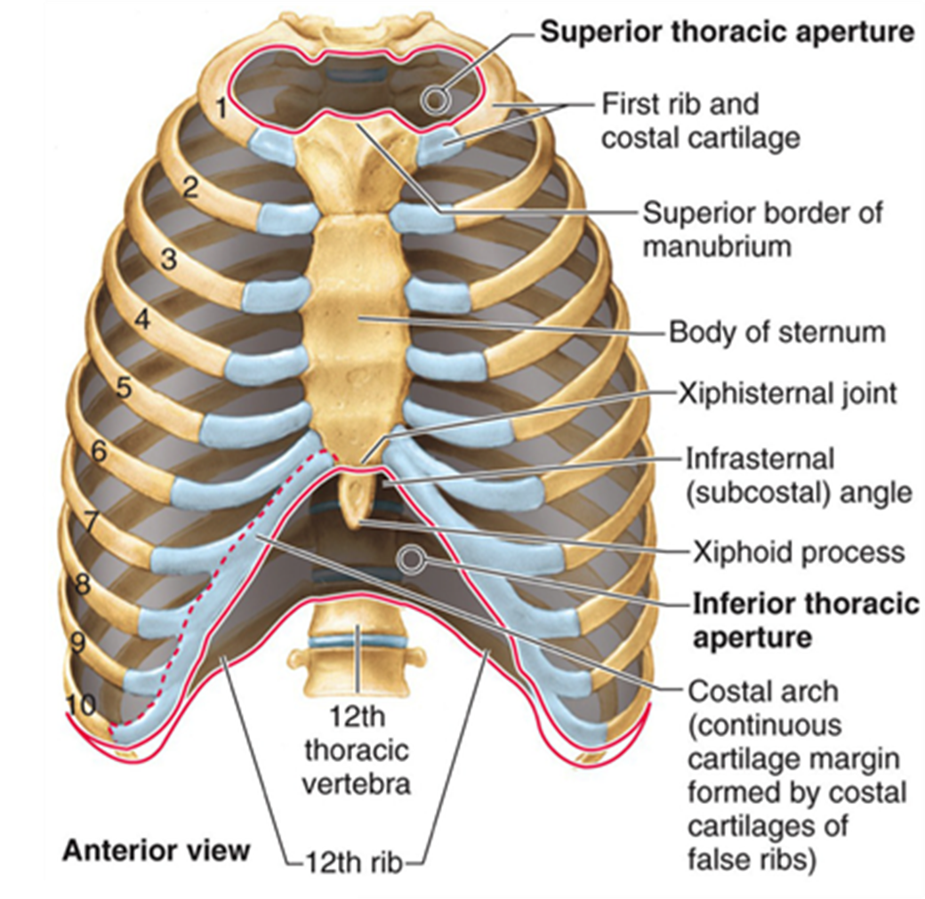
The thoracic cavity is made up of 12 pairs of ribs that connect in the posterior thorax to the vertebral bodies of the spinal column. In the anterior thorax, the first 7 pairs of ribs are attached to the sternum or breastbone by cartilage. The lower 5 ribs do not attach to the sternum.
Is there a limit to how many X rays you can have?
Now we come to the most important question, How much of Xray is safe for a person in his lifetime? Xray is generally measured or compared with normal environment radiation. The American College of Radiology recommends 100mSv in a lifetime which is equal to 10000 chest Xrays or 25ct chest scans.
How many posterior ribs should you see on a chest X-ray?
9 posterior ribsA good inspiration on a PA CXR shows at least 9 posterior ribs.
How do I know if I have anterior or posterior ribs?
If you feel your own rib cage in the front, you can recognize the downward-pointing angle. The posterior ribs are underlined in light blue, and look more horizontal.
How many ribs should you have?
24 ribsThe rib cage surrounds the lungs and the heart, serving as an important means of bony protection for these vital organs.In total, the rib cage consists of the 12 thoracic vertebrae and the 24 ribs, in addition to the sternum.
How much radiation is too much in a lifetime?
Most guidelines are given as annual radiation limits, usually at 20 millisieverts (mSv/y). Some authors have suggested, however, that a lifetime maximum radiation limit of 400 mSv also is appropriate. Guidelines do not specify how much radiation patients may receive from medical procedures.
How much radiation can a person have in a lifetime?
It is "as low as reasonably achievable; however, not to exceed 5,000 millirems." It is recommended that lifetime cumulative exposure is not to exceed the age multiplied by 1,000 millirems. 500-Occupational limit per year for a minor under 18 exposed to radiation.
How much radiation is too much?
Radiation exposure is commonly measured in millisieverts (mSv). The average person in the U.S. can expect to receive no more than 3 mSv of exposure per year from naturally occurring background radiation. An exposure of greater than 20 mSv is considered high, while greater than 3 mSv to 20 mSv is considered moderate.
What is an anterior rib fracture?
Abusive rib fractures usually result from squeezing of the chest. Anterior–posterior compression of the chest often causes the ribs to break where the rib heads articulate with the vertebrae and where the lateral curvature of the ribs occurs. Acute rib fractures are difficult to visualize by radiographs.
How many ribs CXR hyperinflation?
Hyperinflation is defined as nine-rib or greater expansion on an anteroposterior chest x-ray.
What is the difference between AP and PA view?
The erect anteroposterior chest view is an alternative to the PA view when the patient is too unwell to tolerate standing or leaving the bed 1. The AP view examines the lungs, bony thoracic cavity, mediastinum, and great vessels.
What are 3 types of ribs?
According to their attachment to the sternum, the ribs are classified into three groups: true, false, and floating ribs.
Where is the 7th rib located?
0:221:54Structure Of The Rib Cage - How Many Ribs In Human Body - YouTubeYouTubeStart of suggested clipEnd of suggested clipThe sternum also called the breastbone is located in the middle of your chest.MoreThe sternum also called the breastbone is located in the middle of your chest.
Where is the 12th rib located?
It commonly presents as pain that may be felt in the lower back or lower abdominal region as a result of the 11th or 12th mobile rib irritating the surrounding tissues and nervous systems....Twelfth rib syndromeSide view of the rib cageSymptomsFlank, groin, or loin painDiagnostic methodPhysical examination4 more rows
Where is the 11th rib located?
The eleventh rib is categorised as one of the 'floating' ribs. It articulates posteriorly with the eleventh thoracic vertebra. Anteriorly, its free end is ensheathed within muscle and does not articulate with the sternum.
Where are the ribs attached?
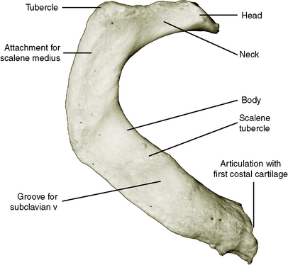
The ribs are attached to corresponding thoracic vertebrae posteriorly. They articulate at the costovertebral joints at the head of the rib and at the costotransverse joints with the tubercle. Anteriorly, most are attached directly to the sternum. They articulate at the costochondral joints with some exceptions.
Where is the internal intercostal muscle located?
The internal intercostal muscle extends inferoposteriorly from the costal groove to the superior border of the rib below
Which muscle extends inferoposteriorly from the costal groove to the superior border of the rib?
Each costal groove accommodates an intercostal vein, artery and nerve in that order downwards. The internal intercostal muscle extends inferoposteriorly from the costal groove to the superior border of the rib below.
How many pairs of ribs are there?
There are 12 pairs of ribs which are separated by intercostal spaces. The first seven ribs progressively increase in length, the lower five ribs then begin to decrease in length. Ribs are highly vascular and trabecular with a thin outer layer of compact bone.
What are the atypical ribs?
The 1 st, 11 th and 12 th ribs are considered atypical ribs due to their anatomical features. The remaining ribs are typical. Ribs can also be divided into true, false and floating ribs: true ribs: the first seven pairs of ribs are true ribs as they are attached to the sternum directly by costal cartilages anteriorly.
How many pairs of ribs are there in the human body?
They may occasionally arise from the seventh cervical ( cervical ribs) and first lumbar ( lumbar ribs ) vertebrae. Up to 5-8% people have 11 pairs of ribs as a normal anatomic variant 6. Much rarer are supernumerary ribs in normals, although it has been documented 6. rib fracture. buckle rib fracture.
What percentage of the population has a floating rib?
In 35-70% of the population, the 10 th rib may also be floating. Additional variation is associated with ethnicity. Bifid or forked ribs are an uncommon variant occurring in ~0.2% of the population. They may be more prevalent in females and the right side. Ribs do not always arise from thoracic vertebrae.
How many ribs should you see for inspiratory effect?
inspiration: count the posterior ribs. You should see 10 to 11 ribs with a good inspiratory effect.
How many posterior ribs are above the diaphragm?
more than 6 anterior or 10 posterior ribs above the diaphragm level on the midclavicular line.
How many anterior ribs are there on a radiograph?
To check for an adequate degree of inspiration, count the anterior ribs on the right. In a good radiograph, 6 anterior ribs should be visible above the right hemidiaphragm. The radiograph used to demonstrate proper alignment also shows evidence of a good degree of inspiration (see Figure 1).
How many ribs do you have?
Most people have 24 ribs, with 12 on each side of the body. The ribs and rib cage are excellent examples of the human body’s multi-faceted and multi-functional design. They are strong enough to support the skeleton and protect the vital organs in the chest cavity, including the heart, lungs, and spleen.
What bone is the rib?
Each rib is a curved, flattened bone that contributes to the wall of the thorax. The ribs articulate posteriorly with the T1–T12 thoracic vertebrae, and most attach anteriorly via their costal cartilages to the sternum. There are 12 pairs of ribs.
Where should rib 10 be exposed?
The exposure should be made at full inspiration and should show rib 10 posteriorly above the diaphragm and rib 6 anteriorly. Both costophrenic angles and the lower parts of the diaphragm should be visible.
What does it mean when a film is taken without full inspiration?
Films taken without a full inspiration are described as having a “ poor inspiratory result ”. This may result from a poor inspiratory effort or any other condition that prevents full inspiration. This patient’s chest x-ray is normal in full inspiration.
How does smoking affect emphysema?from kenhub.com
Patients without the inherited condition usually acquire emphysema from smoking, as the chemicals in cigarette smoke lead to loss of alpha-1 antitrypsin in the lungs. When present and functional, alpha-1 antitrypsin inhibits another enzyme in the lungs called elastase, which breaks down the elastic fibers. When alpha-1 antitrypsin is not present, elastase can go unchecked and continuously break down elastin. Breakdown of elastin results in hyper-compliance of the lung tissue: the alveoli fill with air easily, but they lose their ability to contract back to their original shape. This leads to the alveoli becoming ballooned out, trapping air in the lungs. As you might expect, this air trapping leads to hyperinflation of the lungs, typically giving the patient a “ barrel-chest ” appearance.
Why does my chest look dark on an X-ray?from kenhub.com
On chest X-ray, this widened chest may be observed, as well as flattening of the diaphragm due to the hyperinflated lungs pushing down on it. Because the alveoli are super-filled with air, the patient’s lungs will typically appear darker than normal on chest X-ray (because, like we said, air appears darker than tissue).
What is the purpose of chest X-rays?from healthline.com
Doctors sometimes use chest X-rays to monitor your progress after surgery to the chest area. Doctors can check to see that any implanted materials are in the right place, and they can make sure you’re not experiencing any air leaks or fluid buildup.
Why do doctors do chest xrays?from healthline.com
Chest X-rays can also determine if you have fluid in your lungs, or fluid or air surrounding your lungs. Your doctor could order a chest X-ray for a variety of reasons, including to assess injuries resulting ...
What happens if a patient is rotated more to the right?from kenhub.com
If a patient is rotated more to the right, the distance between the medial margin of the right clavicle and the spinous process will be greater than the distance between the medial margin of the left clavicle and the spinous process.
What does it mean when a chest X-ray shows a pleural effusion?from kenhub.com
In evaluating a chest X-ray, it is also vital to check the costophrenic angles at the peripheral edges of the diaphragm, as blunting of the costophrenic angles (such that they are no longer appear sharp and their borders no longer appear distinct, but rather “grayed-out”) could signify the presence of a pleural effusion (a fluid collection between the pleura and lung tissue). Pleural effusions may not always be obvious, so it is always important to look attentively. A small posterior effusion may be seen more easily via a lateral film rather than a PA film.
What is the first thing you think of when you picture a chest X-ray?from kenhub.com
When you picture a chest X-ray, what is the first thing you think of? If you thought “lungs,” that’s a pretty solid response, because needing to evaluate the lungs is one of the biggest reasons to order a chest X-ray. Air on an X-ray looks dark, so when the lungs are clear and healthy, that’s exactly how they should look: not quite black, because there is still tissue there, but still quite dark.
What are the densest structures on a chest X-ray?
Bones are the densest structures visible on a normal chest X-ray. Despite this it is easy to overlook important abnormalities of the bones which may be very subtle.
Why is the sternum included in a frontal view?
The sternum is also included on a frontal view but it overlies other midline structures and so is obscured. The bones are used as useful markers of chest radiograph quality. They are used to assess patient rotation, adequacy of inspiration and X-ray penetration.
What is the superior edge of a rib used for?
Note: To avoid damaging the subcostal nerves or vessels the superior edge of a rib is used as the landmark during procedures such as chest drain insertion. The spine can be seen through the heart indicating adequate X-ray penetration.
How many ribs are there in the lung?
Less than 5 ribs indicates incomplete inspiration. More than 7 ribs suggests lung hyper-expansion.
Which structures are landmarks for rotation?
The spinous processes of the vertebrae (posterior structures) and the medial ends of the clavicles (anterior structures) are landmarks to assess rotation. The ribs should be checked on every chest X-ray. The right 5th rib is highlighted (roll-over image)
Can you see a fractured rib on a chest xray?
Acute rib fractures are often invisible, therefore chest X-rays are not helpful if there is clinical suspicion of a rib injury unless a complication such as pneumothorax is suspected. Occasionally you will see an important abnormality of the bones on a chest X-ray such as bone metastases.
Where is the subcostal groove on a rib X-ray?
The subcostal grooves are visible on the underside of the ribs ( red highlights) These grooves contain the subcostal nerves and vessels that accompany each rib.
What is ribs PA?
The ribs PA view is a specific projection employed in the assessment of the anterior ribs. Unlike a standard chest radiograph, this projection applies a lower kV higher mAs technique to highlight bony structures.
Why is the chin raised?
the chin is raised as to be out of the image field. shoulders are rotated anteriorly to allow the scapulae to move laterally off the lung fields, and this can be achieved by either: hands placed on the posterior aspect of the hips, elbows partially flexed rolling anterior or.
Where are hands placed around the image receptor?
hands are placed around the image receptor in a hugging motion with a focus on the lateral movement of the scapulae
When should the rib cage be visible?
The entire rib cage should be visible from the 1st to the 12th.
Is the rib series unnecessary?
The rib series is often considered to be an unnecessary, unjustified projection in many radiology departments.
Is rib trauma a referral?
The isolated referral of a rib series in the assessment of rib trauma is controversial 1 however, still widely used. Ensure the projection is not performed under a chest examination AEC setting, this will not achieve the correct exposure to adequately present the bony structures of the thoracic cage.
Is the clavicle horizontal or vertical?
the clavicle is in the same horizontal plane. a minimum of ten posterior ribs is visualized above the diaphragm. the ribs and thoracic cage are seen only faintly over the heart. clear vascular markings of the lungs should be visible.
How to view ribs on X-ray?
In this case, the midclavicular line of the diagnosed side should be located along the median longitudinal line of the couch. The upper limb is extended along the body, the legs are bent at the knees. On the frontal plane, the body should be parallel to the plane of the couch. This position allows you to clearly see the lower ribs, especially against the background of intense darkening of the liver. [ 5]
How to obtain an anterior oblique image?
To obtain an anterior oblique image, which is necessary to study the state of the anterolateral costal regions, a person is placed on the abdomen. The diagnosed half of the breast should fit snugly against the surface of the couch, and the opposite half should be slightly raised. The frontal plane of the body should intersect with the plane of the couch at an angle of 40-45 degrees. The upper limb from the side of the study is extended along the body, adjoining the back surface to the couch. The other arm is bent at the elbow, the palm rests on the table. The criterion for adequate placement is to obtain a clear image of the anterolateral parts of the ribs. [ 6]
Why do doctors do X-rays of the ribs?
Most often, the procedure is prescribed when a rib fracture is suspected. If multiple trauma is detected, the doctor may insist on performing a plain X-ray, which is necessary to obtain more objective and complete information about the damage. Plain X-ray shows the existing damage to the internal organs and the whole chest.
What should a patient inform the radiologist before starting the study?
Before starting the study, the patient should inform the radiologist about the pathologies suffered, surgical interventions on the chest organs, about the presence of foreign objects, implants in the area under study. Women must be sure to report pregnancy.
What is the thoracic skeletal frame?
The thoracic skeletal frame is a reliable protection for the internal organs. An X-ray of the ribs is, in fact, the same X-ray of the chest, during which you can see not only bone structures, but also the heart, lungs, respiratory tract, and the spinal column. During the examination, the doctor may see damage or violation of the shape of the bones, or the development of any pathological process.
What to do before X-ray?
Immediately before the X-ray, the patient is asked to take off his outer clothing, undress to the waist. If there are any jewelry in the neck or chest area, then they must be removed. If a person has long hair, then they need to be gathered up: they should not fall into the image area.
How to eliminate radioactive substances from the body?
The most important thing for accelerating the elimination of radioactive substances from the body is adherence to the drinking regime with the use of at least 2 liters of water per day. In addition to water, it is allowed to drink freshly squeezed juices, fruit drinks, green tea. It is allowed to drink a little red dry wine. Grapes, pomegranates, nuts, and dairy products also have anti-radiation properties. Prunes, flaxseeds, nettle leaves are useful. It is advisable to include sour cream, cottage cheese, carrots, beets, buckwheat, seafood in the diet. But you should avoid products with preservatives, dyes, flavors and flavorings, as well as smoked meats and semi-finished products.