How many peduncles does the cerebellum have?
Cerebellar peduncle. There are six cerebellar peduncles in total, three on each side: Superior cerebellar peduncle is a paired structure of white matter that connects the cerebellum to the midbrain. Middle cerebellar peduncles connect the cerebellum to the pons and are composed entirely of centripetal fibers.
How is the cerebellum connected to the brainstem?
The cerebellum is connected to the brainstem by three pairs of cerebellar peduncles: the superior peduncle with the midbrain, the middle peduncle with the pons, and the inferior peduncle with the medulla oblongata. Afferent and efferent connections that run between the cerebellum, brainstem and spinal cord, travel through the cerebellar peduncles.
What is the function of the superior cerebellar peduncle?
Superior cerebellar peduncle is a paired structure of white matter that connects the cerebellum to the midbrain.
How is the middle cerebellar peduncle formed?
Later, projections of the axons of the corticopontine and the pontocerebellar fibers will develop the middle cerebellar peduncle and connect the cerebral cortex with the cerebellum. The inferior cerebellar peduncle will form mainly by the growth of sensory axons from the spinal cord, the olivary and vestibular nuclei. [9]
How many pairs of peduncles connect the cerebellum to the brain stem?
three pairsThe cerebellum is connected to the rest of the nervous system by three pairs of peduncles, or feet. The cerebellar peduncles anchor the cerebellum to the brainstem. All afferent and efferent fibers of the cerebellum pass through the three peduncles and the pons to the other levels of the nervous system.
How many peduncle does cerebellum have?
three cerebellar pedunclesCerebellar peduncles are the structure connecting the cerebellum to the brain stem and the cerebrum. There exist three cerebellar peduncles.
What are the 3 cerebellar peduncles?
There are three on each side: the inferior cerebellar peduncle (#4025, #6172), the middle cerebellar peduncle (brachium pontis) (#8361, #6553), and the superior cerebellar peduncle (brachium conjunctivum) (#6554). The middle and inferior cerebellar peduncles contain most of the cerebellar afferents.
What structures connect the cerebellum to brainstem?
The cerebellum is connected to the brainstem by three pairs of cerebellar peduncles: the superior peduncle with the midbrain, the middle peduncle with the pons, and the inferior peduncle with the medulla oblongata.
What are the 3 cerebellar peduncles and state their functions?
These peduncles are superior, middle and inferior. Superior cerebellum connects cerebellum to the midbrain, middle to the pons and inferior to the medulla oblongata. Superior cerebellar peduncle (brachium conjunctivum) ascends upward from the anterior cerebellar notch to the tectum of the midbrain.
What are the 3 parts of the cerebellum?
There are three functional areas of the cerebellum – the cerebrocerebellum, the spinocerebellum and the vestibulocerebellum. Cerebrocerebellum – the largest division, formed by the lateral hemispheres. It is involved in planning movements and motor learning.
Where in the brainstem the cerebellar peduncles are connected?
Cerebellar peduncles connect the cerebellum to the brain stem. There are six cerebellar peduncles in total, three on each side: Superior cerebellar peduncle is a paired structure of white matter that connects the cerebellum to the mid-brain....Cerebellar peduncle.Cerebellar penduncleTA25845FMA77791Anatomical terms of neuroanatomy7 more rows
How many cerebral peduncle are there?
two cerebral pedunclesThere are two cerebral peduncles, one on each side of the brainstem. Definitions of what is included in the cerebral peduncles vary slightly, but a common definition is that they consist of the entire portion of the midbrain that lies anterior to the cerebral aqueduct.
Which part of the brainstem has most of the cerebellar peduncles?
Anatomy. The cerebellar peduncles are three pairs of nerve tracts that connect the cerebellum to the brain stem. They all arise from the left and right sides of the brain stem at the level of the pons and enter the anterior cerebellum to form the roots of the arbor vitae, or cerebellar white matter.
What connects the cerebrum to the brainstem?
Pons. Pons and medulla, anterior. An important pathway for tracts that run from the cerebrum down to the medulla and spinal cord, as well as for tracts that travel up into the brain. It also forms important connections with the cerebellum via fibre bundles known as the cerebellar peduncles.
What connects the cerebellum to the brainstem quizlet?
- the three cerebellar peduncles connect the cerebellum to the brainstem.
Which of the cerebellar peduncles connects the cerebellum to the midbrain?
Rostral cerebellar peduncle (brachium conjunctivum) connects the cerebellum to the midbrain and forebrain. Efferent fibers in the peduncle decussate immediately in the midbrain and thus they affect contralateral motor centers.
How many cerebral peduncle are there?
two cerebral pedunclesThere are two cerebral peduncles, one on each side of the brainstem. Definitions of what is included in the cerebral peduncles vary slightly, but a common definition is that they consist of the entire portion of the midbrain that lies anterior to the cerebral aqueduct.
What are the cerebellar peduncles named for?
The peduncles are named for the directions they take in connecting to the brainstem (synonyms are shown below parenthetically). Caudal cerebellar peduncle (restiform body) connects the cerebellum to the medulla oblongata.
Where are peduncles located?
midbrainThe cerebral peduncles are located on either side of the midbrain and are the frontmost part of the midbrain, and act as the connectors between the rest of the midbrain and the thalamic nuclei and thus the cerebrum.
Why is it called a peduncle?
Peduncle comes from ped (Latin for foot) plus -uncle (an Old French diminutive ending) so it literally means tiny foot.
Where do the superior peduncles enter the brain?
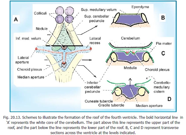
The superior cerebellar peduncles (brachia conjunctiva) emerge from the cerebellum and ascend to form the lateral portion of the roof of the fourth ventricle, where they enter the brainstem below the inferior colliculi. They are bridged by the superior medullary velum. The superior cerebellar peduncles represent the main output route from the cerebellum, and as such, most of their fibers are efferent. A relatively small afferent contribution is present. The efferent pathways include the cerebellorubral, dentatothalamic, and fastigioreticular tracts. All of them emerge from cerebellar nuclei; the cerebellorubral fibers from the globose and emboliform nuclei, the dentatothalamic fibers from the dentate nucleus, and the fastigioreticular fibers from the fastigial nucleus. They emerge together from the various nuclei to ascend in the roof of the fourth ventricle and proceed anteriorly to the midbrain tegmental area medial to the lateral lemniscus. The cerebellorubral fibers cross over at this point to enter the contralateral red nucleus. The dentatothalamic fibers also cross over and ascend to synapse in the ventral intermediate (VI) and ventral anterior (VA) nuclei of the thalamus. The fastigioreticular fibers enter the reticular formation of the midbrain, pons, and medulla oblongata. Afferent pathways include the anterior spinocerebellar and tectocerebellar tracts. The fibers of the anterior spinocerebellar tract originate in Clarke's column of the spinal cord and cross in the anterior white commissure to the lateral funiculus, where they ascend to upper pontine levels before crossing back to enter the cerebellum through the superior peduncle. They terminate in the hind limb region of the cerebellar cortex. The tectocerebellar tracts emerge from the superior and inferior colliculi on both sides, terminating in the intermediate vermis (culmen, declive, folium, tuber, pyramid) and the lobulus simplex. The function of the tectocerebellar tract is not known, but it is widely believed to mediate visual and auditory reflexes.
Where do the fibers of the anterior spinocerebellar tract originate?
The fibers of the anterior spinocerebellar tract originate in Clarke's column of the spinal cord and cross in the anterior white commissure to the lateral funiculus, where they ascend to upper pontine levels before crossing back to enter the cerebellum through the superior peduncle.
What is the middle peduncle?
Middle cerebellar peduncles connect the cerebellum to the pons and are composed entirely of centripetal fibers. Inferior cerebellar peduncle is a thick rope-like strand that occupies the upper part of the posterior district of the medulla oblongata.
Where do tectocerebellar tracts end?
They terminate in the hind limb region of the cerebellar cortex. The tectocerebellar tracts emerge from the superior and inferior colliculi on both sides, terminating in the intermediate vermis (culmen, declive, folium, tuber, pyramid) and the lobulus simplex.
Which part of the thalamus is the dentatothalamic fiber?
The dentatothalamic fibers also cross over and ascend to synapse in the ventral intermediate (VI) and ventral anterior (VA) nuclei of the thalamus. The fastigioreticular fibers enter the reticular formation of the midbrain, pons, and medulla oblongata.
Which ventricle has a diamond?
The peduncles form the lateral border of the fourth ventricle, and form a distinctive diamond – the middle peduncle forming the central corners of the diamond, while the superior and inferior peduncles form the superior and inferior edges, respectively.
How does the cerebellum develop?
The cerebellum develops from the hindbrain vesicle that gives rise to the posterior part of the alar plates of the metencephalon. The cerebellar hemisphere and vermis form by the 12th week. Accordion-like folds gradually start developing from about the fourth month. Neurons of cerebellar cortex form by the neuroblast derived from the matrix cells in the ventricular zone. Other neuroblasts from the ventricular surface differentiate into cerebellar nuclei, which axons grow towards the mesencephalon (midbrain) and create the superior cerebellar peduncle. Later, projections of the axons of the corticopontine and the pontocerebellar fibers will develop the middle cerebellar peduncle and connect the cerebral cortex with the cerebellum. The inferior cerebellar peduncle will form mainly by the growth of sensory axons from the spinal cord, the olivary and vestibular nuclei. [9]
Where is the cerebellum located?
The cerebellum, which is the largest part of the hindbrain, is located in the posterior cranial fossa, behind the fourth ventricle, the pons, and the medulla oblongata. Tentorium cerebelli, an extension of the dura matter, separates the cerebellum from the cerebrum. It is composed of two hemispheres joined by the vermis ...
What is the role of the cerebellum in the brain?
The cerebellum is a vital component in the human brain as it plays a role in motor movement regulation and balance control. The cerebellum coordinates gait and maintains posture, controls muscle tone and voluntary muscle activity but is unable to initiate muscle contraction.
Which layer of the brain contains 80% of the brain's neurons?
The cerebellum is neuron-rich, containing 80% of the brain’s neurons organized in a dense cellular layer. [1][4] The cerebellar cortex is a sheet-like structure, made of a single sheet less than 1mm thick, and accordion-like folds fused at the midline (Essen 2018). Each fold is composed of an inner white matter core that is covered by gray matter. ...
What are the layers of the cortex?
The gray matter of the cortex divides into three layers: an external - the molecular layer; a middle - the Purkinje cell layer; and an internal - the granular layer. The molecular layer contains two types of neurons: the outer stellate cell and the inner basket cell. [4][5]
What is the function of the cerebellum?
The cerebellum coordinates gait and maintains posture, controls muscle tone and voluntary muscle activity but is unable to initiate muscle contraction. Damage to this area in humans results in a loss in the ability to control fine movements, maintain posture, and motor learning. [1][2][3] Structure and Function.
Which nuclei are fastigial?
From medial to lateral, the deep nuclei are the fastigial, interposed (consisting of globose and emboliform nuclei), and dentate nuclei, which is the largest nuclei.[1] Fibers from the dentate, emboliform, and globose nuclei leave the cerebellum through the superior cerebellar peduncle.