What are the functions of microtubules?
What are three functions of microtubules?
- Giving shape to cells and cellular membranes.
- Cell movement, which includes a contraction in muscle cells and more.
- Transportation of specific organelles within the cell via microtubule “roadways” or “conveyor belts.”
What is the function of microtubules in a cell?
- Giving shape to cells and cellular membranes.
- Cell movement, which includes contraction in muscle cells and more.
- Transportation of specific organelles within the cell via microtubule "roadways" or "conveyor belts."
- Mitosis and meiosis: movement of chromosomes during cell division and creation of the mitotic spindle.
What are microtubules associated with?
Microtubules (MTs) play a fundamental role in many vital processes such as cell division and neuronal activity. They are key structural and functional elements in axons, supporting neurite differentiation and growth, as well as transporting motor proteins along the axons, which use MTs as support tracks. Tau is a stabilizing MT associated protein, whose functions are mainly regulated by ...
What do microtubules do?
The four functions of microtubules are:
- Providing structure and support for the cell
- Creating highways for intracellular transport
- Separating chromosomes during cell division
- Allowing for cellular movement through cilia and flagella
What type of cell is microtubule?
eukaryotic cellsMicrotubules are major components of the cytoskeleton. They are found in all eukaryotic cells, and they are involved in mitosis, cell motility, intracellular transport, and maintenance of cell shape. Microtubules are composed of alpha- and beta-tubulin subunits assembled into linear protofilaments.
What organelle makes microtubules?
The centrosomeThe centrosome serves as the initiation site for the assembly of microtubules, which grow outward from the centrosome toward the periphery of the cell.
What is the organelle?
An organelle is a subcellular structure that has one or more specific jobs to perform in the cell, much like an organ does in the body. Among the more important cell organelles are the nuclei, which store genetic information; mitochondria, which produce chemical energy; and ribosomes, which assemble proteins.
What are microtubules?
Microtubules, together with microfilaments and intermediate filaments, form the cell cytoskeleton. The microtubule network is recognized for its role in regulating cell growth and movement as well as key signaling events, which modulate fundamental cellular processes.
What are organelles composed of?
All the cellular organelles are made of macromolecules like carbohydrates, Lipids, Proteins, and Nucleic acids (DNA, RNA). Atoms - To make macromolecules involves even smaller building blocks. You may have heard of atoms before and their parts: neutrons, protons, and electrons.
Is cytoskeleton an organelle?
In this section we will discuss the intracellular components that are not organelles. The cytoskeleton and cytosol are structural elements that help provide the cell with its structure.
What are the 12 organelles in a cell?
Within the cytoplasm, the major organelles and cellular structures include: (1) nucleolus (2) nucleus (3) ribosome (4) vesicle (5) rough endoplasmic reticulum (6) Golgi apparatus (7) cytoskeleton (8) smooth endoplasmic reticulum (9) mitochondria (10) vacuole (11) cytosol (12) lysosome (13) centriole.
What are the 8 types of organelles?
Membranous organellesEndoplasmic reticulum. The endoplasmic reticulum (ER) is a large network of membranes responsible for the production of proteins, metabolism and transportation of lipids, and detoxification of poisons. ... Golgi apparatus. ... Mitochondria. ... Peroxisomes. ... Lysosomes. ... Transport vesicles.
What are not organelles?
In this sense, ribosomes and nucleosomes are not regarded as organelles because they are not bounded by membranes. In the same way, lysosomes and vacuoles, would not qualify as organelle because they are single-membrane bounded cytoplasmic structures.
What is a microtubules and its function?
Microtubules, with intermediate filaments and microfilaments, are the components of the cell skeleton which determinates the shape of a cell. Microtubules are involved in different functions including the assembly of mitotic spindle, in dividing cells, or axon extension, in neurons.
Where are microtubules in the cell?
In cells, the minus ends of microtubules are anchored in structures called microtubule organizing centers (MTOCs). The primary MTOC in a cell is called the centrosome, and it is usually located adjacent to the nucleus. Microtubules tend to grow out from the centrosome to the plasma membrane.
What is a microtubule in an animal cell?
Microtubules are polarized cytoskeletal filaments that serve as tracks for intracellular transport and form a scaffold that positions organelles and other cellular components and modulates cell shape and mechanics.
Where are microtubules formed?
Microtubule nuclear in the mitotic spindle Most of the microtubules that form the mitotic spindle originate from the centrosome.
Where do the microtubules originate?
Microtubules originate from the Golgi with an initial growth preference towards the axon. Their growing plus ends also turn towards and into the axon, adding to the plus-end-out microtubule pool. Any plus ends that reach a dendrite, however, do not readily enter, maintaining minus-end-out polarity.
What produces microtubules in animal cells?
In animal cells, microtubules radiate outwards from an organelle in the center of the cell called a centrosome, which is a microtubule organizing center (MTOC). The cells of plants and fungi do not have centrosomes, and instead the nuclear envelope—the membrane surrounding the cell's nucleus—is an MTOC.
Which organelle produces the microtubules used during cell division?
Definition. Centrioles are paired barrel-shaped organelles located in the cytoplasm of animal cells near the nuclear envelope. Centrioles play a role in organizing microtubules that serve as the cell's skeletal system.
What is a microtubule?
Jump to navigation Jump to search. Polymer of tubulin that forms part of the cytoskeleton. Microtubule and tubulin metrics. Microtubules are polymers of tubulin that form part of the cytoskeleton and provide structure and shape to eukaryotic cells.
How are microtubules formed?
They are formed by the polymerization of a dimer of two globular proteins, alpha and beta tubulin into protofilaments that can then associate laterally to form a hollow tube, the microtubule. The most common form of a microtubule consists of 13 protofilaments in the tubular arrangement.
What is the inner space of a microtubule?
The inner space of the hollow microtubule cylinders is referred to as the lumen . The α and β-tubulin subunits are identical at the amino acid level, and each have a molecular weight of approximately 50 kDa.
What is the role of microtubules in eukaryotic cells?
Microtubules are one of the cytoskeletal filament systems in eukaryotic cells. The microtubule cytoskeleton is involved in the transport of material within cells, carried out by motor proteins that move on the surface of the microtubule. Microtubules are very important in a number of cellular processes.
How to study motor proteins in vitro?
Microtubule in vitro assays for motor proteins such as dynein and kinesin are researched by fluorescently tagging a microtubule and fixing either the microtubule or motor proteins to a microscope slide then visualizing the slide with video-enhanced microscopy to record the travel of the microtubule motor proteins. This allows the movement of the motor proteins along the microtubule or the microtubule moving across the motor proteins. Consequently, some microtubule processes can be determined by kymograph.
How many protofilaments are there in a microtubule?
Typically, microtubules are formed by the parallel association of thirteen protofilaments, although microtubules composed of fewer or more protofilaments have been observed in various species as well as in vitro.
Where do microtubules form in mitosis?
Most of the microtubules that form the mitotic spindle originate from the centrosome. Originally it was thought that all of these microtubules originated from the centrosome via a method called search and capture, described in more detail in a section above, however new research has shown that there are addition means of microtubule nucleation during mitosis. One of the most important of these additional means of microtubule nucleation is the RAN-GTP pathway. RAN-GTP associates with chromatin during mitosis to create a gradient that allows for local nucleation of microtubules near the chromosomes. Furthermore, a second pathway known as the augmin/HAUS complex (some organisms use the more studied augmin complex, while others such as humans use an analogous complex called HAUS) acts an additional means of microtubule nucleation in the mitotic spindle.
Where are microtubules found?
Microtubules are arranged in the form of microtubule-organizing centres. They are structures found in eukaryotic cells. During the interphase, most of the animal cells consist of microtubule-organizing centres. Several proteins are bound to microtubules namely dynein and kinesin.
What are microtubules made of?
What are Microtubules? “Microtubules are microscopic, hollow tubes made of alpha and beta tubulin that are a part of the cell’s cytoskeleton.”.
What is the function of microtubules in the cytoplasm?
Intracellular Organization of Microtubules. In the cytoplasm, microtubules form a structural network. The function of the cytoskeleton in microtubule includes chromosomes segregation, transport, mobility and mechanical support.
What are the structures that give cilia and flagella?
Microtubules give structures to cilia and flagella. They also facilitate the contraction and expansion of the cell helping them to move from one place to another.
How thick are cytoskeletons?
They are the largest structures in the cytoskeleton and are about 24 nm thick. They facilitate cell movement, cell division, and transportation of materials within the cells. They are also involved in the division of chromosomes during the process of mitosis and in locomotion.
Which axis of the cell is responsible for transporting organelles, vesicles, and proteins?
So that it would be easy to facilitate the transportation of organelles, vesicles, and proteins along the apical-basal axis of the cell. They play a vital role in cell migration as well. Also Read: Cytoskeleton.
Is tubulin a heterodimer?
This, tubulin is a heterodimer. Microtubules play a vital role in all eukaryotic cells. These cells release protein tubulin in a normal manner that involves transcription of the gene coding for tubulin, which yields RNA and is followed by transcription of mRNA to produce proteins.
Where are microtubules located in animal cells?
In animal cells, the major microtubule-organizing centeris the centrosome, which is located adjacent to the nucleusnear the center of interphase(nondividing) cells (Figure 11.39). During mitosis, microtubules similarly extend outward from duplicated centrosomes to form the mitotic spindle, which is responsible for the separation and distribution of chromosomesto daughter cells. The centrosomethus plays a key role in determining the intracellular organization of microtubules, although most details of its function remain a mystery.
What is the name of the protein that makes up microtubules?
In contrast to intermediate filaments, which are composed of a variety of different fibrous proteins, microtubules are composed of a single type of globular protein, called tubulin. Tubulin is a dimer consisting of two closely related 55-kd polypeptides, α-tubulinand β-tubulin. Like actin, both α- and β-tubulin are encoded by small families of related genes. In addition, a third type of tubulin (γ-tubulin) is specifically localized to the centrosome, where it plays a critical role in initiating microtubuleassembly (discussed shortly).
How do centrosomes move during mitosis?
The two centrosomes then separate and move to opposite sides of the nucleus, forming the two poles of the mitotic spindle. As the cell enters mitosis, the dynamics of microtubuleassembly and disassembly also change dramatically. First, the rate of microtubule disassembly increases about tenfold, resulting in overall depolymerization and shrinkage of microtubules. At the same time, the number of microtubules emanating from the centrosome increases by five- to tenfold. In combination, these changes result in disassembly of the interphase microtubules and the outgrowth of large numbers of short microtubules from the centrosomes.
What are centrioles in animal cells?
The centrosomes of most animal cells contain a pair of centrioles, oriented perpendicular to each other, surrounded by amorphous pericentriolar material(Figure 11.41). The centrioles are cylindrical structures consisting of nine triplets of microtubules, similar to the basal bodies of cilia and flagella (discussed later in the chapter). Although centrioles are probably the precursors of basal bodies, they appear to be dispensible for the function of the centrosome. Centrioles do not appear to be required for the assembly or organization of microtubules, and they are not found in plant cells, many unicellular eukaryotes, and some animal cells (such as mouse eggs). The microtubules that emanate from the centrosome terminate in the pericentriolar material, not the centrioles, and it is the pericentriolar material that initiates microtubuleassembly.
How many protofilaments are in a tubulin dimer?
Tubulin dimers polymerize to form microtubules, which generally consist of 13 linear protofilaments assembled around a hollow core (Figure 11.37). The protofilaments, which are composed of head-to-tail arrays of tubulindimers, are arranged in parallel. Consequently, microtubules (like actinfilaments) are polar structures with two distinct ends: a fast-growing plus end and a slow-growing minus end. This polarity is an important consideration in determining the direction of movement along microtubules, just as the polarity of actin filaments defines the direction of myosinmovement.
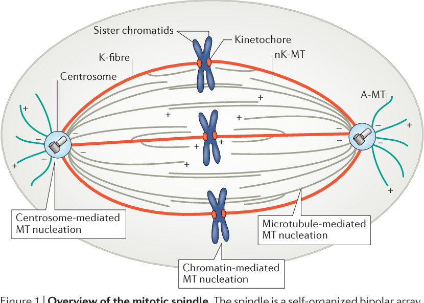
What is the function of the mitotic spindle?
As first proposed by Marc Kirschner and Tim Mitchison in 1986, formation of the mitotic spindleinvolves the selective stabilization of some of the microtubules radiating from the centrosomes. These microtubules are of three types, two of which make up the mitotic spindle. Kinetochore microtubulesattach to the condensed chromosomesof mitotic cells at their centromeres, which are associated with specific proteinsto form the kinetochore(see Figure 4.16). Attachment to the kinetochore stabilizes these microtubules, which, as discussed below, play a critical role in separation of the mitotic chromosomes. The second type of microtubules found in the mitotic spindle (polar microtubules) are not attached to chromosomes. Instead, the polar microtubulesemanating from the two centrosomes are stabilized by overlapping with each other in the center of the cell. Astral microtubulesextend outward from the centrosomes to the cell periphery and have freely exposed plus ends. As discussed later, both the polar and astral microtubulesalso contribute to chromosome movement by pushing the spindle poles apart.
How are microtubules visualized?
Growth of microtubules from the centrosome. Microtubules in mouse fibroblasts are visualized by immunofluorescence microscopy using an antibody against tubulin. (A) The distribution of microtubules in a normal interphase cell. (B) This cell was treated (more...)
What are non-membrane organelles?
Most non-membranous organelles are part of the cytoskeleton, the major support structure of the cell. These include: filaments, microtubules , and centrioles.
What are the functions of organelles?
Organelles are small structures within the cytoplasm that carry out functions necessary to maintain homeostasis in the cell. They are involved in many processes, for example energy production, building proteins and secretions, destroying toxins, and responding to external signals. Organelles are considered either membranous or non-membranous.
What is the function of the endoplasmic reticulum?
The endoplasmic reticulum (ER) is a large network of membranes responsible for the production of proteins, metabolism and transportation of lipids, and detoxification of poisons. There are two types of endoplasmic reticulum with separate functions: smooth endoplasmic reticulum and rough endoplasmic reticulum. The presence or absence of ribosomes in the ER’s plasma membrane determines whether it is classified as smooth or rough ER.
What is the function of transport vesicles?
Transport vesicles are used to move proteins around the cell and to release neurotransmitters into the synaptic space.
What is the function of peroxisomes?
Peroxisomes are single membrane compartments that contain enzym es used to remove hydrogen atoms from substrates. The free hydrogen atoms then bind to oxygen and create hydrogen peroxide.
What is the smallest unit of life?
Cells are the smallest units of life. They are a closed system, can self-replicate, and are the building blocks of our bodies. In order to understand how these tiny organisms work, we will look at a cell’s internal structures. We will focus on eukaryotic cells, cells that contain a nucleus.
What is the function of mitochondria?
Cell (mitochondria in green) Mitochondria are the powerhouses of the cell. Cellular respiration, the generation of energy from sugars and fats, occurs in these organelles. Some of the enzymes that catalyze respiration are found within the matrix. Other proteins involved in these reactions are built into the wall of the inner membrane.
What is the process of a cell that surrounds a large particle with pseudopodia and then engul?
exocytosis and it requires expenditure of ATP. When a cell surrounds a large particle with pseudopodia and then engulfs it, the process is called. phagocytosis. Pinocytosis is the process in which the cell. internalizes ("drinks") a droplet of interstitial fluid.
Which transporter moves two substances across the membrane in the same direction?
Symport active transporters move two substances across the membrane in the same direction.
Which process requires expenditure of ATP?
exocytosis and it requires expenditure of ATP.
What is the function of the selective permeability of the plasma membrane?
The selective permeability of the plasma membrane allows all substances to pass through the membrane, ensuring that the inside of the cell is in constant interaction with its external environment.
What is the role of b. in microtubule assembly?
b. serves as a site for both microtubule and intermediate filament assembly.
Which end of a microtubule can undergo dynamic instability independently?
d. Each end of a microtubule can undergo dynamic instability independently.
Which heterodimer is arranged with mixed orientation in each protofilament?
a. α-β-tubulin heterodimers are arranged with mixed orientation in each protofilament, and protofilaments are arranged with mixed polarity in the microtubule wall.

Overview
Functions
Microtubule plus ends are often localized to particular structures. In polarized interphase cells, microtubules are disproportionately oriented from the MTOC toward the site of polarity, such as the leading edge of migrating fibroblasts. This configuration is thought to help deliver microtubule-bound vesicles from the Golgi to the site of polarity.
Dynamic instability of microtubules is also required for the migration of most mammalian cells t…
History
Tubulin and microtubule-mediated processes, like cell locomotion, were seen by early microscopists, like Leeuwenhoek (1677). However, the fibrous nature of flagella and other structures were discovered two centuries later, with improved light microscopes, and confirmed in the 20th century with the electron microscope and biochemical studies.
In vitro assays for microtubule motor proteins such as dynein and kinesin are researched by fluores…
Structure
In eukaryotes, microtubules are long, hollow cylinders made up of polymerised α- and β-tubulin dimers. The inner space of the hollow microtubule cylinders is referred to as the lumen. The α and β-tubulin subunits are ~50% identical at the amino acid level, and both have a molecular weight of approximately 50 kDa.
These α/β-tubulin dimers polymerize end-to-end into linear protofilaments that a…
Intracellular organization
Microtubules are part of the cytoskeleton, a structural network within the cell's cytoplasm. The roles of the microtubule cytoskeleton include mechanical support, organization of the cytoplasm, transport, motility and chromosome segregation. In developing neurons microtubules are known as neurotubules, and they can modulate the dynamics of actin, another component of the cytos…
Microtubule polymerization
Nucleation is the event that initiates the formation of microtubules from the tubulin dimer. Microtubules are typically nucleated and organized by organelles called microtubule-organizing centres (MTOCs). Contained within the MTOC is another type of tubulin, γ-tubulin, which is distinct from the α- and β-subunits of the microtubules themselves. The γ-tubulin combines with several other associated proteins to form a lock washer-like structure known as the "γ-tubulin ring comp…
Microtubule dynamics
Dynamic instability refers to the coexistence of assembly and disassembly at the ends of a microtubule. The microtubule can dynamically switch between growing and shrinking phases in this region. Tubulin dimers can bind two molecules of GTP, one of which can be hydrolyzed subsequent to assembly. During polymerization, the tubulin dimers are in the GTP-bound state. The GTP bound to α-tubulin is stable and it plays a structural function in this bound state. Howe…
Regulation of microtubule dynamics
Although most microtubules have a half-life of 5–10 minutes, certain microtubules can remain stable for hours. These stabilized microtubules accumulate post-translational modifications on their tubulin subunits by the action of microtubule-bound enzymes. However, once the microtubule depolymerizes, most of these modifications are rapidly reversed by soluble en…
What Are Microtubules?

Microtubules Structure
- Microtubules are arranged in the form of microtubule-organizing centres. They are structures found in eukaryotic cells. During the interphase, most of the animal cellsconsist of microtubule-organizing centres. Several proteins are bound to microtubules namely dynein and kinesin. Microtubules are made of subunits called tubulin. Each tubulin is made of an alpha and a beta-tu…
Intracellular Organization of Microtubules
- In the cytoplasm, microtubules form a structural network. The function of the cytoskeleton in microtubule includes chromosomes segregation, transport, mobility and mechanical support. It can either shrink or grow to generate energy which is due to the presence of motor proteins that allow cellular components and others to be carried along with microtubules. The arrangements i…