Is a goblet cell unicellular or multicellular?
yes, A goblet cell is a unicellular gland. it is a single secretory cell such as mucinogen-secreting cells (goblet cell). There is also multicellular glands like in the gastric area or even ur sweat glands.
Where are goblet cells found in the human body?
goblet cells are an example of this the goblet cells are found in the lining of the of the respiratory and digestive tract unicellular these glands have to be divided into 2 major groups multicellular glands
What is the shape of the goblet cells in the intestine?
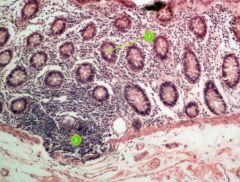
The archetype of such a uni cellular gland is represented by the flask-shaped, mucus-producing and storing intestinal goblet cells shown in the electron micrograph. Mucus-producing single secretory cells exist dispersed in the mucosa of various other organs.
What is the function of goblet cells in bronchitis?
Their role is to protect the surface of epithelium, lubricate it, and catch harmful particles. Although protective, goblet cells may be involved in pathophysiology of certain respiratory diseases, such as chronic bronchitis. This article will discuss goblet cells histology and function.
What type of gland is goblet?
mucous merocrine exocrine glandsGoblet cells are modified epithelial cells that secrete mucus on the surface of mucous membranes of organs, particularly those of the lower digestive tract and airways. Histologically, they are mucous merocrine exocrine glands.
Why goblet cells are unicellular gland?
A goblet cell is known as unicellular gland as because they are made up of single celled secretory cells. It is a glandular simple columnar epithelial cell, whose function is to secrete gel-forming mucins, the major components of mucus.
What are the unicellular gland?
The unicellular glands are scattered single cells, such as goblet cells, found in the mucous membranes of the small and large intestine. The multicellular exocrine glands known as serous glands develop from simple epithelium to form a secretory surface that secretes directly into an inner cavity.
What type of cells have goblet cells?
Goblet cells arise from pluripotent stem cells and derive their name from their goblet, cup-like appearance. The primary function of goblet cells is to secrete mucin and create a protective mucus layer. Goblet cells are also thought to be involved with immunoregulation.
What are multicellular glands?
Multicellular glands are multicelled glands. They are formed by invagination from an epithelial sheet (like the endocrine glands) but will never lose the duct connecting them to the free surface of the epithelial sheet (surface of the skin or lumen of the open body cavities).
What is a goblet cell class 9?
Goblet cell is a mucus - secreting epithelial cell (as of columnar epithelium) that is distended with secretion at the free end.
What are 10 examples of unicellular?
Following are some of the examples of unicellular organisms:Escherichia coli.Diatoms.Protozoa.Protista.Streptococcus.Pneumococci.Dinoflagellates.
What are unicellular cells examples?
Unicellular organisms are organisms consisting of one cell only that performs all vital functions including metabolism, excretion, and reproduction. Unicellular organisms can either be prokaryotes or eukaryotes. Examples of unicellular organisms are bacteria, archaea, unicellular fungi, and unicellular protists.
What are 3 examples of a unicellular organism?
Unicellular organisms include bacteria, protists, and yeast. For example, a paramecium is a slipper-shaped, unicellular organism found in pond water.
Where are goblet cells mainly found?
Goblet cells are mucin-producing cells found scattered among other cells of the intestinal villi and crypts in lesser numbers than the absorptive cells. Overall, they are found in greater numbers in the large intestine and distal ileum than in the rest of the intestine.
Which unicellular gland is responsible for mucus secretion?
The goblet cell is a unicellular gland, which is responsible for the secretion of mucus. The goblet cells are present on the outer lining of the mucous membranes.
Where is goblet cells located?
Goblet cells are found intercalated within the epithelia of the conjunctiva, respiratory epithelium, and gastrointestinal epithelium.
Why does a goblet cell appear empty?
Goblet cell: Dispersed among the columnar absorptive epithelial cells. They appear empty because some mucin is lost during the preparation of the specimen.
What is a unicellular exocrine gland?
Unicellular exocrine glands consist of single cells specialized for secretion scattered amongst other non-secretory epithelial cells of a surface membrane. They have no ducts, of course, but they secrete their products directly on the free surface of open body cavities and thus, are considered exocrine.
Which unicellular gland is responsible for mucus secretion?
The goblet cell is a unicellular gland, which is responsible for the secretion of mucus. The goblet cells are present on the outer lining of the mucous membranes.
What is the name for unicellular exocrine glands?
goblet cellA unicellular exocrine gland is called a goblet cell. Goblet cells are specialized flask-shaped glandular cells that are scattered among the cells of simple columnar epithelium. These goblet cells secrete a protective fluid called mucus onto the free surface of the tissues.
Abstract
Exocrine glands usually are highly complex organs composed of a single or different secretory cell types, which form secretory units, so-called acini. However, there exists secretory cells forming unicellular glands in the mucosa of the respiratory, digestive and urogenital tract as well as in the conjunctiva of the eye and in the gall bladder.
Keywords
These keywords were added by machine and not by the authors. This process is experimental and the keywords may be updated as the learning algorithm improves.
What is the function of goblet cells?
Hint:The goblet cells arise from the pluripotent stem cells. The initial function of the goblet cells is to secrete mucin and to create a protective mucus layer. Clinically the goblet cells are associated with respiratory diseases and inflammatory bowel diseases.
Which glands secrete sweat?
Complete answer:The exocrine glands secrete their products onto the skin or into the body cavities. The unicellular exocrine glands, which is also called the goblet glands do this directly by the exocytosis, while the multicellular glands, which is also called the salivary glands, transport their product through the duct on the epithelial surface. The products secreted by the exocrine glands include sweat, oil, mucus, bile, and more.
Where are goblet cells found?
Note:The goblet cells can be found scattered among other cells in the epithelium of many organs, especially in the intestinal and the respiratory tracts. In some areas, the number of goblet cells is small as compared to the other cell types, while in the tissues such as the colon; goblet cells are much more abundant. They are responsible for the production and maintenance of the protective mucus blanket by synthesizing and secreting the high-molecular-weight glycoprotein called mucins.
What is the function of mucus gel?
The airways mucus gel performs a critical function in defending the respiratory tract against pathogenic and environmental challenges. In normal physiology, the secreted mucins, in particular the polymeric mucins MUC5AC and MUC5B, provide the organizing framework of the airways mucus gel and are major contributors to its rheological properties. However, overproduction of mucins is an important factor in the morbidity and mortality of chronic airways disease (e.g., asthma, cystic fibrosis, and chronic obstructive pulmonary disease). The roles of these enormous, multifunctional, O-linked glycoproteins in health and disease are discussed.
What is compound exocytosis?
It represents a specialized form of secretion in which vesicles undergo fusion with each other as well as with the plasma membrane. In most cases, compound exocytosis occurs sequentially, with deeper-lying vesicles fusing, after a delay, with vesicles that have already fused with the plasma membrane. However, in some cells, vesicles can also apparently fuse with each other intracellularly before any interaction with the plasma membrane. In this review, we discuss the general features of compound exocytosis, and the features that are specific to particular cells. We consider mechanisms that might impose the requirement for vesicles to fuse with the plasma membrane before they become able to fuse with each other, the possibility that there are biochemical differences between vesicle-plasma membrane fusion events and subsequent secondary homotypic vesicle fusion events, and the role that cytoskeletal elements might play in the stabilization of fused vesicles, in order to permit secondary fusion events. Finally, we discuss the likely physiological significance of compound exocytosis in the various cell types in which it exists.
What is exocytosis of secretory granules?
Regulated exocytosis of secretory granules or dense-core granules has been examined in many well-characterized cell types including neurons, neuroendocrine, endocrine, exocrine, and hemopoietic cells and also in other less well-studied cell types. Secretory granule exocytosis occurs through mechanisms with many aspects in common with synaptic vesicle exocytosis and most likely uses the same basic protein components. Despite the widespread expression and conservation of a core exocytotic machinery, many variations occur in the control of secretory granule exocytosis that are related to the specialized physiological role of particular cell types. In this review we describe the wide range of cell types in which regulated secretory granule exocytosis occurs and assess the evidence for the expression of the conserved fusion machinery in these cells. The signals that trigger and regulate exocytosis are reviewed. Aspects of the control of exocytosis that are specific for secretory granules compared with synaptic vesicles or for particular cell types are described and compared to define the range of accessory control mechanisms that exert their effects on the core exocytotic machinery.
What are mucins in the body?
Mucins are glycoproteins that are common on the surfaces of many epithelial cells; they are deemed to mediate many interactions between these cells and their milieu. Several of these mucins form the mucus layer that is found in many hollow organs. The biophysical properties of mucins are related to their extensive O-linked glycosylation rather than directly to their polypeptide sequences. Despite the frequent absence of sequence homology, many human genes encoding mucins have been named MUC followed by a number, unjustly suggesting the existence of one large gene family. In this article, it is suggested that the mucin genes be renamed according to their sequence homologies.
What are mucin glycoproteins?
Mucin glycoproteins are major constituents of the glycocalyx that covers mucosal epithelium. Two broad classes of mucins exist: membrane-associated and secretory. Of the secreted mucins, those with cysteine-rich regions are thought to polymerize through disulphide bonds. Among these gel-forming mucins are MUC2, MUC5AC, MUC5B and possibly MUC6. MUC7 lacks cysteine-rich domains and is thought to be secreted as a soluble monomer. Incomplete sequence information prevents classification of other mucins. Tandem repeats of amino acids rich in serine, threonine and proline are a common element in mucin core proteins, giving rise to relatively rigid, linear molecules with great potential for glycosylation. Ten distinct mucin genes have been identified in humans so far. Patterns of expression vary greatly. While MUC9, or oviductin, appears to be restricted to oviduct, the transmembrane mucin MUC1 is widely expressed. Proven functions for the different mucins are largely unknown, although potential functions are addressed in this review. Genetic and protein sequence information and expression profiles are also summarized, followed by a description of mucin assembly. Special attention is given to mucin expression in male and female reproductive tracts.
What is exocytosis and compensatory endocytosis?
In secretory cells, exocytosis and compensatory endocytosis are tightly coupled membrane trafficking processes that control the surface area and composition of the plasma membrane. While exocytic and endocytic processes have been studied independently in great detail, at present there is much interest in understanding the mode of their coupling. This review discusses emerging insights into the coupling of these processes, both in the chemical synapses of neurons and in non-neuronal cells.
What are exocrine glands?
Exocrine glands usually are highly complex organs composed of a single or different secretory cell types, which form secretory units, so-called acini. However, there exists secretory cells forming unicellular glands in the mucosa of the respiratory, digestive and urogenital tract as well as in the conjunctiva of the eye and in the gall bladder. The archetype of such a uni cellular gland is represented by the flask-shaped, mucus-producing and storing intestinal goblet cells shown in the electron micrograph. Mucus-producing single secretory cells exist dispersed in the mucosa of various other organs. Mucus, their major secretory product together with water, ions and other glycoproteins forms a highly hydrated, visco-elastic blanket at the surface of the mucosa. This mucus layer not only protects against mechanical and chemical irritants as well as bacterial infection but also prevents dehydration of the mucosa and is a regulator of the specific organ flora.
What are sheets of tissue called?
Terms in this set (150) a group of cells with a similar structure and function is called a. tissue. sheets of tissue are called. membranes. these tissues are found on the surface as either coverings or linings. epithelial. these cells are capable of secretion and may be called glands. epithelial tissue.
What are the functions of nerve tissue?
the functions of nerve tissue are. feeling and interpreting sensation, initiation of movement, the rapid regulation of body functions such as heart rate and breathing, organization of information for learning and memory. these are sheets of tissue that cover or line surfaces or that separate organs from one another.
What is the muscle called that contains neurons?
cardiac. this muscle as a whole is called myocardium. cardiac. this tissue consists of nerve cells called neurons and some specialized cells found only in this tissue. nerve. this system has two divisions called central nervous system and peripheral nervous tissue.
Which muscle has one nucleus?
cardiac. this muscle is branched and has one nucleus and have faint striations. cardiac. intercalated discs in this muscle permit the electrical impulses of muscle contraction to pass swiftly from cell to cell.
Which muscle tissue controls the amount of light that strikes the retina?
smooth. the iris of the eye has two sets of this muscle in fibers to constrict and dilate the pupil which regulates the amount of light that strikes the retina. smooth. the cells of the heart are known as this muscle tissue. cardiac. this muscle is branched and has one nucleus and have faint striations. cardiac.
Where are goblet cells found?
goblet cells are an example of this the goblet cells are found in the lining of the of the respiratory and digestive tract. unicellular. these glands have to be divided into 2 major groups. multicellular glands. these glands have ducts to take the secretion away from the gland to the site of it's function.
Which type of tissue makes up the functional units of the thyroid gland and salivary glands?
simple cuboidal epithelium. this type of tissue makes up the functional units of the thyroid gland and salivary glands. simple cuboidal epithelium. in the thyroid gland the cuboidal epithelium secretes this hormone. thyroxine. these cells are taller than they are wide and are specialized for secretion and absorption.
