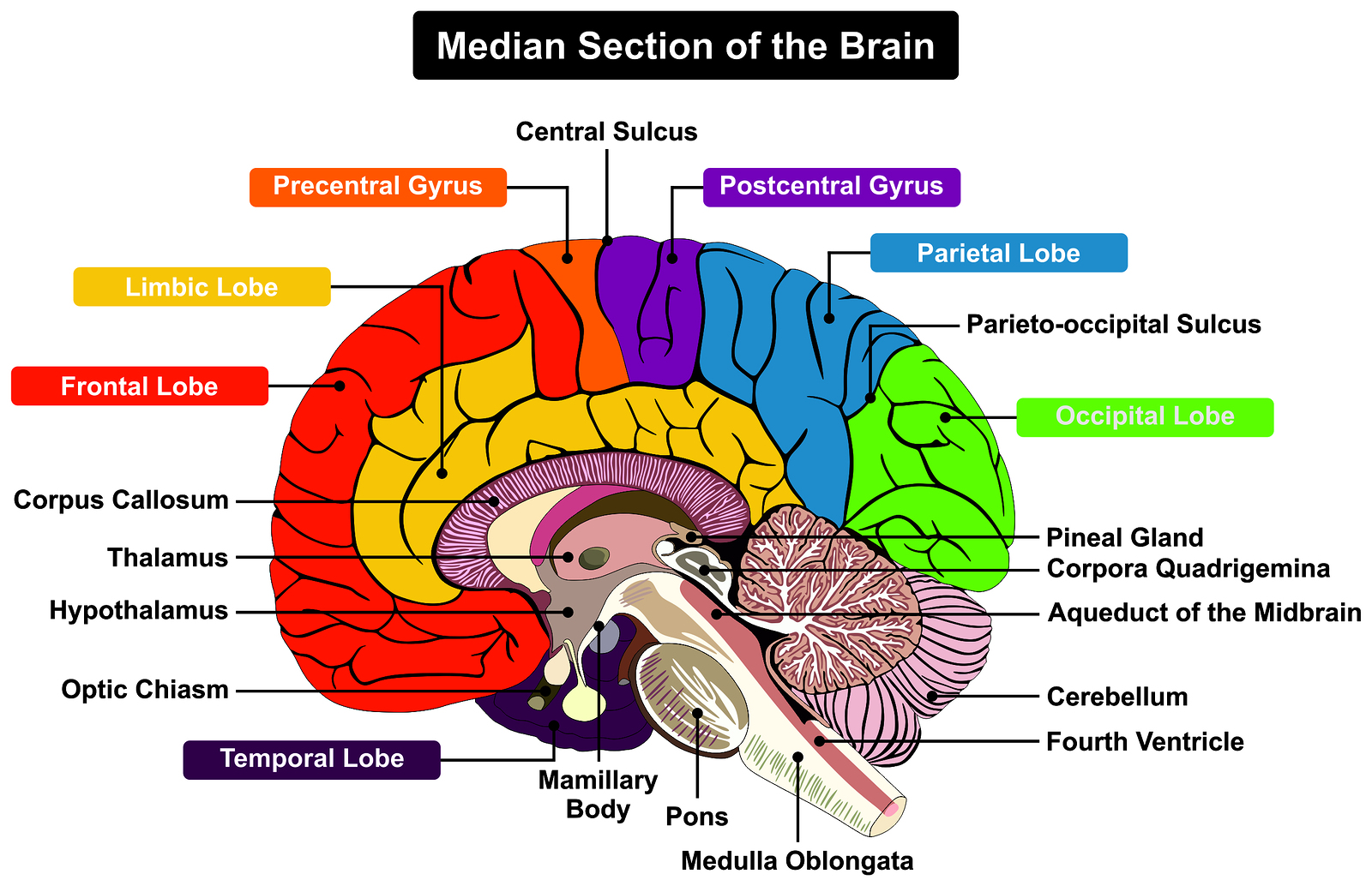
The thalamus is a small structure within the brain located just above the brain stem between the cerebral cortex and the midbrain and has extensive nerve connections to both. The main function of the thalamus is to relay motor and sensory signals to the cerebral cortex.
What is the orientation of the thalamus in the brain?
The thalamus in a 360° rotation. The thalamus is a paired structure of gray matter located in the forebrain which is superior to the midbrain, near the center of the brain, with nerve fibers projecting out to the cerebral cortex in all directions.
What is the thalamus?
The thalamus is a paired structure of gray matter located in the forebrain which is superior to the midbrain, near the center of the brain, with nerve fibers projecting out to the cerebral cortex in all directions.
How many thalami are there in the brain?
There are two thalami, one in each hemisphere of the brain. They lie above the brain stem and the midbrain (or mesencephalon), which allows for connections of nerve fibers to reach the cerebral cortex in all directions.
What is the function of the medial surface of the thalamus?
The medial surface of the thalamus comprises the upper portion of the lateral wall of the third ventricle of the brain and is lined by ependyma (remember that ependyma is the layer of ependymal cells that create cerebrospinal fluid, CSF). The medial surface serves to connect the two thalami by an interthalamic adhesion.
See more
Is the thalamus in the midbrain or forebrain?
forebrainThe structures in the forebrain include the cerebrum, thalamus, hypothalamus, pituitary gland, limbic system, and the olfactory bulb. The midbrain consists of various cranial nerve nuclei, tectum, tegmentum, colliculi, and crura cerebi.
What is included in the midbrain?
There are three main parts of the midbrain - the colliculi, the tegmentum, and the cerebral peduncles. Of the 12 cranial nerves, two thread directly from the midbrain - the oculomotor and trochlear nerves, responsible for eye and eyelid movement.
Is the thalamus in the middle of the brain?
Your thalamus lies above your brainstem in the middle of your brain. Although it may look like a single structure, you actually have two, side-by-side thalami, one in each hemisphere (side) of your brain.
What are the two parts of midbrain?
midbrain, also called mesencephalon, region of the developing vertebrate brain that is composed of the tectum and tegmentum. The midbrain serves important functions in motor movement, particularly movements of the eye, and in auditory and visual processing.
Which of the following is related to midbrain?
The midbrain or mesencephalon is the forward-most portion of the brainstem and is associated with vision, hearing, motor control, sleep and wakefulness, arousal (alertness), and temperature regulation. The name comes from the Greek mesos, "middle", and enkephalos, "brain".
Where in the brain is the thalamus located?
diencephalonThe thalamus is a paired gray matter structure of the diencephalon located near the center of the brain. It is above the midbrain or mesencephalon, allowing for nerve fiber connections to the cerebral cortex in all directions — each thalamus connects to the other via the interthalamic adhesion.
Where is the thalamus and hypothalamus located?
diencephalonThalamus and hypothalamus are both parts of the brain. Along with the epithalamus and perithalamus, they are both located in the region of the brain called the diencephalon.
What lobe is the thalamus located?
The thalamus is a small structure within the brain located just above the brain stem between the cerebral cortex and the midbrain and has extensive nerve connections to both. The primary function of the thalamus is to relay motor and sensory signals to the cerebral cortex.
Which structure is part of the midbrain quizlet?
The midbrain comprises the tectum, tegmentum, the cerebral aqueduct, and the cerebral peduncles, as well as several nuclei and fasciculi.
Is pons part of midbrain?
The pons (from Latin pons, "bridge") is part of the brainstem that in humans and other bipeds lies inferior to the midbrain, superior to the medulla oblongata and anterior to the cerebellum.
Is the amygdala in the midbrain?
The midbrain is the smallest region of the brain, and is located most centrally within the cranial cavity. Limbic System – the limbic system is often referred to as our “emotional brain”, or 'childish brain'. It is found buried within the cerebrum and contains the thalamus, hypothalamus, amygdala and hippocampus.
Is basal ganglia in midbrain?
The basal ganglia are situated at the base of the forebrain and top of the midbrain. Basal ganglia are strongly interconnected with the cerebral cortex, thalamus, and brainstem, as well as several other brain areas.
Where is the midbrain located?
Your midbrain (derived from the mesencephalon of the neural tube) is a part of the central nervous system, located below your cerebral cortex and at the topmost part of your brainstem . This tiny, but mighty, structure plays a crucial role in processing information related to hearing, vision, movement, pain, sleep, and arousal.
How long is the midbrain?
The midbrain measures around 1.5 centimeters in length and is sandwiched between the diencephalon (which includes the thalamus and hypothalamus) and the pons. 2
What is the role of the brainstem in sleep?
Your brainstem also plays a critical role in sleep and consciousness. Your midbrain can then be broken down into two main parts: 1 . Tegmentum: This anterior surface of the midbrain contains numerous structures including the reticular formation, the periaqueductal gray (PAG) matter, certain cranial nerve nuclei, ...
What causes the eye to move in the opposite direction?
Oculomotor (Third) Nerve Palsy. Any lesion within the midbrain (stroke, tumor, inflammation, infection) may damage the oculomotor nerve, resulting in an eye that is positioned in a downward and outward direction. Other symptoms of an oculomotor nerve palsy include: 1 . A dilated pupil.
What is the name of the condition that results from a stroke within the dorsal (upper side)?
Claude's syndrome: This condition results from a stroke within the dorsal (upper side) tegmentum of the midbrain. It results in ipsilateral oculomotor nerve palsy with contralateral cerebellar ataxia (incoordinated movements).
Which lobe of the brain is responsible for generating eye movements and neck muscle activity?
Nerve cells within the superior colliculi process vision signals from the retina of the eye before channeling them on to the occipital lobe located at the back of the head. The superior colliculi of the midbrain is also responsible for generating eye movements and neck muscle activity. 3
What is the treatment for a brain tumor?
For example, patients with a brain tumor that affects the midbrain may require surgery, radiation, and/or chemotherapy.
Which part of the thalamus is connected to the thalamus?
The medial surface of the thalamus constitutes the upper part of the lateral wall of the third ventricle, and is connected to the corresponding surface of the opposite thalamus by a flattened gray band, the interthalamic adhesion.
What is the blood supply of the thalamus?
The thalamus derives its blood supply from a number of arteries: the polar artery ( posterior communicating artery ), paramedian thalamic-subthalamic arteries, inferolateral (thalamogeniculate) arteries, and posterior (medial and lateral) choroidal arteries. These are all branches of the posterior cerebral artery.
What are the derivatives of the diencephalon?
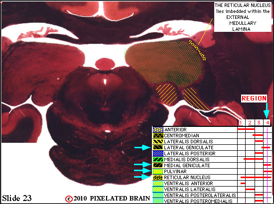
Derivatives of the diencephalon include the dorsally-located epithalamus (essentially the habenula and annexes) and the perithalamus (pre thalamus) containing the zona incerta and the thalamic reticular nucleus. Due to their different ontogenetic origins, the epithalamus and the perithalamus are formally distinguished from the thalamus proper. The metathalamus is made up of the lateral geniculate and medial geniculate nuclei.
What is the thalamus made of?
The thalamus comprises a system of lamellae (made up of myelinated fibers) separating different thalamic subparts. Other areas are defined by distinct clusters of neurons, such as the periventricular nucleus, the intralaminar elements, the "nucleus limitans", and others.
Where is SHH signaling in the thalamus?
SHH signaling from the MDO induces a posterior-to-anterior wave of expression the proneural gene Neurogenin1 in the major (caudal) part of the thalamus , and Ascl1 (formerly Mash1) in the remaining narrow stripe of rostral thalamic cells immediately adjacent to the MDO, and in the prethalamus.
Where is the mid-diencephalic organizer induced?
At the interface between the expression domains of Fez and Otx, the mid-diencephalic organizer (MDO, also called the ZLI organiser) is induced within the thalamic anlage. The MDO is the central signalling organizer in the thalamus. A lack of the organizer leads to the absence of the thalamus. The MDO matures from ventral to dorsal during development. Members of the SHH family and of the Wnt family are the main principal signals emitted by the MDO.
What is the sensory pathway that transmits pain, temperature, and itch?
The spinothalamic tract is a sensory pathway originating in the spinal cord. It transmits information to the thalamus about pain, temperature, itch and crude touch. There are two main parts: the lateral spinothalamic tract, which transmits pain and temperature, and the anterior (or ventral) spinothalamic tract, which transmits crude touch and pressure.
What are the parts of the thalamus?
The anterior, mediodorsal, and centromedian nuclei of the thalamus are the primary parts that play a role in this emotional regulation: 1 Anterior: involved in the storage of memory and emotion. 2 Mediodorsal: responsible for motivation, enthusiasm, and emotions related to inspiration. 3 Centromedian: governs the emotional component of pain.
Why is it important to understand the anatomy of the thalamus?
Understanding the anatomy of the thalamus will help you in comprehending the specific regulatory mechanisms of this structure.
Why is the thalamus important?
The thalamus is extremely important to the regulation of the human nervous system. It is the center of information processing, and is what maintains consciousness, organizes subconscious information and regulates the very survival of the human being. There is still much to be learned about this structure and it poses quite the challenge due to its countless neuronal connections to structures within the central nervous system, limbic system, and more.
How many ends does the thalamus have?
The thalamus has two ends, the anterior and posterior poles, and four surfaces: medial, lateral, superior, and inferior. Nuclei in a given pole or surface regulate specific functions or processing of sensory information and maintain particular connections with parts of the nervous and limbic system. Understanding the anatomy of the thalamus will ...
What is the limbic system?
Limbic System. Miscellaneous Functions of the Thalamus. The thalamus, or the dorsal and ventral thalamus collectively, are two oval structures made up of gray matter at the base of the cerebrum. This structure’s primary function is as a relay center through which sensory nerves transmit signals from the spinal cord and brainstem on the way to ...
What is the role of the thalamus in the body?
The thalamus also plays a significant role in sensory perception and movement. Certain areas of the thalamus are dedicated to specific parts of the body and where the sensations are meant to travel toward the cerebral cortex.
What is the lateral surface of the thalamus?
The lateral surface of the thalamus is covered by a layer of myelinated fibers called the external medullary lamina which separates the lateral surface from the reticular nuclei.
What is the difference between the thalamus and the cerebral cortex?
Whereas the connections between the thalamus and the cerebral cortex are ipsilateral, meaning they communicate on the same side of the brain.
What is the purpose of the thalamus?
The thalamus is made up of different types of nuclei, each of which serve a unique purpose, from relaying sensory and motor signals to the regulation of consciousness and alertness.
Why is the thalamus important for sleep?
Due to the thalamus being important for generating normal sleep thalamocortical rhythms, sleep disorders may result from damage such as insomnia. Language deficits because of thalamic damage, known as thalamic aphasia, can result in difficulties with lexical semantics.
Why does my thalamus feel tingly after a stroke?
Thalamic pain syndrome can occur when there are disturbances in one of the pathways of the thalamus which affects the sensation of temperature following a stroke. This can result in tingling or burning pain, as well as discomfort with temperature changes.
What is the outer covering of the thalamus?
Reticular nucleus. The reticular nucleus forms a sheet that makes the outer covering of the thalamus and can influence the activity of other nuclei within the thalamus. The reticular nucleus receives input from the cerebral cortex as well as the dorsal thalamic nuclei.
What is the thalamus made of?
The thalamus is mostly comprised of grey matter but is also surrounded by two layers of white matter. They are oval shaped in appearance, almost looking like eggs, with two protuberances on the surface. One of these is known as the medial geniculate bodies, which are important for the processing of auditory information.
What are the functions of the brain?
Below are a list of some of the associated functions: 1 Contributions to perception 2 Relaying motor information 3 Relaying sensory information 4 Role in memory 5 Alertness and attention 6 Consciousness and awareness 7 Role in cognition
What are the two parts of the midbrain?
The midbrain consists of two major parts: cerebral peduncles and tectum. The cerebral peduncles consist of the crura cerebri and tegmentum. They are separated from each other by a darkened stripe called the substantia nigra. The dorsal part of the tegmentum is traversed by the cerebral aqueduct, which connects the third and fourth ventricles of the brain. The tectum lies dorsal to the tegmentum and cerebral aqueduct, and it contains the nuclei of the superior and inferior colliculi.
What is the posterior surface of the midbrain called?
The posterior surface of the midbrain is called the tectum , or roof, of the midbrain. The tectum features four tubercles on its surface which lie inferior to the pineal gland . The upper pair of tubercles are the left and right superior colliculi, while the lower pair are the left and right inferior colliculi.
How many sensory pathways are there in the midbrain?
The midbrain contains five sensory pathways; one fasciculus and four lemnisci. Namely, they are the medial longitudinal fasciculus (MLF) and the medial, trigeminal, spinal, and lateral lemnisci. The medial longitudinal fasciculus is located just dorsal to the decussation of the superior cerebellar peduncle.
What is the most rostral part of the brainstem that connects the pons and cerebellum?
Midbrain (Mesencephalon) The midbrain, or mesencephalon , is the most rostral part of the brainstem that connects the pons and cerebellum with the forebrain. For most of its part, the midbrain sits in the posterior cranial fossa, traversing the hiatus of the tentorium cerebelli. The midbrain is the shortest part of the brainstem.
Where are the nuclei of the oculomotor and trigeminal nerves located?
The nuclei of the oculomotor (CN III), trochlear (CN IV), and trigeminal nerves (CN V) are located near the periaqueductal gray matter.
What are the peduncles of the midbrain?
Cerebral peduncles. On the cross-section of the midbrain, we can see that the cerebral peduncles consist of the ventral and dorsal regions. The ventral region of each crus is called the crus cerebri, and contains the white matter from the cortex.
Which part of the brain is the shortest?
The midbrain is the shortest part of the brainstem. However, it contains many important structures that make it essential for the proper functioning of the body: It contains the relay nuclei involved in the processing of auditory and visual information.
Which part of the midbrain is composed of the superior and inferior colliculi?
Tectum: The dorsal portion of the midbrain that is composed of the superior and inferior colliculi. These colliculi are rounded bulges that are involved in visual and auditory reflexes. The superior colliculus processes visual signals and relays them to the occipital lobes.
What are the cranial nerves located in the midbrain?
The oculomotor and trochlear cranial nerves are located in the midbrain. These nerves control eye and eyelid movement. The cerebral aqueduct, a canal that connects the third and fourth cerebral ventricles, is also located in the midbrain. Other components of the midbrain include:
What is the area of the brain that connects the forebrain to the hindbrain?
MediaForMedical / Getty Images. The midbrain is the area of the brain that connects the forebrain to the hindbrain. The midbrain and hindbrain together compose the brainstem. The brainstem connects the spinal cord with the cerebrum.
What is the function of the diencephalon?
The diencephalon is the region of the brain that relays sensory information and connects components of the endocrine system with the nervous system. The diencephalon regulates a number of functions including autonomic, endocrine, and motor functions. It also plays a major role in sensory perception. Components of the diencephalon include: 1 Thalamus: A limbic system structure that connects areas of the cerebral cortex that are involved in sensory perception and movement with other parts of the brain and spinal cord. The thalamus also plays a role in the control of sleep and wake cycles. 2 Hypothalamus: Acts as the control center for many autonomic functions including respiration, blood pressure, and body temperature regulation. This endocrine structure secretes hormones that act on the pituitary gland to regulate biological processes including metabolism, growth, and the development of reproductive system organs. As a component of the limbic system, the hypothalamus influences various emotional responses through its influence on the pituitary gland, skeletal muscular system, and autonomic nervous system. 3 Pineal Gland: This small endocrine gland produces the hormone melatonin. Production of this hormone is vital to the regulation of sleep-wake cycles and also influences sexual development. The pineal gland converts nerve signals from the sympathetic component of the peripheral nervous system into hormone signals, thereby linking the nervous and endocrine systems.
What are the components of the diencephalon?
Components of the diencephalon include: Thalamus: A limbic system structure that connects areas of the cerebral cortex that are involved in sensory perception and movement with other parts of the brain and spinal cord. The thalamus also plays a role in the control of sleep and wake cycles.
What are the four lobes of the telencephalon?
These lobes include the frontal lobes, parietal lobes, occipital lobes, and temporal lobes . The cerebral cortex contains folded bulges called gyri that create indentations in the brain.
Which brain division is the largest?
The forebrain is by far the largest brain division. It includes the cerebrum, which accounts for about two-thirds of the brain's mass and covers most other brain structures. The forebrain consists of two subdivisions called the telencephalon and diencephalon.
Overview
Anatomy
The thalamus is a paired structure of gray matter located in the forebrain which is superior to the midbrain, near the center of the brain, with nerve fibers projecting out to the cerebral cortex in all directions. The medial surface of the thalamus constitutes the upper part of the lateral wall of the third ventricle, and is connected to the corresponding surface of the opposite thalamus by a flattene…
Function
The thalamus has multiple functions, generally believed to act as a relay station, or hub, relaying information between different subcortical areas and the cerebral cortex. In particular, every sensory system (with the exception of the olfactory system) includes a thalamic nucleus that receives sensory signals and sends them to the associated primary cortical area. For the visual system, for example, inputs from the retina are sent to the lateral geniculate nucleus of the thalam…
Development
The thalamic complex is composed of the perithalamus (or prethalamus, previously also known as ventral thalamus), the mid-diencephalic organiser (which forms later the zona limitans intrathalamica (ZLI) ) and the thalamus (dorsal thalamus). The development of the thalamus can be subdivided into three steps. The thalamus is the largest structure deriving from the embryonic diencephalon, the posterior part of the forebrain situated between the midbrain and the cerebrum.
Clinical significance
A thalamus damaged by a stroke can lead to thalamic pain syndrome, which involves a one-sided burning or aching sensation often accompanied by mood swings. Bilateral ischemia of the area supplied by the paramedian artery can cause serious problems including akinetic mutism, and be accompanied by oculomotor problems. A related concept is thalamocortical dysrhythmia. The occlusion of the artery of Percheron can lead to a bilateral thalamus infarction.
Additional images
• Human brain dissection, showing the thalamus.
• Human thalamus along with other subcortical structures, in glass brain.
• Lateral group of the thalamic nuclei.
• Medial group of the thalamic nuclei.
See also
• 5-HT7 receptor
• Krista and Tatiana Hogan - conjoined twins with joined thalami
• List of regions in the human brain
• Nonmotor region of the ventral nuclear group of the thalamus
External links
• Stained brain slice images which include the "thalamus" at the BrainMaps project