Boundaries of thorax:
- At the back –12 thoracic vertebra forming the part of back bone
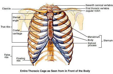
- At the front – breast bone or sternum
- Enclosed by ribs which starts from the vertebral column at the back to the sternum in the front on both sides and the muscles between the ribs (intercostal muscles)
What are the boundaries of the thoracic cavity Quizlet?
Jan 17, 2020 · The boundaries of the Thoracic Cavity are the Ribs (and Sternum), Vertebral Column, and the Diaphragm. The Diaphragm seperates the Thoracic Cavity from the Abdominal Cavity. Mediastinum - Space between the left and right Pleural Cavities.
What is the thoracic cavity in vertebrates?
From posterior boundary of the thoracic cavity. Ribs. Lateral boundary of thoracic cavity. Intercostal muscles. Lateral boundary of thoracic cavity. Sets with similar terms. PT 731 Kines Practical #2. 70 terms. rickianderson. Muscles of Forced Expiration. 25 terms. jmh1352. BIOS214 lab quiz 6. 83 terms.
What are the bony boundaries of the thorax?
The boundaries of the Thoracic Cavity are the Ribs (and Sternum), Vertebral Column, and the Diaphragm. The Diaphragm seperates the Thoracic Cavity from the Abdominal Cavity. Mediastinum - Space between the left and right Pleural Cavities. Contains the Pericardial Cavity which surrounds the Heart, Trachea, Esophagus, Thymus, and Blood Vessals.
What are the boundaries of the canine thoracic cavity?
Jan 01, 2008 · The boundaries of the canine and feline thoracic cavity consist of the thoracic skeleton, the cranial and caudal thoracic apertures and the covering soft tissue structures. The rib cage is covered by thoracic, pectoral, spinal and other musculature, subcutaneous fat and skin, and serves as an attachment for the thoracic extremities.
What are the boundaries for the abdominal cavity?
abdominal cavity, largest hollow space of the body. Its upper boundary is the diaphragm, a sheet of muscle and connective tissue that separates it from the chest cavity; its lower boundary is the upper plane of the pelvic cavity. Vertically it is enclosed by the vertebral column and the abdominal and other muscles.
What 3 areas are bounded to the thoracic cavity?
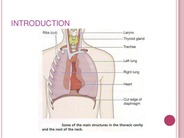
The thoracic cavity is actually composed of three spaces each lined with mesothelium, a special film-like tissue that separates vital organs. The pleural cavities surround the lungs, while the pericardial cavity surrounds and protects the heart. These tissues in the thoracic cavity can be seen in the image below.Apr 28, 2017
What is the boundary between the thoracic and the abdominopelvic cavity?
The diaphragm forms the boundary between the superior thoracic cavity and the inferior abdominopelvic cavity.
What is thoracic cavity Class 10?
Thoracic cavity, also called chest cavity, is the second largest hollow space of the body. It is enclosed by the ribs, vertebral column and the sternum. The lungs lie in the chest cavity or thoracic cavity which is separated from abdominal cavity by a muscular partition called diaphragm.Apr 14, 2020
Where is the thoracic region?
Your thoracic spine is located in the center of your upper and middle back. It begins at the base of your neck (cervical spine) and ends around the bottom of your rib cage, just above your lower back (lumbar spine).
What structure divides the thoracic cavity into right and left parts?
The diaphragmThe diaphragm forms the floor of the thoracic cavity and separates it from the more inferior abdominopelvic cavity.
Which body cavity is superior to the thoracic cavity?
Ventral body cavityVentral body cavity The superior mediastinum is a wedge-shaped cavity located between the superior regions of the two thoracic cavities.
What are the four cavities of the body?
Ventral body cavity–the thoracic cavity, the abdominal cavity, and the pelvic cavity in combination. Thoracic cavity–the space occupied by the ventral internal organs superior to the diaphragm. Abdominopelvic cavity–the abdominal cavity and the pelvic cavity in combination.
What is the thoracic wall?
Thoracic wall. The first step in understanding thorax anatomy is to find out its boundaries. The thoracic, or chest wall, consists of a skeletal framework, fascia, muscles, and neurovasculature – all connected together to form a strong and protective yet flexible cage.
What is the inferior thoracic aperture?
The inferior thoracic aperture is almost completely covered by the diaphragm, separating it from the abdominal cavity. Moving forward with the skeletal scaffold of the thorax, we have the thoracic skeleton. It is made up of the sternum, twelve pairs of ribs, twelve thoracic vertebrae, and interconnecting joints.
What is the chest?
The chest, properly called the thorax, is the superior part of the trunk located between the neck and abdomen. It consists of several components: 1 Thoracic wall 2 Several cavities 3 Neurovasculature and lymphatics 4 Internal organs 5 Breasts
Which nerve provides innervation?
Innervation is provided by the recurrent laryngeal nerve, sympathetic trunk, and esophageal nervous plexus. Esophagus in situ (anterior view) If you want to master the anatomy of the esophagus, including its arteries, veins, and the thoracic nerves supplying it, jump into the following study unit.
What is the chest called?
The chest, properly called the thorax, is the superior part of the trunk located between the neck and abdomen. It consists of several components: Thoracic wall. Several cavities. Neurovasculature and lymphatics. Internal organs. Breasts.
Where is the mediastinum located?
The mediastinum is located centrally and bordered by two pleural cavities laterally. The mediastinum consists of superior and inferior mediastinal cavities. The inferior mediastinal cavity is comprised of anterior, middle and posterior compartments. Neurovasculature.
What is the space between the ribs called?
Running between every two adjacent ribs are anatomical spaces called intercostal spaces . There are eleven in total, each one containing the intercostal muscles ( external, internal, and innermost) together with the intercostal neurovascular bundle. This consists of the intercostal vein, artery, and nerve.
What is the boundary of the thoracic cavity?
The boundaries of the thoracic cavity are unit of the Ribs (and Sternum ), Vertebral Column , and therefore the Diaphragm. The Diaphragm separates the thoracic cavity from the abdominal cavity.
What is the central compartment of the thoracic cavity?
The central compartment of the thoracic cavity is the mediastinum. There is a unit of 2 openings of the thoracic cavity, a superior pectoral aperture called the pectoral recess and a lower inferior pectoral aperture called the pectoral outlet.
What is the dorsal cavity?
As its name implies, it contains organs lying a lot of posterior within the body. The dorsal cavity, again, is often divided into 2 parts. The higher portion, or the cavity, home the brain, and therefore the lower portion or canalis vertebralis homes the medulla spinalis. 2. Thoracic Cavity.
Where is the thorax located?
In mammals, the thorax is that the region of the body fashioned by the sternum, the pectoral vertebrae, and therefore the ribs. It extends from the neck to the diaphragm and doesn’t embody the higher limbs. The center and therefore the lungs reside within the thoracic cavi ty, also as several blood vessels.
What is the effect of inhalation on the respiratory system?
Throughout the method of inhalation, the respiratory organ volume expands as a result of the contraction of the diaphragm and intercostal muscles (the muscles that area of the unit connected to the rib cage ), therefore increasing the Thoracic cavity.
What happens to the diaphragm during exhalation?
Throughout exhalation, the diaphragm conjointly relaxes, moving higher into the thoracic cavity. This will increase the pressure among the thoracic cavity relative to the surroundings. Air rushes out of the lungs thanks to the pressure gradient between the thoracic cavity and therefore the atmosphere.
What are the symptoms of a symtom?
Common symptoms are a unit of pain, fever, and shortness of breath. Treatment is directed toward evacuation of fluid and alleviation of the underlying condition, typically associate degree infected respiratory organ however additional seldom a diffuse inflammatory condition like atrophic arthritis.
What is the thoracic cavity?
The thoracic cavity also contains the esophagus, the channel through which food is passed from the throat to the stomach. The chest cavity is lined with a serous membrane, which exudes a thin fluid.
Where is the sternum located?
Sternum, in the anatomy of tetrapods (four-limbed vertebrates), elongated bone in the centre of the chest that articulates with and provides support for the clavicles (collarbones) of the shoulder girdle and for the ribs. Its origin in evolution is unclear. A sternum appears in certain salamanders; it….
What is the pleura?
Encyclopædia Britannica, Inc. The pleura is a continuous sheet of endothelial, or lining, cells supported by a thin base of loose connective tissue. The membrane is well supplied with blood vessels, nerves, and lymph channels.
What is the second largest hollow space in the body?
Full Article. Thoracic cavity, also called chest cavity, the second largest hollow space of the body. It is enclosed by the ribs, the vertebral column, and the sternum, or breastbone, and is separated from the abdominal cavity (the body’s largest hollow space) by a muscular and membranous partition, the diaphragm.
What is the term for fluid accumulation in the pleural cavity?
Subscribe Now. Accumulation of fluid in the pleural cavity is called hydrothorax. If the fluid is bloody, the condition is described as hemothorax; if it contains pus, pyothorax. The accumulation of fluid may or may not be accompanied by air.
What are the symptoms of a rheumatoid arthritis?
Common symptoms are pain, shortness of breath, and fever. Treatment is directed toward evacuation of fluid and alleviation of the underlying condition, often an infected lung but more rarely a diffuse inflammatory condition such as rheumatoid arthritis.
What is the rib?
rib. Rib, any of several pairs of narrow, curved strips of bone (sometimes cartilage) attached dorsally to the vertebrae and, in higher vertebra tes, to the breastbone ventrally, to form the bony skeleton, or rib cage, of the chest. The ribs help to protect the internal organs that they enclose and lend…. vertebral column.
What is the procedure to access the thoracic cavity?
Access to the thoracic cavity is required for treatment of a variety of conditions in dogs and cats and ranges from simple tube thoracostomy to complex approaches involving a combination of sternotomy, thoracotomy, rib pivot, and celiotomy. In the majority of cases, however, the thorax is approached in one of three ways: intercostal thoracotomy (or thoracostomy), median sternotomy, or transdiaphragmatic incision. Because the thoracic cavity is deep and narrow on a transverse plane in cats and many breeds of dog, intercostal thoracotomy provides good access for the majority of clinical procedures. Sternotomy, however, is required for major surgery of the cranial mediastinum and for access to both sides of the thorax.
What is the skin of the thoracic wall?
Skin of the thoracic wall possesses some anatomic features that may be useful for reconstruction. Skin of the dorsolateral thorax is supplied mainly by the thoracodorsal artery, which arises from the subscapular artery and supplies the latissimus dorsi muscle and the skin. Flaps that can be based on the thoracodorsal artery include the thoracodorsal axial pattern flap and a composite musculocutaneous flap incorporating the latissimus dorsi muscle. This flap is particularly useful because it provides a means of reestablishing integrity of the thoracic wall as well as skin coverage. 12 The double-layered elbow fold may be separated from the upper foreleg and unfolded to produce a subdermal plexus flap capable of closing defects of the sternum or lateral thorax.
How many vertebrae do dogs have?
Dogs and cats have 13 thoracic vertebrae, 13 ribs, and 9 sternebrae (Figures 104-1 and 104-2 ). Ribs one to nine articulate with the sternebrae via cartilaginous extensions from the costochondral junctions. The sternebrae are connected to one another by fibrocartilage. The cranialmost sternebra, the manubrium, provides the point of attachment for the sternocephalicus muscle. The caudalmost sternebra, the xiphoid, is positioned dorsal to the linea alba and ventral to the falciform ligament. The xiphoid does not have any direct muscular attachments other than the diaphragm; the fascial connection between the paired rectus abdominus muscles (linea alba) merges with the fascia of the deep pectoral muscle ventral to the xiphoid. 12
Where do intercostal nerves pass?
Intercostal nerves (Figure 104-5) arise as ventral branches of the thoracic spinal nerves and pass ventrally along the caudal edge of each rib in association with the intercostal arteries and veins. There are 12 intercostal arteries on each side of the thorax.
What is the musculoskeletal structure of the thorax?
The complex musculoskeletal structure of the thorax facilitates complete resection of tumors with adequate margins. Natural tissue planes allow separation of muscle bellies to reduce postoperative morbidity, and anatomic landmarks allow intraoperative delivery of local anesthesia. 12
What muscles are involved in the thorax?
Muscle groups (Figures 104-3 and 104-4) associated with the thorax include the intrinsic and extrinsic muscles of respiration, muscles of the abdominal wall, and locomotor musculature. 12 The locomotor muscles attach the forelimb to the trunk. Most significant for thoracic surgeons are the latissimus dorsi, the serratus ventralis thoracis group, and the superficial and deep pectoral muscles. The latissimus dorsi muscle originates from the lumbodorsal fascia and thoracolumbar vertebrae and converges cranioventrally to insert on the proximal humerus. It draws the scapula and hence the forelimb caudally. The latissimus dorsi muscle may either be divided or elevated to gain access to the intercostal muscles for intercostal thoracotomy. Limb function does not seem to be unduly impaired if this muscle is separated or relocated. Elevation of the muscle has been suggested to cause less postoperative pain but can also reduce surgical access or necessitate more active retraction to maintain surgical exposure. The thoracolumbar attachments of the latissimus dorsi muscle can be incised and the muscle rotated ventrally to close large defects in the thoracic wall.
What is intercostal thoracotomy?
A, Intercostal thoracotomy is performed with the animal in lateral recumbency. B, An incision in the skin, subcutaneous tissues, and cutaneous trunci muscle is made parallel to the ribs and extends from the costovertebral junction to the sternum.