What are the three layers of the meninges?
They’re known as:
- Dura mater: This is the outer layer, closest to your skull.
- Arachnoid mater: This is the middle layer.
- Pia mater: This is the inner layer, closest to your brain tissue.
What is the Order of the layers of the meninges?
Meninges Layers
- Dura Mater. This outer layer connects the meninges to the skull and vertebral column. ...
- Arachnoid Mater. This middle layer of the meninges connects the dura mater and pia mater. ...
- Pia Mater. This thin inner layer of the meninges is in direct contact with and closely covers the cerebral cortex and spinal cord.
What does the word meninges mean?
The primary function of the meninges and of the cerebrospinal fluid is to protect the central nervous system. Meninges is the plural of meninx, from Ancient Greek: μῆνιγξ mēninx "membrane".
What are the three meninges of the brain?
Structure of Meninges
- Pia Mater. The meningeal envelope that tightly adheres to the surface of the spinal cord and brain is known as the pia mater.
- Dura Mater. The outermost of the three meninges is known as the dura mater (also called pachymeninx), which is a thick, strong, and dense membrane.
- Dura Mater Infolding. ...
- Arachnoid Mater. ...
- Leptomeninges. ...
What is meninges composed of?
Three layers of membranes known as meninges protect the brain and spinal cord. The delicate inner layer is the pia mater. The middle layer is the arachnoid, a web-like structure filled with fluid that cushions the brain. The tough outer layer is called the dura mater.
What cells are meninges made of?
The principal cell types in meninges are fibroblasts within the stroma and the vascular endothelium. The extent of collagen production is greater in the thicker outer dura mater and relatively less in the two thinner leptomeningeal layers, the arachnoid and pia mater.
What are meninges filled with?
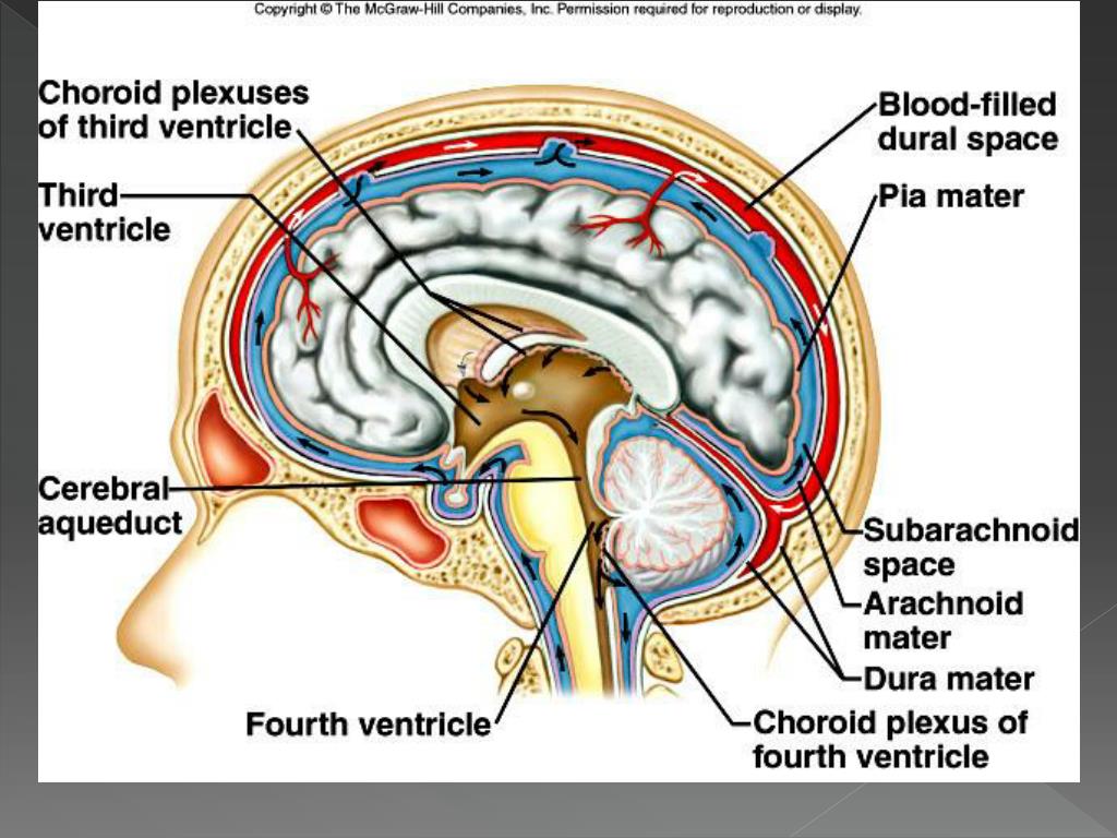
The subarachnoid space is a space between your arachnoid mater and pia mater. It's filled with cerebrospinal fluid. Cerebrospinal fluid cushions and protects your brain and spinal cord.
How are the meninges formed?
The cranial meninges originate from a mesenchymal sheath on the surface of the developing brain, called primary meninx, and undergo differentiation into three layers with distinct histological characteristics: the dura mater, the arachnoid mater, and the pia mater.
What is dura mater made of?
The dura mater is composed of two layers: the periosteal/endosteal layer and the meningeal layer. The dural venous sinuses are between these two layers. The dura folds to form septa that create the falx cerebri, tentorium cerebelli, falx cerebelli, and diaphragma sellae.
What are the 3 functions of the meninges?
The function of the meninges is to protect the brain and spinal cord from mechanical trauma, to support the blood vessels and to form a continuous cavity through which the cerebrospinal fluid (CSF) passes.
What specific type of animal tissue are meninges made up of?
The outer layer, the dura mater, is tough, white fibrous connective tissue. The middle layer of meninges is arachnoid, a thin layer resembling a cobweb with numerous threadlike strands attaching it to the innermost layer.
What is the role of the meninges?
The primary function commonly attributed to meninges and CSF is to protect the central nervous system (CNS). This is mainly because meninges provide a tight anchoring of the CNS to the surrounding bones able to prevent side-to-side movement and providing stability.
What specific type of animal tissue are meninges made up of?
The outer layer, the dura mater, is tough, white fibrous connective tissue. The middle layer of meninges is arachnoid, a thin layer resembling a cobweb with numerous threadlike strands attaching it to the innermost layer.
What is meninges in biology?
(meh-NIN-jeez) The three thin layers of tissue that cover and protect the brain and spinal cord.
What are the meninges quizlet?
Thick, tough outer covering of the brain. It consists of an outer periosteal layer and an inner meningeal layer. The two layers of dura separate from eachother at the dural partitions, which project inward and incompletely separate parts of the brain and the intracranial venous structures.
What 3 structures make up the brainstem?
The brainstem is the structure that connects the cerebrum of the brain to the spinal cord and cerebellum. It is composed of three sections in descending order: the midbrain, pons, and medulla oblongata.
What is the meninges?
Updated July 02, 2019. The meninges is a layered unit of membranous connective tissue that covers the brain and spinal cord. These coverings encase central nervous system structures so that they are not in direct contact with the bones of the spinal column or skull.
How many layers are there in the meninges?
The meninges can be generally separated into three distinct layers, each with its own specific function and traits.
What are the three membranes of the meninges?
The meninges are composed of three membrane layers known as the dura mater, arachnoid mater, and pia mater . Each layer of the meninges serves a vital role in the proper maintenance and function of the central nervous system.
How does the arachnoid membrane get its name?
The arachnoid membrane loosely covers the brain and spinal cord and gets its name from its web-like appearance. The arachnoid mater is connected to the pia mater through tiny fibrous extensions that span the subarachnoid space between the two layers.
Which layer of the brain connects the dura mater to the skull?
The outer periosteal layer firmly connects the dura mater to the skull and covers the meningeal layer. The meningeal layer is considered the actual dura mater. Located between these two layers are channels called dural venous sinuses.
Which granulations remove cerebrospinal fluid from the subarachnoid space and send it to?
Arachnoid granulations remove cerebrospinal fluid from the subarachnoid space and send it to the dural venous sinuses, where it is reabsorbed into the venous system. Pia Mater: This thin inner layer of the meninges is in direct contact with and closely covers the cerebral cortex and spinal cord.
Which layer of the spinal column is composed of the meningeal layer and does not contain a perio?
The dura mater of the spinal column is composed of the meningeal layer and does not contain a periosteal layer. Arachnoid Mater: This middle layer of the meninges connects the dura mater and pia mater. The arachnoid membrane loosely covers the brain and spinal cord and gets its name from its web-like appearance.
What is the outermost of the meninges?
The outermost of the three meninges is the dura mater (or pachymeninx), a strong, thick, and dense membrane. It is composed of dense fibrous tissue, and its inner surface is covered by flattened cells like those present on the surfaces of the pia mater and arachnoid.
What is the function of the meninges and cerebrospinal fluid?
The primary function of the meninges and of the cerebrospinal fluid is to protect the central nervous system. The pia mater is the meningeal envelope that firmly adheres to the surface of the brain and spinal cord.
What is the process of arachnoid villi?
These fingerlike processes of the arachnoid, called arachnoid villi or arachnoid granulations, are involved in the passage of cerebrospinal fluid from the subarachnoid space to the dural sinuses . Spinal anesthetics are often introduced into the subarachnoid space.
What are the trabeculae of the arachnoid?
The arachnoid trabeculae are embryologic remnants of the common origin of the arachnoid and pia mater, and they have the frail structure characteristic of these two of the meninges. The pia mater and arachnoid together are called the leptomeninges.
What is the arachnoid space?
Over the pia mater and separated from it by a space called the subarachnoid space is the arachnoid, a thin, transparent membrane. It is composed of fibrous tissue and, like the pia mater, is covered by flat cells also thought to be impermeable to fluid.
Which part of the brain is the outer part of the dura mater?
The outer portion of the dura mater over the brain serves as a covering, or periosteum, of the inner surfaces of the skull bones .
What is the pia mater?
The pia mater is pierced by blood vessels that travel to the brain and spinal cord. Britannica Quiz. The Human Body.
What are the three meninges?
There are three layers of meninges, known as the dura mater, arachnoid mater and pia mater. These coverings have two major functions: Provide a supportive framework for the cerebral and cranial vasculature. Acting with cerebrospinal fluid to protect the CNS from mechanical damage.
What is the outermost layer of the meninges?
The dura mater is the outermost layer of the meninges and is located directly underneath the bones of the skull and vertebral column. It is thick, tough, and inextensible. The dura mater consists of two layered sheets of connective tissue: Periosteal layer – lines the inner surface of the bones of the cranium.
What is the causative vessel of brain trauma?
The causative vessel is usually the middle meningeal artery, tearing as a consequence of brain trauma. Subdural – venous blood collects between the dura and the arachnoid mater. It results from damage to cerebral veins as they empty into the dural venous sinuses.
What is the function of cerebrospinal fluid?
It contains cerebrospinal fluid, which acts to cushion the brain. Small projections of arachnoid mater into the dura (known as arachnoid granulations) allow CSF to re-enter the circulation via the dural venous sinuses. By TeachMeSeries Ltd (2021) Fig 3 – Coronal section of the skull, meninges and cerebrum.
What is the role of the meninges in the CNS?
Acting with cerebrospinal fluid to protect the CNS from mechanical damage. The meninges are often involved cerebral pathology , as a common site of infection (meningitis), and intracranial bleeds. In this article, we shall look at the anatomy of the three layers, and their clinical correlations.
What is the name of the gland that covers the hypophysial fossa of the sphen?
Diaphagma sellae – covers the hypophysial fossa of the sphenoid bone. It contains a small opening for passage of the stalk of the pituitary gland.
Where does the dura mater get its vascular supply?
The dura mater receives its own vascular supply - primarily from the middle meningeal artery and vein. It is innervated by the trigeminal nerve (V1, V2 and V3).
What are the three meninges?
The brain membranes or meninges. surround and protect the brain. There are three different meninges. Those are. the dura mater, pia mater, and arachnoidea mater. The soft envelope, or pia, is. located directly on the glia. The arachnoidea mater is located on top of it, and the exterior, hard envelope or dura mater is located above the arachnoidea.
Where are the brain meninges located?
They are located between the brain and the skull and between the spinal cord and spinal vertebrae and are constructed of loose and dense connective tissues. These are three membranes that.
What are the two layers of the brain?
The most external membrane is dura mater. It is made up of solid connective tissue and completely encloses the brain and spinal cord. It consists of two sheets: 1 The outer, periosteal layer lining#N#the inner side of the bones of the skull and spinal canal, richly supplied with#N#blood vessels and nerves, 2 The inner, meningeal layer whose#N#interior is covered with a monolayer mesenchymal epithelium ( 2 ).
What is formed after brain mass removal?
After removal of brain masses from the skull, thick partitions are observed, which build up duplications of the dura mater, and are formed by protrusion of the meningeal layer of hard membrane into the cranial cavity. These compartments separate the individual parts of the brain, support them and thus ensure their permanent position during various movements and head positions.
How is the brain separated from the hard sheath?
It is separated from the hard sheath by the subdurale cavum that is filled with liquor. The hard cover is on the outside. It is a rigid fibrous sack whose task is to cover the brain for its protection. It is very rigid and of different thicknesses.
How many nootropics are in Mind Lab Pro?
Boost Your Brain with Mind Lab Pro. Your brain is incredibly complex. Mind Lab Pro has 11 different nootropics all working together to increase your cognition and brainpower to help you live a better life.
What are the three membranes that surround the brain and spinal cord?
These are three membranes that. surround the brain and spinal cord: the outer hard membrane (dura mater), the. soft membrane (pia mater) which is rich in blood vessels, and the internal. connective membrane (arachnoidea mater) that keeps the brain immersed in the. cerebrospinal fluid ( 1 ).
What are the middle layers of the meninges?
Middle layers (arachnoid): Resembling spider webs, the arachnoid or middle layers of the meninges project arachnoid trabeculae, which are strands of connective tissue to the innermost layer. This is membrane is marked by granulations, which are protrusions at areas of contact with the outermost layer. Unlike the innermost layer, the cranial middle layers straddle the edges of the cortical sulci, or depressions in the brain.
What is the spine meninge?
Spinal meninges also encase the cauda equina, the bundle of nerves and nerve roots at the base of the spine. This includes nerves corresponding to lumbar (low back) vertebrae, as well as the sacrum (the bony, triangular structure at the base of the spine). 2
What are the health issues that affect the meninges?
Given their critical role, it’s little wonder that anatomical variations and health issues in the meninges can have a serious impact. Birth defects, such as spina bifida and anencephaly, as well as infection ( meningitis) and bleeding (as in cerebral hematoma or hemorrhage ), can lead to chronic disability or become fatal. 1
What are the effects of genetic mutations on the meninges?
These coding errors lead to neural tube defects, in which the meninges never fully forms. These potentially very severe diseases of the meninges include:
What are the three layers of protective tissue that surround the brain and spinal cord?
Surrounding the brain and spinal cord are three layers of protective tissue, collectively called the meninges . Meninges are a necessary cushion between these vital organs and the cranium (or skull) and vertebrae (spine). They also prevent cerebrospinal fluid (CSF), the clear fluid the brain and spinal cord sit in, from leaking, while providing structural support for important blood vessels and nerves. 1
Why are meninges important?
These layers are tasked with the essential job of protecting and nourishing the brain and spine. No doubt the burden due to congenital conditions, infections, injuries, or other disorders of these crucial membranes can be heavy.
Where does the cranial meninges get blood?
The cranial meninges primarily receive blood from by the middle meningeal artery (MMA), which is a branch of the internal carotid artery, which progresses up the neck. On each side, the MMA enters the skull through an opening in its side called the foramen spinosum and continues through the epidural space. 3
What are the layers of meninges?
There are three layers to the meninges. From inside to outside they are the pia mater, arachnoid mater, and dura mater.
What are the meninges of the brain?
The meninges of the brain are similar to the meninges of the spinal cord. The meninges function to provide several important services in the body. In this lesson, investigate the three types of meninges, each meninges' anatomy, where the meninges occur in the layers of the brain, and various disorders that may arise when these important structures are damaged.
What is the outermost layer of arachnoid mater?
The dura mater is the outermost, thickest layer, and it sounds like "durable." The arachnoid mater is web-like in appearance and thus can be remembered by thinking of "arachnids" (spiders). The pia mater is the innermost, thinnest layer, and it can be remembered as "pliable."
What is the thinnest meninge of the brain?
The pia mater , which translates to "tender matter," is the thinnest and deepest meninge of the brain. This thin, clear layer adheres to the surface of the brain and follows the natural hills and valleys (called gyri and hillocks) of the surface of the brain. The pia mater functions to contain cerebrospinal fluid within its proper pathway. Additionally, about 30% of the total CSF is produced by the pia mater .
What are granulations in the arachnoid mater?
Arachnoid granulations are structures within the arachno id mater. They are small growths that occur on the membrane of the arachnoid mater and extend into the dural sinuses. These growths are typically harmless but may cause issues when they become too large. A large arachnoid granulation may result in the occlusion of a blood vessel or block important CSF pathways.
What is the difference between arachnoid mater and pia mater?
The deeper lower layer (the pia mater) follows the brain closely and adheres to the deep grooves and hills . This results in spaces that exist within the arachnoid mater and pia mater that are quite large , such as areas where the pia mater dips deeply with a groove in the brain while the arachnoid mater is held up closer to the skull. These large grooves are called subarachnoid cisterns. The subarachnoid cisterns provide space that is critical for nerves and vessels to pass through the layers of the brain. They also contain CSF. There are 9 commonly observed subarachnoid cisterns in the body, which are named based on their location:
What is the arachnoid mater?
The arachnoid mater is just below the dura mater. The term "arachnoid" comes from the word "arachnid," meaning "spider." This name is in reference to the appearance of the arachnoid mater, which resembles the stringy, white fibers of a spider web. The most important function of the arachnoid mater is to house the arachnoid space. This space is created by the crisscrossing fibers called arachnoid trabeculae which connect the layer to the pia mater while creating a passageway.
What are the three potential spaces that are bound by the meninges?
These layers bound three clinically important potential spaces: the epidural, subdural, and subarachnoid spaces. The function of the meninges is to protect the brain and spinal cord from mechanical trauma, to support the blood vessels and to form a continuous cavity through which the cerebrospinal fluid (CSF) passes.
What is the name of the compartment in which the meningeal dura mater is located?
The meningeal dura mater overlies the trigeminal ganglion, enclosing it in a compartment known as the trigeminal cave (Meckel’s cave).
What is the cranial arachnoid mater?
Arachnoid mater. The cranial arachnoid mater is a spiderweb-like meningeal layer, interposed between the dura and pia. The potential space between the arachnoid and dura is called the subdural space and according to some authors, it contains a very thin layer of fluid.
How is CSF reabsorbed into the dural sinuses?
Finally, the CSF is reabsorbed into the dural venous sinuses by diffusing through the subarachnoid granulations in the cranial subarachnoid space.
What is the outermost layer of the cranial dura?
The cranial dura mater is the outermost meningeal layer , consisting of dense irregular connective tissue. It is composed of two layers; The superficial layer is the periosteal cranial dura. It overlies the inner table of the cranial vault bones, acting like the periosteal layer of the cranium.
Which cranial dura lies superficial to the arachnoid mater?
The meningeal cranial dura, which lies superficial to the arachnoid mater.
What are the three membranes that separate the brain and spinal cord?
The meninges are the three membranes that envelop the brain and spinal cord and separate them from the walls of their bony cases ( skull and vertebral column ). Based on their location, meninges are referred to as the cranial meninges which envelop the brain, and spinal meninges which envelop the spinal cord.
How many percent of meningiomas are convex?
Accounting for approximately 20 percent of meningiomas, convexity meningiomas may not present symptoms until the tumor has become large enough to push on the brain. Falcine and parasagittal meningioma forms in or next to the falx, a very thin layer of tissue between the two sides of the brain. Intraventricular meningioma forms within ...
Where do meningiomas grow?
Meningiomas grow out of the middle layer of the meninges called the arachnoid. They grow slowly and may exist for years before being detected. Sometimes doctors will discover a meningioma incidentally on a magnetic resonance imaging ( MRI) scan of the head or spinal cord.
Where does sphenoid wing meningioma form?
Sphenoid wing meningioma forms on the skull base behind the eyes. Approximately 20 percent of meningiomas are sphenoid wing. Olfactory groove meningioma forms along the nerves that run between the brain and the nose and account for around 10 percent of meningiomas. This type of tumor can cause a loss of smell, and can grow large enough ...
Where does convexity meningioma grow?
Convexity meningioma grows on the surface of the brain directly under the skull. Accounting for approximately 20 percent of meningiomas, convexity meningiomas may not present symptoms until the tumor has become large enough to push on the brain.
What percentage of brain tumors are meningioma?
Meningioma is the most common type of primary brain tumor, accounting for approximately 30 percent of all brain tumors.
Can meningioma come back?
Tumors in this area can cause visual problems and dysfunction of the pituitary gland. Recurrent meningioma: Any meningioma may come back. When a meningioma does recur, it may be the same grade or a more aggressive or malignant form.
Can intraventricular meningioma cause hydrocephalus?
An intraventricular meningioma may cause a blockage of CSF flow, leading to hydrocephalus. Skull base meningioma grows in the bones that form the bottom of the skull and in the bony ridge in the back of the eyes. These are more difficult to remove surgically than convexity meningiomas.
What causes meningioma?
Doctors know that something alters some cells in your meninges to make them multiply out of control, leading to a meningioma tumor.
Why are meningiomas not visible?
In many cases, because meningiomas do not cause any noticeable signs or symptoms, they are only discovered as a result of imaging scans done for reasons that turn out to be unrelated to the tumor, such as a head injury, stroke or headaches.
How do you know if you have a meningioma?
Signs and symptoms of a meningioma typically begin gradually and may be very subtle at first. Depending on where in the brain or, rarely, spine the tumor is situated, signs and symptoms may include: Changes in vision, such as seeing double or blurriness. Headaches, especially those that are worse in the morning.
What is the middle layer of the brain?
The middle layer is the arachnoid, a web-like structure filled with fluid that cushions the brain. The tough outer layer is called the dura mater. A meningioma is a tumor that arises from the meninges — the membranes that surround your brain and spinal cord.
Does radiation cause meningioma?
Radiation treatment. Radiation therapy that involves radiation to the head may increase the risk of a meningioma.
Can meningiomas cause disability?
Most meningiomas grow very slowly, often over many years without causing symptoms. But sometimes , their effects on nearby brain tissue, nerves or vessels may cause serious disability. Meningiomas occur more commonly in women and are often discovered at older ages, but may occur at any age.