What are the three layers of the heart?
Three distinct layers comprise the heart walls, from inner to outer: 1 Endocardium 2 Myocardium 3 Epicardium (inner layer of the pericardium)
How many layers are there in the heart?
Three distinct layers comprise the heart walls, from inner to outer:
How does aging affect heart muscle?
Normal aging processes alter heart muscle structurally and physiologically. Arteries become less compliant and stiffen over time, and in older years, this results in an increase in afterload due to greater pressure against which the heart muscle must contract. As a compensatory response, left ventricular thickness increases from cardiomyocyte hypertrophy. Over time, cardiomyocytes diminish with age from apoptosis, necrosis, or autophagy leading to an overall decrease in cardiomyocyte number in aged heart muscle.[7] As a compensatory mechanism, the remaining cardiomyocytes may hypertrophy or undergo pathological remodeling. These changes result in a decrease in cardiac compliance and an increase in wall stiffness. Within the heart muscle cells, age-related changes induce a shift from alpha-myosin heavy chain to the beta-myosin heavy chain with reduced cross-bridge cycling activity.[7] This condition ultimately leads to contractile decline and diastolic dysfunction in the aging heart muscle. Calcium homeostasis is also affected during aging processes due to a reduced ability of SERCA pumps and the sodium-calcium pumps to effectively restore resting membrane potential calcium levels.[7] Disrupted calcium homeostasis affects heart muscle relaxation mechanics and thus leads to diastolic dysfunction.
What are the cells that make up the heart muscle?
Histologically, heart muscles are composed of cells called cardiomyocytes that have unique structures and properties correlating to their contractile function.[1] Cardiomyocytes are striated, uninucleate muscle cells found exclusively in the heart muscle.
Where does blood come from in the heart?
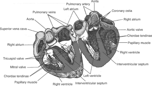
The heart muscles’ blood supply comes directly from the system of coronary arteries that runs within the epicardial layer. Two main coronary arteries, the left coronary artery (LCA) and the right coronary artery (RCA), branch directly off the aorta via the coronary Ostia. These arteries and their branches supply tributary arteries that run perpendicular to the heart surface and transverse from the epicardium, through the myocardium, and down to the endocardium.[3] The LCA quickly branches into the left anterior descending (LAD) coronary artery and the left circumflex (LCX) coronary artery. The LAD runs vertically down the interventricular groove towards the apex and supplies blood to the anterior left ventricular myocardium, the anterior two-thirds of the interventricular septal myocardium, and the anterolateral papillary muscle connecting the mitral valves. The LCX courses horizontally along the atrioventricular groove and gives rise to the left obtuse marginal coronary artery, together supplying the lateral and posterior left ventricular myocardium. The RCA runs horizontally along the right atrioventricular groove and gives rise to the right acute marginal coronary artery, which supplies the right ventricular myocardium. The RCA also gives rise to the posterior descending artery (PDA) in about 90% of the human population (the PDA comes from the LCX in the other approximately 10%), which supplies the posterior myocardium of both ventricles, the posterior one-third of the interventricular septal myocardium, and the posteromedial papillary muscle of the mitral valves.[3] Blood flow via the coronary arteries to the myocardium occurs during diastole and ventricular relaxation via the passive flow of blood into the aortic Ostia. During systole and ventricular contraction, the coronary arteries become compressed, and thus impedes myocardial blood flow.
What is the function of the heart muscle?
Thus, the heart muscle acts as a functional syncytium with rapid synchronized contractions that are responsible for pumping blood throughout the body. Functionally, the heart muscles rely on electrochemical gradients and the potentials to generate contractile force for each heartbeat.
Which veins drain blood from the left ventricular myocardium?
Venous drainage of the left ventricular myocardium is completed by the interventricular vein and the great cardiac vein, which drains into the coronary sinus, found in the posterior right atrioventricular groove, which then drains into the right atrium.[3] The anterior cardiac veins are responsible for draining blood from the right ventricular myocardium directly into the right atrium. [3]
What are the layers of the heart?
There are three layers that make up the heart muscle. They include: Epicardium, Myocardium and the Endocardium.
Which layer of the heart contracts and squeezes the heart?
The Myocardium is the layers of Muscle that contracts and squeezes the heart so blood can travel through the heart with force. The intercalated discs are specific in this layer which controls the calcium which is needed for contraction of the hearts and pressures in the ventricles. The Left side of the heart has greater pressure than the right.
What is the sac around the heart called?
There is also a pericardium which is a sac around the heart as a cover. This protects the surface of the heart.
What are the layers of the heart wall?
It is the cardiac muscle that enables the heart to contract and allows for the synchronization of the heartbeat. The heart wall is divided into three layers: epicardium, myocardium, and endocardium.
Which layer of the heart is in direct contact with the myocardium?
Also found in this heart layer are the coronary blood vessels, which supply the heart wall with blood. The inner layer of the epicardium is in direct contact with the myocardium.
Which layer of the heart is covered by the heart valves?
This layer lines the inner heart chambers, covers heart valves, and is continuous with the endothelium of large blood vessels. The endocardium of heart atria consists of smooth muscle, as well as elastic fibers. An infection of the endocardium can lead to a condition known as endocarditis.
Which layer of the heart is the thickest?
The myocardium is the thickest layer of the heart wall, with its thickness varying in different parts of the heart. The myocardium of the left ventricle is the thickest, as this ventricle is responsible for generating the power needed to pump oxygenated blood from the heart to the rest of the body.
What is the outer protective layer of the heart?
Epicardium: the outer protective layer of the heart.
What is the heart?
The heart is an extraordinary organ. It is about the size of a clenched fist, weighs about 10.5 ounces and is shaped like a cone. Along with the circulatory system, the heart works to supply blood and oxygen to all parts of the body. The heart is located in the chest cavity just posterior to the breastbone, between the lungs, ...
Which muscle fibers are responsible for cardiac conduction?
Cardiac conduction is made possible by specialized myocardial muscle fibers. These fiber bundles, consisting of the atrioventricular bundle and Purkinje fibers, carry electrical impulses down the center of the heart to the ventricles. These impulses trigger the muscle fibers in the ventricles to contract.
What Are the Layers of the Heart?
The heart is partially so efficient because of its shape. The orientation of the different compartments of the heart allows for deoxygenated blood to enter, pass to the lungs to receive oxygen, and exit back to the body. As the heart pumps, the alignment of the valves and compartments of the heart prevent the oxygen-rich blood and oxygen-depleted blood from mixing and flowing backward.
What is the middle layer of the heart?
The myocardium is the middle layer of the heart that is composed of contractile cardiac muscle. It is innervated by the Purkinje fibers, which stimulate it to contract. These contractions are controlled by cardiomyocytes, which act as pacemakers within the heart. The innermost layer of the heart is the endocardium, which is smooth and composes the valves and inner walls of the heart. The innermost surface of the endocardium is the endothelium. The middle layer contains smooth muscle and vessels and is called the elastic tissue layer. The subendocardial is the layer that connects the endocardium to the myocardium. Together, the pericardium and layers of the heart wall support the structure and position of the heart.
What is the innermost layer of the heart?
The innermost layer of the heart is referred to as the endocardium . "Endo" means "inner," thus endocardium refers to the inner portion of the heart. The endocardium is a smooth layer of tissue that lines the inside chambers of the heart. It works to separate blood within the heart from the cardiac muscle composing the myocardium and allows for a slick surface for blood to pass over without getting stuck. The endocardium also contains blood vessels like the epicardium, thus it assists in delivering nutrients and oxygen to tissues of the heart. The valves of the heart are also composed of the endocardium and function to prevent the backflow of fluid through the heart as it pumps.
What is the outer surface of the heart called?
"Epi" refers to outside, and "cardium" refers to the heart. Thus, the epicardium is the outside of the heart. The epicardium is sometimes referred to as the visceral pericardium. This is because it is composed of connective tissue that, in part, connects to the pericardium and forms its second layer.
Which layer of the heart contains blood vessels?
The epicardium is the outer layer that contains blood vessels. The myocardium is the middle layer that is composed of contractile tissues. The endocardium is the innermost layer and composes the valves, inner lining of the chambers, and contains vessels and nerves.
Which layer of the heart is responsible for the passage of material from the heart to the myocardium?
The endothelium is composed of special endothelial cells which assist in the passage of material from the inside of the heart to the myocardium. The elastic layer is composed of another involuntary muscle known as smooth muscle. This is the same type of muscle found within the intestines and provides involuntary contractions. These contractions constrict blood vessels within the endocardium and force blood to move. The subendocardial layer is the layer closest to the myocardium. It acts as a layer of connective tissue to adhere the endocardium to the myocardium.
What are the three layers of the heart wall?
Learn about the pericardium and the three layers of the heart wall. Discover distinctive features of the pericardium, epicardium, myocardium, and endocardium. Updated: 09/23/2021