
The Human Eye (Eyeball) Diagram, Parts and Pictures
- Eyeball. The eyeball is a round gelatinous organ that contains the actual optical apparatus. ...
- Orbit. The orbit is the bony hollow region in the skull that houses the eyeball and is also referred to as the socket.
- Appendages. ...
- Nerves of the Eye. ...
What are the basic parts of the eye?
BASIC PARTS OF THE EYEThis video describes the 8 most common basic anatomy parts of the eyeball. It includes the cornea, lens, iris, pupil, vitreous, retin...
What is this part of the eyelid called?
The orbital septum is an extension of periosteum from the orbital roof (upper eyelid) and orbital floor (lower eyelid). It serves as a barrier for preventing infections/blood/inflammation from spilling over between the anterior eyelid and the orbit.
What are the structures of the eyelid?
- Skin
- Subcutaneous tissue
- Muscle - orbital part of the orbicularis oculi muscle
- Orbital septum - extensions of the periosteum from the orbital margin, that extends through both eyelids and supports them;
- Tarsus - plates of the dense connective tissue present in both eyelids. ...
What are the parts and functions of the eye?
To understand how the eye sees, it helps to know the eye structures and functions:
- Cornea: Light enters through the cornea, the transparent outer covering of the eye. ...
- Aqueous Humor: The fluid beneath the cornea has a composition similar to that of blood plasma. ...
- Iris and Pupil: Light passes through the cornea and aqueous humor through an opening called the pupil. ...

What are the 4 layers of the eyelid?
Upper eyelid (Fig. 2–6) (mnemonic: 4-5-7 rule): The lower 5 mm has 4 layers: skin, orbicularis, tarsus, and conjunctiva.
What are the components of the eyelid?
Each eyelid contains a fibrous plate, called a tarsus, that gives it structure and shape; muscles, which move the eyelids; and meibomian (or tarsal) glands, which secrete lubricating fluids. The lids are covered with skin, lined with mucous membrane, and bordered with a fringe of hairs, the eyelashes.
What are the three layers of the eyelid?
Upper eyelid anatomy: It is clinically useful to divide the anatomical structures in the upper lid into three layers - the superficial anterior lamella, middle lamella and the posterior lamella.
What are the eyelids called?
There are two points at which the upper and lower eyelids meet. The one on the inner aspect is called the medial canthus and the one on the outer aspect is called the lateral canthus.
What is the bottom part of the eyelid called?
The lower eyelid is formed by three lamellae: the anterior, middle, and posterior lamella. The anterior lamella is the skin and orbicularis oculi muscle, the middle lamella is the tarsal plate and orbital septum, and the posterior lamella is the conjunctiva and the lower lid retractors.
How many types of eyelids are there?
The upper lid crease can be divided into three types: (1) a single eyelid (no visible lid crease), (2) a low eyelid crease (low-seated, nasally tapered, including hidden fold), and (3) a double eyelid (well-formed supratarsal crease).
What is the area between the eyelid and eyebrow called?
This space is also called the palpebral fissure. Typically the palpebral fissure measures between 28 to 30 mm wide and around 9 to 10 mm in height.
What is the upper eyelid?
The upper eyelid extends superiorly to the eyebrow, which separates it from the forehead. The lower lid extends below the inferior orbital rim to join the cheek, forming folds where the loose connective tissue of the eyelid is juxtaposed with the denser tissue of the cheek. (See the images below.) Upper eyelid anatomy.
What is eyelid fat called?
Fat Pads. There are a number of different fat pads that are present within and around the eyelid. One layer of fat called the pre-aponeurotic fat is found right behind the orbital septum and in front of the levator aponeurosis.
Do humans have 4 eyelids?
You know that little pink thing nestled in the corner of your eye? It's actually the remnant of a third eyelid. In humans, it's vestigial, meaning it no longer serves its original purpose. There are several other vestigial structures in the human body, quietly riding along from one of our ancestor species to the next.
What are the layers of the eyelid?
The layers are: Subcutaneous connective tissue (the Oculoplastics BCSC book lumps the skin and subcutaneous tissue into one layer, as clinically they are fairly indistinct) Levator palpebrae superioris muscle (not present in the lower eyelid) Müller muscle (inferior tarsal muscle in the lower eyelid)
How many structures are there in the upper eyelid?
There are several ways to mentally organize the multiple layers of the upper eyelid. The Fundamentals BCSC book lists 9 structures, while the Oculoplastics BCSC book lists 7 structures; they are essentially the same lists so there’s no need to fret over which list to memorize.
How many sections are there in the orbicularis oculi muscle?
There are 2 sections of the orbicularis oculi muscle:
Why do my eyelids swell?
The significant eyelid swelling seen in conditions such as preseptal cellulitis (shown above) is caused by the accumulation of fluid in the loose connective tissue.
Which muscle is absent in the superior eyelid?
Eyelid Folds. In non-Asians, the levator palpebrae superioris muscle has some attachments to the upper border of the tarsus, which forms a superior eyelid fold. In non-Asians, the levator palpebrae superioris muscle does not have these attachments, so the superior eyelid fold is minimal or absent.
Why is it important to know the orientation of the eyelid margin?
Knowing the orientation and position of the margin structures is especially important with trauma, where restoration of the anatomy as best as possible is critical.
Why is the right pupil smaller than the left?
The right pupil is smaller than the left pupil (miosis), as a result of loss of the sympathetic tone to the right pupil.
What is the layer of the eyelid made of?
Microscopically, this layer of the eyelid is made of epidermis and dermis. The epidermis has 6-7 layers of stratified squamous epithelium and the dermis is a layer of dense connective tissue. The dermis has elastic fibers, blood vessels, melanocytes, lymphatics, and nerves.
What is the medical term for the eyelid?
The common medical term for eyelid is palpebra. The medical term for eyelids is palpebrae ( plural of palpebra). Any medical words related to eyelid has a prefix palpebra- , e.g., palpebral fissure, levator palpebrae superioris, palpebral glands.
How many eyelids are there in the human eye?
Based on the anatomical position, there are 2 eyelids in an eye: the upper eyelid and the lower eyelid. The superior palpebral sulcus divides the upper lid into a tarsal plate and orbital plate.
What is the difference between the upper and lower eyelids?
In primary gaze position, the lower lid just touches the cornea, and the upper lid covers one-sixth of the cornea. The upper eyelid extends from the superior boundary of the palpebral fissure to the eyebrow. Similarly, the lower eyelid extends from the inferior boundary of the palpebral fissure to merge into the cheek.
How wide is the eyelid margin?
Each eyelid margin is 2 mm wide and is divided into the lacrimal portion and the ciliary portion by the lacrimal papilla. The lacrimal portion covers a one-sixth area of margin medial to the punctum while the ciliary portion covers the five-sixth part of the lid margin, lateral to the punctum.
What is the function of the striated muscle in the upper eyelid?
The main function of LPS is to raise the upper eyelid.
What is the role of the eyelid?
The eyelids play an important role to distribute tears over the anterior surface of the eyeball (cornea and conjunctiva) and contribute to the drainage of the tear. These act as shutters thereby providing protection to the eye from excessive light, foreign particles, chemicals, and injuries.
What is the function of the eyelid?
This can be either voluntarily or involuntarily. The human eyelid features a row of eyelashes along the eyelid margin, which serve to heighten the protection of the eye from dust and foreign debris, as well as from perspiration. "Palpebral" (and "blepharal") means relating to the eyelids. Its key function is to regularly spread the tears and other secretions on the eye surface to keep it moist, since the cor nea must be continuously moist. They keep the eyes from drying out when asleep. Moreover, the blink reflex protects the eye from foreign bodies.
What is the meaning of the eyelid?
The human eyelid features a row of eyelashes along the eyelid margin, which serve to heighten the protection of the eye from dust and foreign debris, as well as from perspiration. "Palpebral" (and "blepharal") means relating to the eyelids.
Why do people have eyelid surgery?
Most of the cosmetic eyelid surgeries are aimed to enhance the look of the face and to boost self-confidence by restoring a youthful eyelid appearance. They are intended to remove fat and excess skin that may be found on the eyelids after a certain age.
Why does my eyelid turn inward?
Entropion usually results from aging, but sometimes can be due to a congenital defect, a spastic eyelid muscle, or a scar on the inside of the lid that could be from surgery, injury, or disease. It is an asymptomatic condition that can, rarely, lead to trichiasis, which requires surgery. It mostly affects the lower lid, and is characterized by the turning inward of the lid, toward the globe.
Why is my eyelid so dry?
Laxity is also another aging-related eyelid condition that can lead to dryness and irritation. Surgery may be necessary to repair the eyelid to its natural position. In certain instances, excessive lower lid laxity creates the Fornix of Reiss – a pocket between the lower eyelid and globe – which is the ideal location to administer topical ophthalmic medications.
How to treat crusty eyelids?
Treatment normally consists in maintaining a good hygiene of the eye and holding warm compresses on the affected eyelid to remove the crusts. Gently scrubbing the eyelid with the warm compress is recommended as it eases the healing process. In more serious cases, antibiotics may be prescribed.
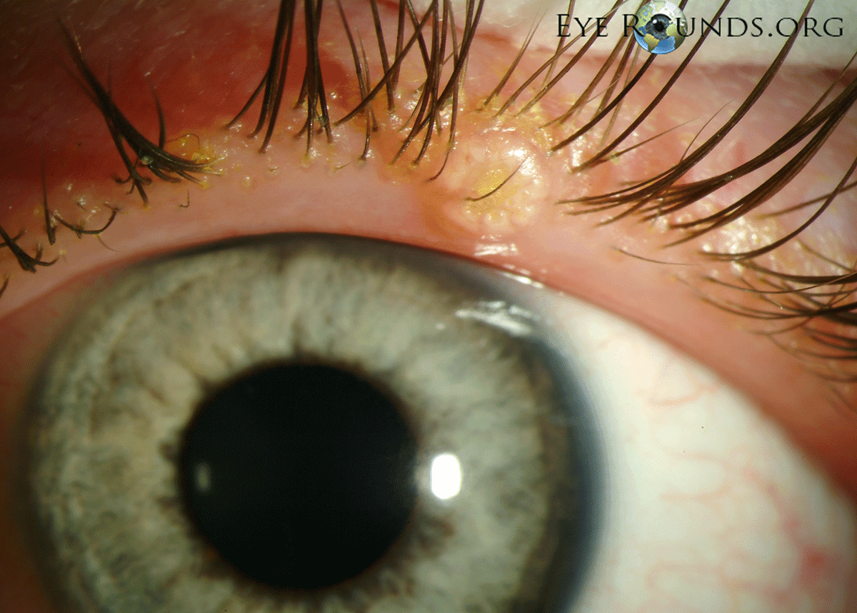
What is a stye eye?
Eyelid affected by stye. Hordeolum ( stye) is an infection of the sebaceous glands of Zeis usually caused by Staphylococcus aureus bacteria, similar to the more common condition Acne vulgaris. It is characterized by an acute onset of symptoms and it appears similar to a red bump placed underneath the eyelid.
What is the eyelid?
Eyelid Anatomy. Our eyes are probably the most important vital structures we have in our body. They discovered on the surface by a thin layer of skin and soft tissue called the eyelids. The eyelids serve multiple purposes including protecting the eyeball from injury, controlling the amount of light that enters the eye and also constantly ...
Where are the lower eyelids located?
Similarly, the lower eyelid lies at the border of the lower limbus. There are two points at which the upper and lower eyelids meet. The one on the inner aspect is called the medial canthus while that at the outer aspect is called the lateral canthus.
What is the function of the eyelid?
All these functions together help maintain the structural integrity of the eyeball and protect them from external influences. From an anatomical point of view, the eyelid consists primarily of skin, underlying soft tissue also called a subcutaneous tissue and a thin layer of muscle called the orbicularis oculi.
What is the sagittal section of the eyelid?
If one were to look at the eyelid in a more detailed manner, a sagittal section taken across the eyelid will offer a clear view of the various structures that form it. Of course, it must be borne in mind that the structures that are visualised depend on the plane at which the sections are taken.
Which branch of the internal carotid artery passes through the orbital septum?
Another branch of the internal carotid artery is the lacrimal artery that passes through the orbital septum along each eyelid and ultimately joins the marginal arcade.
What is the tissue that helps the eyelids open?
Finally, there also exists a small amount of fat tissue as well. The eyeball is covered by a thin layer of tissue called the conjunctiva.
Where does the upper eyelid start?
The upper eyelid starts at the eye and extends up words joined the skin of the forehead. It is distinguished from the forehead skin by the presence of eyebrows. Similarly, the lower eyelid starts at the eye and extends to join the skin of the cheek.
What is the surface of the eye called?
The Surface of the Eye. The surface of the eye and the inner surface of the eyelids are covered with a clear membrane called the conjunctiva. The layers of the tear film keep the front of the eye lubricated. Tears lubricate the eye and are made up of three layers. These three layers together are called the tear film.
What part of the eye is the orbit?
Eye Anatomy: Parts of the Eye Outside the Eyeball. The eye sits in a protective bony socket called the orbit. Six extraocular muscles in the orbit are attached to the eye. These muscles move the eye up and down, side to side, and rotate the eye. The extraocular muscles are attached to the white part of the eye called the sclera.
What is the role of the cornea and lens in the eye?
By helping to focus light as it enters the eye, the cornea and the lens both play important roles in giving us clear vision. In fact, 70% of the eye's focusing power comes from the cornea and 30% from the lens.
What is the muscle that controls the movement of the eyeball?
This is a strong layer of tissue that covers nearly the entire surface of the eyeball. This illustration shows eye muscles , which control eye movement.
What part of the retina is responsible for the transmission of light?
The retina has special cells called photoreceptors. These cells change light into energy that is transmitted to the brain. There are two types of photoreceptors : rods and cones.
What is the function of the pupil?
Directly behind the pupil sits the lens. The lens focuses light toward the back of the eye. The lens changes shape to help the eye focus on objects up close.
What is the drainage angle of the eye?
The eye is always producing aqueous humor. To maintain a constant eye pressure, aqueous humor also drains from the eye in an area called the drainage angle. Behind the anterior chamber is the eye’s iris (the colored part of the eye) and the dark hole in the middle called the pupil. Muscles in the iris dilate (widen) or constrict (narrow) ...
How many muscles are there in the eye?
The eye has six muscles. These muscles arise from the eye socket (orbit) and work to move the eye up and down, side to side, or in a circular motion.
What is the white part of the eye?
The sclera is sometimes known as the "whites" of the eye. It covers more than 80% of the eyeball's surface. 2
What is the membrane that covers the sclera?
The conjunctiva is the membrane covering the sclera (white portion of your eye). The conjunctiva also covers the interior of your eyelids.
How many fibers are in the optic nerve?
The optic nerve is a bundle of about 1.2 million nerve fibers that transmit visual information to the central nervous system (brain). 7
How do eyes work?
The eyes work in the same way as cameras. When you focus on an object, light is reflected and enters the eye through the cornea. As the light passes through, the dome-shaped nature of the cornea bends light , enabling the eye to focus on fine details.
Where are light rays focused?
Light rays are focused on the macula lutea when an eye is looking directly at an object.
Which organ is the source of life that keeps the retina functioning effectively?
In short, the choroid is the source of life that keeps the retina functioning effectively.
What is the eyelid?
Updated on May 23, 2020. An eyelid is a thin layer of skin that covers and protects the eye. The eye contains a muscle that retracts the eyelid to "open" the eye either voluntarily or involuntarily. Human eyelids contain a row of eyelashes that protect the eye from dust particles, foreign bodies, and perspiration. The Eyelid.
What glands are located under the eyelid?
The eyelid contains several different types of glands including sebaceous glands, sweat glands, tear glands, and meibomian glands. Tear glands that give us our every day lubricating tears are small and located throughout the lid. 1 The lacrimal gland, which is located up under the upper eyelid and under the body orbit, secretes reflex tears. The lacrimal gland secretes tears created when we cry emotionally or when we get something in our eye. The lacrimal gland attempts to wash the debris away.
What is the term for the extra eyelid skin that develops in people over 50 years old?
Dermatochalasis : Dermatochalasis is extra eyelid skin that develops in people over 50 years of age. Dermatochalasis develops as a part of the normal aging process. It is caused by fat prolapsing or moving forward and eyelid tissue losing its tone as we get older. Dermatochalasis can be so severe that it blocks your upper visual field. A surgery, known as a blepharoplasty, can be performed to remove this tissue and restore full vision function. 3
Why does my eyelid flip outward?
A long-standing facial paralysis can also cause ectropion. When the eyelid tone becomes weak, simply rolling over on your pillow at night can cause the eyelid to flip outward. Myokymia: Myokymia is the medical term of an eyelid twitch. The skin of the eyelid moves involuntarily.
What is the name of the abnormal, forceful contraction of the eyelid muscles?
Blepharospasm: Blepharospasm is the abnormal, forceful contraction of the eyelid muscles. 7 The exact cause is unknown and it does not seem to be linked to other diseases. Symptoms usually begin slowly but increase over time, and contractions can become forceful and involve both eyelids.
Why does my eyelid move involuntarily?
Myokymia can usually be felt and seen by the sufferer. It is caused by extreme fatigue, stress, anxiety, consuming excess caffeine and spending too much time on the computer. Rest and relaxation usually are all that is needed for myokymia to resolve. 6 .
What is the function of the eyelid?
One of the main functions of the eyelid is to protect the eye and keep out foreign bodies. Another important function of the eyelid is to regularly spread tears on the surface of the eye to keep it moist. With every blink, there is a slight pumping or squeezing mechanism that expresses tears over your eye. Also, there is a slight horizontal movement that pushes tears toward the puncta, the drain pipe for the tears for proper disposable and drainage.
Overview
Clinical significance
Any condition that affects the eyelid is called eyelid disorder. The most common eyelid disorders, their causes, symptoms and treatments are the following:
• Hordeolum (stye) is an infection of the sebaceous glands of Zeis usually caused by Staphylococcus aureus bacteria, similar to the more common condition Acne vulgaris. It is characterized by an acute onset of symptoms and it appears sim…
Structure
The eyelid is made up of several layers; from superficial to deep, these are: skin, subcutaneous tissue, orbicularis oculi, orbital septum and tarsal plates, and palpebral conjunctiva. The meibomian glands lie within the eyelid and secrete the lipid part of the tear film.
The skin is similar to areas elsewhere, but is relatively thin and has more pigment cells. In diseased persons these may wander and cause a discoloration of the lids. It contains sweat gla…
Function
The human eyelid features a row of eyelashes along the eyelid margin, which serve to heighten the protection of the eye from dust and foreign debris.
Anatomical variation
An anatomical variation in humans occurs in the creases and folds of the upper eyelid.
An epicanthic fold, the skin fold of the upper eyelid covering the inner corner (medial canthus) of the eye, may be present based on various factors, including ancestry, age, and certain medical conditions. In some populations the trait is …
Society and culture
Blepharoplasty is a cosmetic surgical procedure performed to correct deformities and improve or modify the appearance of the eyelids. With 1.43 million people undergoing the procedure in 2014, blepharoplasty is the second most popular cosmetic procedure in the world (Botulinum toxin injection is first), and the most frequently performed cosmetic surgical procedure in the world.
East Asian blepharoplasty, or "double eyelid surgery", has been reported to be the most commo…
Additional images
• Horizontal section through the eye of an eighteen days' embryo rabbit. X 30
• Sagittal section of right orbital cavity
• Sagittal section through the upper eyelid
• The tarsi and their ligaments. Right eye; front view
See also
• Cellulitis – a bacterial infection involving the inner layers of the skin
• Dermatochalasis – an excess of eyelid skin that may obstruct vision
• Gland of Moll – a modified sweat gland at the base of the eyelashes