What causes semilunar valves to open and close?
What causes the closing of the aortic Semilunar valve? As the ventricles contract, ventricular pressure exceeds arterial pressure, the semilunar valves open and blood is pumped into the major arteries. This is due to the elevated pressures in the aorta and the pulmonary artery pushing the blood back toward the ventricles to close the semilunar valves.
How many valves are located in the heart?
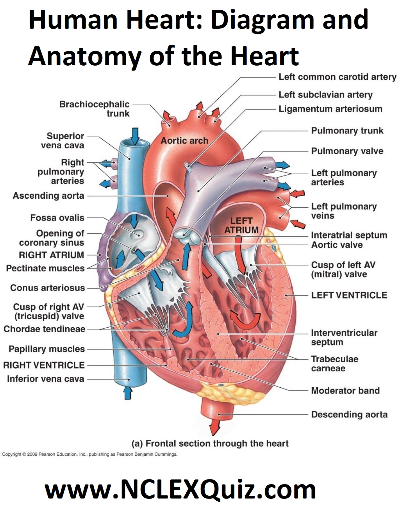
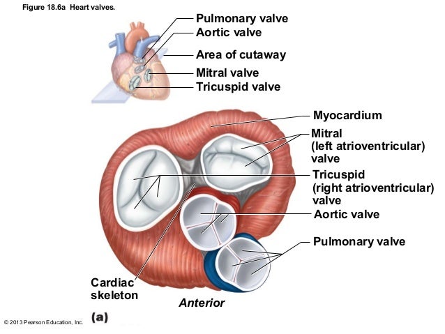
There are four valves within your heart. They are the mitral, tricuspid, aortic and pulmonic valves. The valves make sure blood flows in only one direction through the heart.
What is the function of the valves in the heart?
What are heart valves?
- The process begins when oxygen-depleted blood (from the arms, legs, body, and head) enters the right atrium. ...
- The blood then flows through the tricuspid valve into the right ventricle, which is the lower right pumping chamber.
- The ventricle pumps this blood through the pulmonary valve to the pulmonary artery, where it enters the lungs for oxygenation.
What valves are also known as the semilunar valves?
Things to Remember
- A human heart has four valves; two atrioventricular valves and two semilunar valves.
- The atrioventricular valves, also known as AV valves, are a connector between the ventricles and the atria.
- The two atrioventricular valves are the tricuspid valve and the bicuspid valve.
- Semilunar Valves are a connector between the ventricles and arteries.

How many semilunar valves are in the heart?
two semilunar valvesThere are four valves associated with the heart. The two atrioventricular valves, and the two semilunar valves. the two semilunar valves, the aortic and pulmonary, have a similar design, each consisting of a fibrous valve ring at the base of the vessel, with three leaflets, each occupying a third of closed valve.
What are the semilunar valves and what are their functions?
The semilunar valves are half-moon-shaped leaflets of endocardium and connective tissues, situated between the aorta and the left ventricle and between the pulmonary artery and the right ventricle. These valves permit blood to be forced into the arteries, but prevent backflow from the arteries into the ventricles.
What are the types of semilunar valves?
There are two types of semilunar valves: (1) the aortic valves and (2) the pulmonary valve. The aortic valves have three cusps and lies in between the left ventricle and the aorta. When open, the aortic valves allow the passage of blood from the ventricleinto the arteries.
What are the 2 semilunar valves and where are they located be specific?
The valve between the right ventricle and pulmonary trunk is the pulmonary semilunar valve. The valve between the left ventricle and the aorta is the aortic semilunar valve.
What is the function of semilunar valves quizlet?
What is the main function of the semilunar valves? prevent backflow from the main artery back into the associated ventricle during ventricular diastole.
What are the semilunar valves quizlet?
What are the two names of the semilunar valves? Aortic and pulmonary(semilunar, SL) valves.
What are the 4 types of heart valves?
What are heart valves?Tricuspid valve. Located between the right atrium and the right ventricle.Pulmonary valve. Located between the right ventricle and the pulmonary artery.Mitral valve. Located between the left atrium and the left ventricle.Aortic valve. Located between the left ventricle and the aorta.
Why are they called the semilunar valves?
Semilunar valves connect your heart ventricles (lower chambers) and arteries. Semilunar valves get their name from the crescent moon shape of the flaps that make up the valve.
What is the right semilunar valve called?
The pulmonary valveThe pulmonary valve can also be referred to as the pulmonic valve, the right semilunar valve, and the right arterial valve. Its three leaflets, or cusps, are difficult to name because of the oblique angle of the valve.
Is the aortic valve a semilunar valve?
The aortic valve is one of the four heart valves. It is also called aortic semilunar due to its semilunar shape and is between the left ventricle and the aorta with the function of ensuring that oxygen-rich blood does not go back in its path.
What are the 2 major valves in the heart?
pulmonary valve: located between the right ventricle and the pulmonary artery. mitral valve: located between the left atrium and the left ventricle.
What are the 3 aortic valves?
Structure. The aortic valve normally has three cusps however there is some discrepancy in their naming. They may be called the left coronary, right coronary and non-coronary cusp. Some sources also advocate they be named as a left, right and posterior cusp.
What is the function of valves What are the two atrioventricular and two semilunar valves and where are they located?
The heart has two kinds of valves, atrioventricular and semilunar valves. These valves open and close during the cardiac cycle to direct the flow of blood through the heart chambers and out to the rest of the body.
What is the function of valves What are the two atrioventricular and two semilunar valves and where are they located quizlet?
what are the two atrioventricular and two semilunar valves and where are they located? the valves in the heart keep the blood flowing in one direction. the two atrioventricular valves are located between the atria and the ventricles on each side of the heart.
1. What are the 2 Semilunar Valves?
We all know that there are 4 different valves that are present in the human heart. These are further divided into 2 categories known as the atriove...
2. Where are the Semilunar Valves Located?
There are 2 important valves that make up the entire semilunar valves section of the heart and these are known as the pulmonary and the aortic semi...
3. What is the function of a semilunar valve?
The function of a semilunar valve is to carry the blood to the distant parts of the body. It also prevents the flow of blood from the aorta and pul...
4. What are the differences in semilunar valves between humans and other animals?
The shape and structure of semilunar valves of humans and other animals are slightly different. The muscles in the valves are lesser in humans, as...
5. What is the Atrioventricular valve? How is it different from a semilunar valve?
The atrioventricular valves divide into the two atria and ventricles. They don’t let the backflow of blood from ventricles to atria. Semilunar valv...
What is a semilunar valve?
Semilunar Valves. The semilunar valves are flaps of endocardium and connective tissue reinforced by fibers which prevent the valves from turning inside out. They are shaped like a half moon, hence the name semilunar (semi-, -lunar).
What Are Heart Valves?
Heart valves are vital to the proper circulation of blood in the body. The heart has two kinds of valves, atrioventricular and semilunar valves. These valves open and close during the cardiac cycle to direct the flow of blood through the heart chambers and out to the rest of the body. Heart valves are formed from elastic connective tissue which provides the flexibility needed to open and close properly. Malfunctioning heart valves inhibit the heart's ability to pump blood and life giving oxygen and nutrients to the cells of the body.
What is the heart valve located between the right ventricle and the pulmonary artery?
Pulmonary Valve: This heart valve is located between the right ventricle and pulmonary artery. When closed, it prevents the back flow of blood as it is pumped from the right ventricle to the pulmonary artery. When open, it allows oxygen-depleted blood to be pumped from the right ventricle to the pulmonary artery.
What is the valve that allows blood to flow into the right ventricle?
When open, it allows blood from the right atrium to flow into the right ventricle. Mitral Valve: This heart valve is located between the left atrium and left ventricle. When closed, it allows the left atrium to fill with oxygen-rich blood returning to the heart from the pulmonary veins.
Why do valves regurgitate?
Valve regurgitation occurs when valves don't close correctly allowing blood to flow backward into the heart. In valve stenosis, valve openings become narrow due to enlarged or thickened valve flaps.
What is the audible sound made by the closing of the heart valves?
The audible sounds that can be heard from the heart are made by the closing of the heart valves. These sounds are referred to as the "lub-dupp" sounds. The "lub" sound is made by the contraction of the ventricles and the closing of the atrioventricular valves. The "dupp" sound is made by the semilunar valves closing.
Where are the AV valves located?
The atrioventricular valves are thin structures that are composed of endocardium and connective tissue. They are located between the atria and the ventricles . Tricuspid Valve: This heart valve is located between the right atrium and the right ventricle.
What are the two semilunar valves in the heart?
The aortic semilunar valve, which protects the point of connection between the aorta and the left ventricle of the heart, and the pulmonary semilunar valve, which protects the point of attachment between the pulmonary artery and the right ventricle of the heart, are the two semilunar valves. Only when the semilunar valves are closed do the ventricles fill with blood.
What is Semilunar Valve?
A mammal’s heart includes four valves: The atria and ventricles are separated by the two atrioventricular valves (AV valves). They restrict blood from returning from the ventricles to the atria. The bicuspid (mitral) and tricuspid valves are the two kinds.
What are the two types of semilunar valves?
The aortic and pulmonary valves are the two types of semilunar valves. The aortic valves, which are located between the left ventricle and the aorta, contain three cusps. The aortic valves allow blood to flow from the ventricle into the arteries when they are open. It stops blood from returning to the ventricle when it is closed. The cardiac valve that connects the right ventricle to the pulmonary artery is known as the pulmonary valve. The pulmonary valve, like the aortic valve, contains three cusps. It also opens at the start of ventricular systole and shuts at the conclusion. The second heart sound is caused by the closing of these two valves.
How many cusps are in the pulmonary and aortic valves?
Both the aortic and pulmonary valves are made up of three cusps. Endocardium foldings cover the cusps of the valves of the heart. Semilunar valve cusps are thinner than atrioventricular valve leaflets, but their structure is identical, with the exception that they lack chordae tendineae. The aortic valve allows blood to flow from the left ventricle into the arteries in one direction.
What are the leaflets of the heart called?
Flaps or leaflets make up the semilunar valves in the heart. Leaflets are termed “semilunar ” because their edges are linked to the artery wall in a half-moon shape. Each aortic and semilunar valve leaflet contains a fibrous tissue core and is lined by an endothelial tissue with an elastin coating. As a result, it’s known as the backbone, and it’s made up of a collagen layer that makes it looser.
Why do pulmonary arteries make a noise when they close?
When the aortic and pulmonary arteries close quickly, the second sound is produced by the movement of blood inside elastic large vessels (such as the aorta and pulmonary artery) as well as the two ventricles. Valves do not make a sound when they open. As a result, the noises are only created when the valves close, not when they open.
Which valves are connected to the ventricle walls?
The atrioventricular valves are connected to the ventricle walls via the chordae tendineae. This attachment keeps valves from inverting. When the heart muscles contract, the pressure created by the flow of blood affects the valve’s opening and closing completely. The mitral valve, which is made up of two leaflets, the aortic and posterior leaflets, is positioned on the left side and allows blood to flow from the left atrium to the left ventricle.
Which valve in the left ventricle pumps blood into the aorta?
Next, the left ventricle pumps blood through the aortic semilunar valve into the aorta. The aorta is the largest blood vessel in your body and carries blood with oxygen from the heart to the body. The semilunar valve prevents blood from flowing back into the left ventricle and keeps it moving towards the body.
Which valve pumps blood to the lungs?
The right ventricle pumps the blood to the lungs through another valve, called the pulmonary semilunar valve, and then it returns to the left atrium. The blood flows through the bicuspid valve into the left ventricle.
How Does the Heart Work?
To learn more about the semilunar valve, let's take a look at how blood flows through the heart. Blood enters the heart through the inferior and superior vena cava. These vessels bring blood that has been used by the body back to the heart so that it can get more oxygen. The blood flows into the right atrium, the first chamber of the heart. The blood collects there until the tricuspid valve opens, allowing it to flow to a larger chamber called the right ventricle. After all the blood enters the ventricle, the tricuspid valve slams shut, preventing blood from flowing back into the right atrium. This is very important because it causes blood to flow in only one direction through the heart and blood vessels.
How do valves work?
All valves function to prevent blood backflow into the heart, acting like doors between the chambers. Imagine if you had a tube of toothpaste. The bottom end is sealed, right? This is analogous to the valve in the heart. It seals one chamber from another. Then imagine you squeeze the toothpaste. The toothpaste only comes out the cap end, right? Right! But, if we cut the seal off of the bottom and squeezed; toothpaste would seep from both ends, and we would have a mess!
Why is the tricuspid valve important?
This is very important because it causes blood to flow in only one direction through the heart and blood vessels.
What are the problems with the heart valves?
There are two main problems that can occur with the valves in the heart: stenosis and regurgitation. During stenosis, the valves become hardened through excess plaque or from scarring due to trauma of the heart. This means that the blood cannot flow as easily through the valve, and less blood gets to the body.
What happens if the valve doesn't shut?
If the valve didn't shut once the blood enters the next chamber, as the chamber squeezed, or contracted, to pump the blood, the blood would go in both directions. This would push the blood backwards.
How many valves are there in the heart?
The heart has four valves - one for each chamber of the heart. The valves keep blood moving through the heart in the right direction. The mitral valve and tricuspid valve are located between the atria (upper heart chambers) and the ventricles (lower heart chambers). The aortic valve and pulmonic valve are located between the ventricles and ...
What valves close when the right ventricle is full?
Closed tricuspid and mitral valves. When the right ventricle is full, the tricuspid valve closes and keeps blood from flowing backward into the right atrium when the ventricle contracts (squeezes). When the left ventricle is full, the mitral valve closes and keeps blood from flowing backward into the left atrium when the ventricle contracts.
How does blood flow from the right atrium to the left ventricle?
1. Open tricuspid and mitral valves. Blood flows from the right atrium into the right ventricle through the open tricuspid valve, and from the left atrium into the left ventricle through the open mitral valve. 2. Closed tricuspid and mitral valves.
What is the tissue that supports the leaflets of the mitral valve?
The leaflets are attached to and supported by a ring of tough, fibrous tissue called the annulus. The annulus helps to maintain the proper shape of the valve. The leaflets of the mitral and tricuspid valves are also supported by: Chordae tendineae: tough, fibrous strings. These are similar to the strings supporting a parachute.
How do valves work?
How Valves Work. The four valves are to open and close to let blood flow through the heart. The steps below show how the blood flows through the heart and describes how each valve works to keep blood moving. 1.
Where is blood pumped out of the lungs?
Blood is pumped out of the right ventricle through the pulmonic valve into the pulmonary artery to the lungs. As the left ventricle begins to contract, the aortic valve is forced open. Blood is pumped out of the left ventricle through the aortic valve into the aorta.
Where is the aortic valve located?
The aortic valve and pulmonic valve are located between the ventricles and the major blood vessels leaving the heart.
What are the valves of the heart?
Valve anatomy is complex (Figure 1). The mitral and tricuspid atrioventricular ( AV) valves separate the atria from the ventricles, while the aortic and pulmonary semilunar (SL) valves separate the ventricles from the great arteries. AV valves have leaflets and SL valves have cusps. There is a specialized support structure specific to AV valves, while the distinct shape of SL valves creates a unique self-contained support structure within the arterial roots (9, 10). In contrast to the aorta, the aortic root is made up of the fibrous valve annulus region and the arterial tissue within the sinuses of Valsalva. The AV valves are characterized by large asymmetric leaflets hinged to ring shaped annuli on the secured end and tethered to the ventricles by an elaborate apparatus made up of the chordae tendineae and papillary muscles on the mobile end. The fibrous skeleton of the heart is continuous with the annulus fibrosa that constitutes the interconnected fibrous cartilage-like support apparatus of the tricuspid, mitral, and aortic valves. The annulus fibrosa is connected to the muscle of the heart in a manner that is analogous to the attachment of tendon to skeletal muscle (11, 12). The pulmonary valve is separated from the other valves by a muscular sleeve and has a poorly defined, less substantial annulus structure. The annuli of the AV valves are ring-shaped; however, the annulus of the aortic valve is crown-shaped resulting in the “semilunar” shape of the individual cusps (13, 14).
How does the heart valve function?
The heart valves function essentially to maintain unobstructed unidirectional blood flow. The hemodynamics of the normal mature heart are well established (62). Blood flows from low pressure atria to higher pressure ventricles, which in turn supply the great arteries. The left side of the heart maintains significantly higher pressures than the right side. As a result, the impact of various physiologic forces depends on the position and hemodynamic environment of the valve. Valve composition and biomechanics reflect underlying hemodynamics. There are three basic loading states that affect valve tissue during the cardiac cycle: flexure, shear and tension. Flexure occurs when the valve is actively opening or closing, shear occurs when blood is passing through the open valve, and tension occurs when the valve is closed (4, 63). Shear, compressive, and longitudinal stresses contribute to valve deformation, or displacement of the valve tissue during the constant motion of the cardiac cycle (64). Valve tissue has exceptionally high strain because the tissue cycles to a completely unloaded state with each heart beat (49). These deformation forces result in a compensatory balance in cell matrix composition. For example, comparison of porcine aortic and pulmonary valves demonstrates that the left sided aortic valve is thicker, predominantly as a result of increased collagen expression and increased thickness of the fibrosa layer (Alfieri, Carruthers, Yutzey, and Sacks, unpublished data). The heart beats more than 100,000 times per day handling approximately 5 liters of blood per minute. Over the average lifetime, there are greater than 3 billion heart beats, or cardiac cycles. Valve failure may result from an underlying predisposing genotype and valve malformation that alters the response to physiologic stresses. The long held appreciation of age-related degeneration (“wear and tear”) and latent valve disease may in fact represent subtle defects in valve tissue maintenance as regulated by developmental pathways.
What are the SL and AV valves?
SL and AV valves with distinct structural and functional features are present in the human heart (A). The mitral valve (MV) is an AV valve and connects the left atrium (LA) to the left ventricle (LV). The MV consists of an annulus (A, blue line), leaflets and chordae tendineae (CT) that insert into papillary muscles (PM) in the myocardial wall. The aortic valve (AoV) is a SL valve and connects the LV to the aorta (Ao). The AoV consists of an annulus (A, red line) and cusps anchored within the aortic root (Root). Pentachrome staining shows valve ECM structure and composition in human (B,C) and mouse (D,E) aortic valves. At low magnification, SL valve tissue demonstrates cusp and annulus regions in human and mouse (B,D). At high magnification, aortic valve cusp architecture demonstrates similar ECM organization in human and mouse (C,E). The collagen-rich fibrosa layer (F) is oriented on the arterial aspect of the cusp, while the elastin-rich ventricularis layer (V) is oriented on the ventricular aspect of the cusp. The proteoglycan-rich spongiosa layer (S) interconnects the collagen and elastin fibers. IVS interventricular septum. (Panel A from reference (115), with permission.)
Where do heart valve cells come from?
Heart valve cells come from multiple sources in the developing embryo. The endothelial cells that surround the valve leaflets form a continuous epithelial cell layer with the endocardium (2). In the OFT, both the endocardial and myocardial precursors arise from the secondary heart field (26). During the early stages of endocardial cushion formation, the mesenchymal cells of the AV and OFT cushions are derived from endothelial cells, as determined by Tie2-Cre;ROSA26Rreporter lineage tracing in mice (27). In the mature AV valves, the VICs also are derived primarily, if not entirely, from Tie2-Creexpressing endothelial cells (20, 21). In mice, there is little if any contribution of VICs in the AV valves from epicardially-derived cells, as indicated by Wilms Tumor 1 (WT1)-Crelineage analysis (28). However, chick-quail chimera studies in avian embryos have reported significant contributions of epicardium-derived cells in the developing AV valves (27, 29). In the developing OFT cushions, there are significant numbers of neural crest-derived cells, as demonstrated by Wnt1-Crelineage studies in mice (27). In the mature SL valves, the cells of neural crest origin persist and are concentrated in individual cusps of the pulmonary and aortic valves (30)(Mead and Yutzey, unpublished). Overall, lineage tracing studies in mice demonstrate that the majority of VICs arise from endothelially-derived progenitors in the endocardial cushions. However, there is increasing evidence that specific subpopulations in individual valve leaflets arise from distinct embryonic sources. It is not known if these cells from diverse embryonic origins represent different subpopulations of VICs with specific contributions to mature valve structure and function.
How many people die from valve disease annually?
Valve disease results in approximately 20,000 deaths annually (65). The prevalence of aortic valve disease is 2.5% in the United States, corrected for age (66). Aortic valve sclerosis, a marker of valve disease and cardiovascular risk, is present in more than 25% of the aged (67). The actual direct cost for valve disease in the United States alone has been estimated at 1 billion dollars per year (68). Taken together, the public health impact of valve disease and burden to society is underappreciated. Valve disease may manifest as stenosis, an obstruction to outflow, or regurgitation, a defective closure resulting in backward flow. Valve disease tends to progress. Ultimately, ventricular function can be compromised. Aortic valve stenosis is the most common form of valve disease and classically manifests as angina, syncope and heart failure. The diagnosis can be made clinically and confirmed by echocardiography, which quantifies the severity, and, over time, the progression of disease (62). The majority of valve disease at any age has an underlying valve malformation suggesting a genetic basis (8).
Which heart valve is the most common site of disease?
Any one of the four heart valves can be affected; however, the aortic valve is the most common site of disease (7). Aortic valve malformation, including bicuspid aortic valve (BAV), is present in 1–2% of the general population suggesting a developmental origin (8).
What is the annulus fibrosa?
The annulus fibrosa is connected to the muscle of the heart in a manner that is analogous to the attachment of tendon to skeletal muscle (11, 12). The pulmonary valve is separated from the other valves by a muscular sleeve and has a poorly defined, less substantial annulus structure.
Which ventricle forms a complete circle in cross section?
A) The right ventricle forms a complete circle in cross section.
When does ventricular filling begin?
As soon as ventricular pressure falls below atrial pressure, the atrioventricular valve will open and ventricular filling will begin.
Which fibers initiate spontaneous action potentials, which cause the ventricles to contract early?
C) Purkinje fibers initiate spontaneous action potentials, which cause the ventricles to contract early.
Can ventricles depolarize on their own?
C) Yes, because the ventricles will depolarize on their own without nodal stimulation at a rate of 50 times per minute.