Moving from outside the bone to inside the bone, here are the layers:
- Periosteum
- Cortical, or Hard Bone
- Cancellous, or Spongy Bone
- Bone Marrow
How many layers are there in a bone?
The two layers of compact bone and the interior spongy bone work together to protect the internal organs. If the outer layer of a cranial bone fractures, the brain is still protected by the intact inner layer. Figure 6.3.3 – Anatomy of a Flat Bone: This cross-section of a flat bone shows the spongy bone (diploë) covered on either side by a ...
What are the three layers of the bone?
- The outer surface of bone is called the periosteum (say: pare-ee-OSS-tee-um).
- The next layer is made up of compact bone.
- Within the compact bone are many layers of cancellous (say: KAN-sell-us) bone, which looks a bit like a sponge.
What is the innermost layer component of a bone?
There are 3 types of bone tissue:
- Compact tissue. This is the harder, outer tissue of bones.
- Cancellous tissue. This is the sponge-like tissue inside bones.
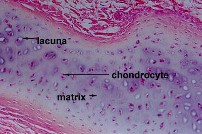
- Subchondral tissue. This is the smooth tissue at the ends of bones, which is covered with another type of tissue called cartilage. Cartilage is a specialized, rubbery connective tissue.
What are the layers of bone tissue?
covering of bone made of two layers; the outer fibrous layer made of dense irregular connective tissue, and the inner osteogenic layer made of elastic fibers and bone cells. This is involved in bone growth (in width) and also serves as a point of attachment for tendons and ligaments. medullary cavity (marrow cavity)
What are the two layers of bone called?
Introduction. Types of Bones: Histologically, bones categorize into two types (1) cortical or compact bone and (2) cancellous bone or spongy bone.
What are layers of bone?
Bones are composed of two layers: a tough outer layer and a spongy inner layer. The outer layer, known as cortical or compact bone, is strong and dense. The inner layer, known as trabecular or cancellous bone, features a light network of connective tissue.
What are the 3 layers of bone?
Bone tissuePeriosteum – the dense, tough outer shell that contains blood vessels and nerves.Compact or dense tissue – the hard, smooth layer that protects the tissue within.Spongy or cancellous tissue – the porous, honeycombed material found inside most bones, which allows the bone to be strong yet lightweight.More items...
What is the outer layer of bone?
The tough, thin outer membrane covering the bones is called the periosteum. Under the hard outer shell of the periosteum are tunnels and canals. Through these, blood and lymphatic vessels carry nourishment for the bone. Muscles, ligaments, and tendons may attach to the periosteum.
What are the two parts of a long bone?
The structure of a long bone allows for the best visualization of all of the parts of a bone ( [link] ). A long bone has two parts: the diaphysis and the epiphysis. The diaphysis is the tubular shaft that runs between the proximal and distal ends of the bone. The hollow region in the diaphysis is called the medullary cavity, which is filled with yellow marrow. The walls of the diaphysis are composed of dense and hard compact bone.
What is the anatomical structure of a long bone?
Anatomy of a Long Bone. A typical long bone shows the gross anatomical characteristics of bone. The wider section at each end of the bone is called the epiphysis (plural = epiphyses), which is filled with spongy bone. Red marrow fills the spaces in the spongy bone.
What type of cells are undifferentiated and develop into osteoblasts?
Osteogenic cells are undifferentiated and develop into osteoblasts. When osteoblasts get trapped within the calcified matrix, their structure and function changes, and they become osteocytes. Osteoclasts develop from monocytes and macrophages and differ in appearance from other bone cells.
What is the red marrow in the bone?
Red marrow fills the spaces in the spongy bone. Each epiphysis meets the diaphysis at the metaphysis, the narrow area that contains the epiphyseal plate (growth plate), a layer of hyaline (transparent) cartilage in a growing bone.
What is the medullary cavity?
The medullary cavity has a delicate membranous lining called the endosteum (end- = “inside”; oste- = “bone”), where bone growth, repair, and remodeling occur. The outer surface of the bone is covered with a fibrous membrane called the periosteum (peri – = “around” or “surrounding”).
Where are osteogenic cells found?
Immature osteogenic cells are found in the deep layers of the periosteum and the marrow.
Which part of the bone is the outer surface of the bone?
The periosteum forms the outer surface of bone, and the endosteum lines the medullary cavity. Flat bones, like those of the cranium, consist of a layer of diploë (spongy bone), lined on either side by a layer of compact bone ( [link] ).
What is the outer layer of bone called?
These cells are part of the outer double layered structure called the periosteum (peri – = “around” or “surrounding”).
What is bone tissue?
function? Bone tissue (osseous tissue) differs greatly from other tissues in the body. Bone is hard and many of its functions depend on that characteristic hardness. Later discussions in this chapter will show that bone is also dynamic in that its shape adjusts to accommodate stresses. This section will examine the gross anatomy ...
What is the wider section of the bone called?
The wider section at each end of the bone is called the epiphysis (plural = epiphyses), which is filled internally with spongy bone, another type of osseous tissue. Red bone marrow fills the spaces between the spongy bone in some long bones. Each epiphysis meets the diaphysis at the metaphysis.
Where do osteocytes get their nourishment?
The spongy bone and medullary cavity receive nourishment from arteries that pass through the compact bone. The arteries enter through the nutrient foramen (plural = foramina), small openings in the diaphysis ( Figure 6.3.10 ). The osteocytes in spongy bone are nourished by blood vessels of the periosteum that penetrate spongy bone and blood that circulates in the marrow cavities. As the blood passes through the marrow cavities, it is collected by veins, which then pass out of the bone through the foramina.
What is the epiphysis covered with?
In this region, the epiphyses are covered with articular cartilage, a thin layer of hyaline cartilage that reduces friction and acts as a shock absorber. Figure 6.32 – Periosteum and Endosteum: The periosteum forms the outer surface of bone, and the endosteum lines the medullary cavity.
What is the diaphysis of a long bone?
The diaphysis is the hollow, tubular shaft that runs between the proximal and distal ends of the bone. Inside the diaphysis is the medullary cavity, which is filled with yellow bone marrow in an adult.
What is inside the diaphysis?
Inside the diaphysis is the medullary cavity, which is filled with yellow bone marrow in an adult. The outer walls of the diaphysis (cortex, cortical bone) are composed of dense and hard compact bone, a form of osseous tissue. Figure 6.3.1 – Anatomy of a Long Bone: A typical long bone showing gross anatomical features.
What are the two types of bone?
Share on Pinterest. Bones are composed of two types of tissue: 1. Compact (cortical) bone: A hard outer layer that is dense, strong, and durable. It makes up around 80 percent of adult bone mass. 2. Cancellous (trabecular or spongy) bone: This consists of a network of trabeculae or rod-like structures.
What are the two things that bones provide?
Bones provide a frame to support the body. Muscle s, tendons, and ligaments attach to bones. Without anchoring to bones, muscles could not move the body. Some bones protect the body’s internal organs. For instance, the skull protects the brain, and the ribs protect the heart and lungs.
What are the components of collagen?
Organic components, being mostly type 1 collagen. Inorganic components, including hydroxyapatite and other salts, such as calcium and phosphate. Collagen gives bone its tensile strength, namely the resistance to being pulled apart. Hydroxyapatite gives the bones compressive strength or resistance to being compressed.
How many types of bones are there in the human body?
There are five types of bones in the human body: Long bones: These are mostly compacted bone with little marrow and include most of the bones in the limbs. These bones tend to support weight and help movement. Short bones: Only a thin layer of compact bone, these include bones of the wrist and ankle.
What is the mineral that gives bones strength?
The mineral calcium phosphate hardens this framework, giving it strength. More than 99 percent of our body’s calcium is held in our bones and teeth. Bones have an internal structure similar to a honeycomb, which makes them rigid yet relatively light.
What is the largest bone in the human body?
The largest bone in the human body is the thighbone or femur, and the smallest is the stapes in the middle ear, which are just 3 millimeters (mm) long. Bones are mostly made of the protein collagen, which forms a soft framework. The mineral calcium phosphate hardens this framework, giving it strength.
Why are bones important?
Although they get less attention than other body parts, bones are more than just a protective scaffold on which the human body is built. Bones also maintain appropriate levels of many compounds and regulate hormonal pathways. Bones are the unsung heroes of anatomy.
What is the smooth tissue at the ends of bones?
The smooth tissue at the ends of bones, which is covered with another type of tissue called cartilage. Cartilage is the specialized, gristly connective tissue that is present in adults. It is also the tissue from which most bones develop in children.
How many bones are there in the human body?
Bones are classified by their shape—as long, short, flat, and irregular. Primarily, they are referred to as long or short. There are 206 bones in the human skeleton, not including teeth and sesamoid bones (small bones found within cartilage): 80 axial bones.
What is the tissue that makes up the body's skeleton?
What is bone? Bone is living tissue that makes up the body's skeleton. There are 3 types of bone tissue, including the following: Compact tissue. The harder, outer tissue of bones. Cancellous tissue. The sponge-like tissue inside bones. Subchondral tissue. The smooth tissue at the ends of bones, which is covered with another type ...
What is the outer membrane of the bone called?
The tough, thin outer membrane covering the bones is called the periosteum. Beneath the hard outer shell of the periosteum are tunnels and canals through which blood and lymphatic vessels run to carry nourishment for the bone. Muscles, ligaments, and tendons may attach to the periosteum.
What is the function of a bone cell?
Found within the bone, its function is to form new bone tissue. Osteoclast. A very large cell formed in bone marrow, its function is to absorb and remove unwanted tissue. Osteocyte. Found within the bone, its function is to help maintain bone as living tissue. Hematopoietic.
What is the role of bone in the body?
Bone provides shape and support for the body, as well as protection for some organs . Bone also serves as a storage site for minerals and provides the medium—m arrow—for the development and storage of blood cells.
Why are bones important?
Because of the complexities of a bone's function, from providing strength and support for the body, to serving as a site for development and storage of blood cells, there are many disorders and diseases that can affect bone.
What is compact bone?
Bone Structure. Compact bone is the hard material that makes up the shaft of long bones and the outside surfaces of other bones. Compact bone consists of cylindrical units called osteons. Each osteon contains concentric lamellae (layers) of hard, calcified matrix with osteocytes (bone cells) lodged in lacunae (spaces) between the lamellae.
What is the medullary cavity?
The medullary cavity, or marrow cavity, is the open area within the diaphysis. The adipose tissue inside the cavity stores lipids and forms the yellow marrow. Articular cartilage covers the epiphysis where joints occur.
What is the membrane covering the outside of the diaphysis?
The periosteum is the membrane covering the outside of the diaphysis (and epiphyses where articular cartilage is absent). It contains osteoblasts (bone‐forming cells), osteoclasts (bone‐destroying cells), nerve fibers, and blood and lymphatic vessels. Ligaments and tendons attach to the periosteum.
What is the membrane that lines the marrow cavity?
The endosteum is the membrane that lines the marrow cavity. Here are the main features of short, flat, and irregular bones: In short and irregular bones, spongy bone tissue is encircled by a thin layer of compact bone tissue. In flat bones, the spongy bone tissue is sandwiched between two layers of compact bone tissue.
Where are red blood cells formed?
It is in the epiphyses where red blood cells are formed. The metaphysis is the area where the diaphysis meets the epiphysis. It includes the epiphyseal line, a remnant of cartilage from growing bones. The medullary cavity, or marrow cavity, is the open area within the diaphysis.
Is a central canal necessary for a long bone?
Thus, no central canal is necessary. Figure 1.Main features of a long bone. The diaphysis, or shaft, is the long tubular portion of long bones. It is composed of compact bone tissue. The epiphysis (plural, epiphyses) is the expanded end of a long bone. It is in the epiphyses where red blood cells are formed.
Which layer of the periosteum contains osteoblasts?
The inner layer of the periosteum is also referred to as the cambrium. It contains osteoblast cells. Osteoblasts are bone-forming cells. They’re very important during the fetal and childhood phases of life when bone tissue is still developing. As a result, the inner layer of the periosteum is thick and rich in osteoblasts in ...
Where do blood vessels enter the bone?
They can pass into the dense and compact layer of bone tissue below, called the bone cortex. Blood vessels enter the bone through channels called Volkmann canals that lie perpendicular to the bone. From there, the blood vessels enter another group of channels called Haversian canals, which run along the length of the bone.
What is the inner layer of the periosteum?
As a result, the inner layer of the periosteum is thick and rich in osteoblasts in the fetus and during early childhood. The inner layer of the periosteum becomes thinner with age. This thinning begins in childhood and continues through adulthood. In many cases, the inner layer becomes so thin that it’s hard to distinguish from the outer layer ...
What is the periosteum?
— Written by Jill Seladi-Schulman, Ph.D. — Updated on September 27, 2018. The periosteum is a membranous tissue that covers the surfaces of your bones. The only areas it doesn’t cover are those surrounded by cartilage and where tendons and ligaments attach to bone.
Can osteoblasts repair fractures?
In many cases, the inner layer becomes so thin that it’s hard to distinguish from the outer layer of the periosteum. If a fracture occurs in adult bone, osteoblasts can still be stimulated to repair the injury. But the rate of regeneration will be slower than it is in a child.