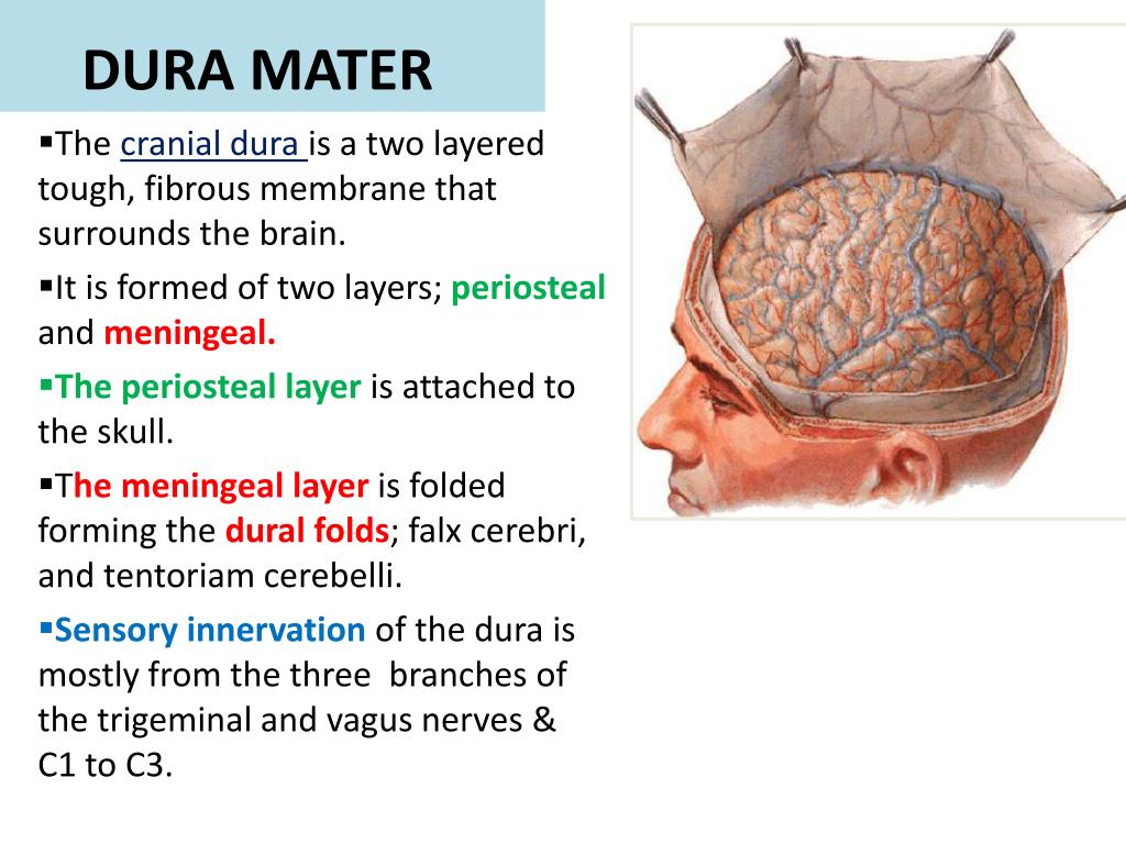
- Macroscopic anatomy. The dura mater is comprised of two distinct layers, the periosteal (superficial) and meningeal (deep) layers.
- Dura mater. The dura mater receives its own blood supply, primarily from the middle meningeal artery (MMA). ...
- Arachnoid mater. The arachnoid mater is a transparent, bi-layered structure found directly below the meningeal layer of the dura mater.
- Pia mater. Meningitis is a life-threatening condition in which the pia and arachnoid mater become acutely inflamed.
- Spinal cord. At the level of the spinal cord, it is important to note that you will also encounter the same three meningeal layers along the full length.
- Key points. There are three distinct meningeal layers, the dura, arachnoid and pia mater, that serve key structural and homeostatic functions for the brain.
- References. Geeky Medics. Cerebrospinal Fluid (CSF) Interpretation. Published in 2017. Geeky Medics. Lumbar Puncture (LP) – OSCE Guide.
What is the structure of the dura mater?
The dura mater is made up of fibroblasts and large amounts of extracellular collagen. The dura mater is composed of two layers: the periosteal/endosteal layer and the meningeal layer. The dural venous sinuses are between these two layers.
What is the periosteal layer of the dura mater?
The periosteal or endosteal layer of the dura mater is simply a layer of periosteum that covers the inner surface of the skull. The layer does not extend beyond the foramen magnum to become contiguous with the dura mater of the spinal cord. The spinal cord dura mater has no periosteal layer.
Where is the dura mater located in the skull?
The dura mater is the outermost layer of the meninges and is located directly underneath the bones of the skull and vertebral column. It is thick, tough, and inextensible. The dura mater consists of two layered sheets of connective tissue: Periosteal layer – lines the inner surface of the bones of the cranium.
What is the potential space between the dura mater and arachnoid mater?
The potential space between the dura mater and the arachnoid mater, which is the second membrane that is located in the middle, is referred to as the subdural space. The pia mater is the innermost membrane that adheres to the surface of the brain.
What is the inner layer of the dura mater?
Cranial dura mater has two layers called lamellae, a superficial layer (also called the periosteal layer), which serves as the skull's inner periosteum, called the endocranium and a deep layer called the meningeal layer.
What are the 3 layers of the dura?
Three layers of membranes known as meninges protect the brain and spinal cord. The delicate inner layer is the pia mater. The middle layer is the arachnoid, a web-like structure filled with fluid that cushions the brain. The tough outer layer is called the dura mater.
Does the spinal cord have two layers of dura mater?
Your brain and spinal cord are protected and supported by three meningeal layers. These membrane layers are the dura mater, arachnoid mater and pia mater.
What occurs at the places where the two layers of dura mater are separated and form pockets or sinuses?
intracranial venous sinusesNormal Anatomy of the Cerebrospinal Fluid Compartment The two layers of dura mater are ordinarily adherent to each other, but they become separated in places to form the walls of the intracranial venous sinuses where CSF is eventually absorbed.
What is dura mater quizlet?
dura mater. Thick, outermost layer of the meninges surrounding and protecting the brain and spinal cord.
Which of the following meninges has two layers?
The outermost mater of the meninges, the dura, is composed of two layers: the periosteal layer that lies closest to the calvarium and the meningeal layer that lies closest to the brain tissue. These together contribute to the dura being a thick, dense, fibrous membrane that is quite inelastic.
How many dural folds are there?
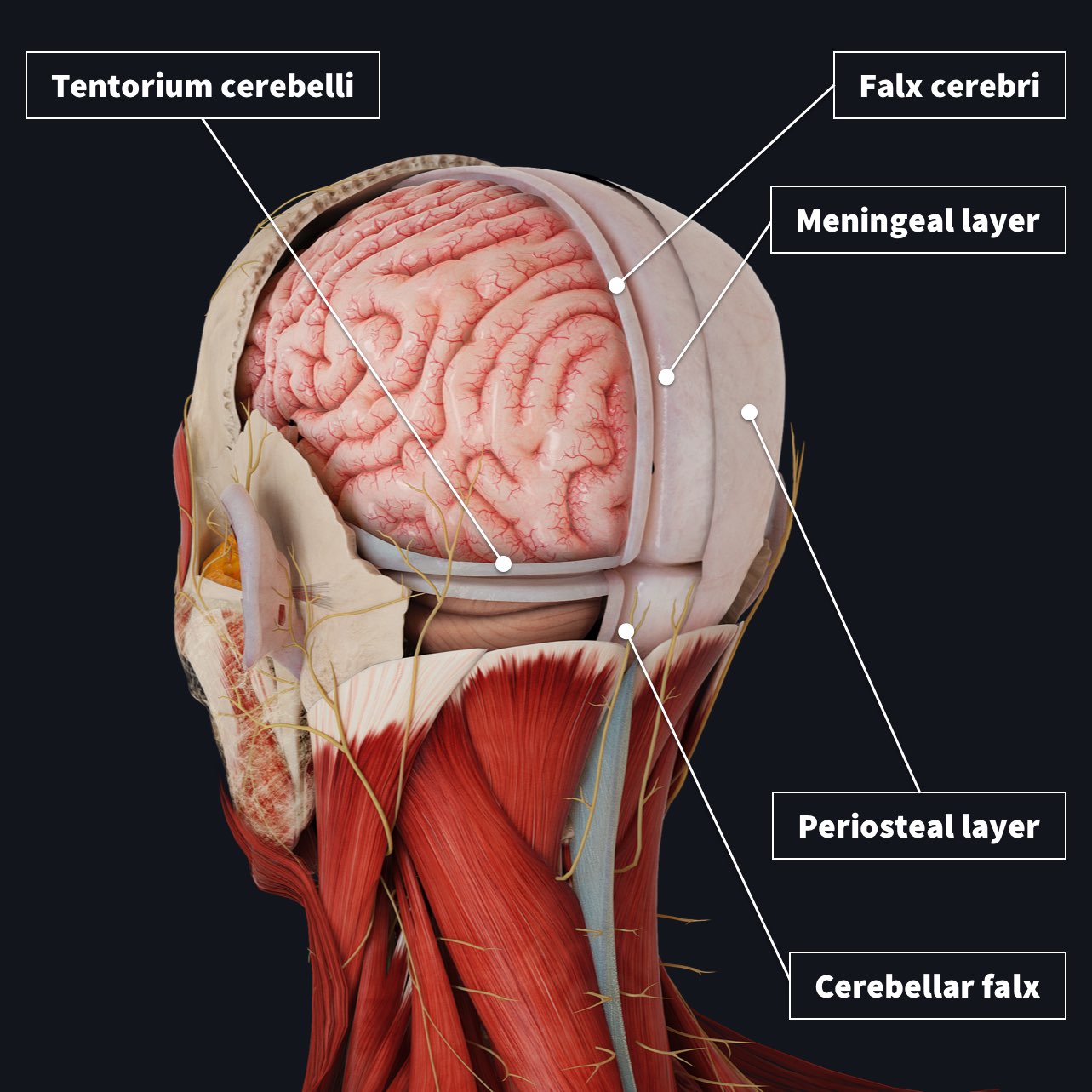
From these, the meningeal layer extends into the cranial cavity and forms four main dural folds, also known as dural reflections (Rea, 2015): The largest one is named the falx cerebri and attaches anteriorly at the crista galli and follows the longitudinal fissure.
What is the space outside the dura mater called?
The space between the spinal dura mater and the periosteum of the vertebral column is called the epidural space. It is filled with loose connective and adipose tissues, and traversed by the anterior and posterior internal vertebral venous plexuses.
What is the main function of the dura mater?
Dura mater encircles and continues to support the large venous channels (called dural sinuses) that help in carrying the blood from the brain to the heart. The structure and position of the dura mater make it a very reliable protective envelop, which is one of its important functions.
What are the 3 layers of meninges and where is each located which one contains the CSF fluid?
The middle layer of meninges is arachnoid, a thin layer resembling a cobweb with numerous threadlike strands attaching it to the innermost layer. The space under the arachnoid, the subarachnoid space, is filled with cerebrospinal fluid and contains blood vessels. The pia mater is the innermost layer of meninges.
What is found inside the dural sinuses?
The walls of the dural venous sinuses are composed of dura mater lined with endothelium, a specialized layer of flattened cells found in blood vessels. They differ from other blood vessels in that they lack a full set of vessel layers (e.g. tunica media) characteristic of arteries and veins.
How many layers does the brain have?
Protecting the brain There are 3 layers of tissue called meninges that help protect the brain. The outer covering of tissue (called the dura mater), closely lines the inside of the skull. The second layer is the arachnoid mater, and the third layer, the pia mater, hugs the surface of the brain.
Why do we need three meningeal layers?
These layers bound three clinically important potential spaces: the epidural, subdural, and subarachnoid spaces. The function of the meninges is to protect the brain and spinal cord from mechanical trauma, to support the blood vessels and to form a continuous cavity through which the cerebrospinal fluid (CSF) passes.
What is the dura of the spinal cord?
The spinal dura mater is a fibrous, non-adherent, tough layer surrounding the spinal cord. It is separated from the wall of the vertebral canal by the epidural space. This space contains loose areolar tissue and a network of internal vertebral venous plexuses.
What is the dura?
The dura mater often gets referred to as merely the dura. It is one of the layers of connective tissue that make up the meninges of the brain (pia, arachnoid, and dura, from inside to outside). It is the outermost layer of the three meninges that surround and protect the brain and spinal cord.
Which layer is the dura mater quizlet?
1) Dura mater - the dura mater layer is the thickest, outermost layer. 2) Arachnoid mater - this layer has a weblike appearance. 3) Pia mater - this is the innermost layer and is very thin at only one cell thick.
What are the two layers of the dura mater?
The dura mater is composed of two layers: the periosteal/endosteal layer and the meningeal layer. The dural venous sinuses are between these two layers. The dura folds to form septa that create the falx cerebri, tentorium cerebelli, falx cerebelli, and diaphragma sellae.
What is the dura mater?
The dura mater often gets referred to as merely the dura. It is one of the layers of connective tissue that make up the meninges of the brain (pia, arachnoid, and dura, from inside to outside). It is the outermost layer of the three meninges that surround and protect the brain and spinal cord. The dura mater is made up of fibroblasts and large amounts of extracellular collagen.[1]
What is the function of the meningeal layer of the dura mater?
The meningeal layer of the dura mater creates several dural folds that divide the cranial cavity into freely communicating spaces. The function of the dural folds is to limit the rotational displacement of the brain.
Why do subdural hematomas occur?
Subdural hematoma arises from the rupture of bridging veins, usually from head trauma. Because the venous pressure in the veins is low, the hematoma is not typically large, and progression is not rapid. An increase in intracranial pressure can increase the rate of bleeding from the ruptured bridging veins.[13] The anatomy of the bridging vein makes it susceptible to tearing within the border cell layer of the dura mater. [13]
What is the periosteal layer of the dura mater?
The periosteal or endosteal layer of the dura mater is simply a layer of periosteum that covers the inner surface of the skull. The layer does not extend beyond the foramen magnum to become contiguous with the dura mater of the spinal cord. The spinal cord dura mater has no periosteal layer.
Where does the venous drain into the dura matter?
Venous drainage of the dura matter is via the meningeal veins that are present in the periosteal layer. These veins follow the branches of the middle meningeal artery and drain into the sphenopalatine sinus or the pterygoid venous plexus. The dural venous sinuses are between the periosteal and meningeal layers. These sinuses are responsible for the venous vasculature of the cranium. The sinuses converge and drain into the internal jugular vein. [8]
What is the epidural space?
Epidural space: The epidural space is a possible space between the dura mater and skull containing fat and blood vessels. Damage to these blood vessels leads to the rapid accumulation of blood in the epidural space forming an epidural hematoma. [3]
How many layers does the dura have?
The dura separates into two layers at dural reflections (also known as dural folds ), places where the inner dural layer is reflected as sheet-like protrusions into the cranial cavity. There are two main dural reflections:
What is the Dura Mater?
FMA. 9592. Anatomical terminology. Dura mater is a thick membrane made of dense irregular connective tissue that surrounds the brain and spinal cord. It is the outermost of the three layers of membrane called the meninges that protect the central nervous system. The other two meningeal layers are the arachnoid mater and the pia mater.
What is the difference between a subdural and an epidural?
Many medical conditions involve the dura mater. A subdural hematoma occurs when there is an abnormal collection of blood between the dura and the arachnoid , usually as a result of torn bridging veins secondary to head trauma. An epidural hematoma is a collection of blood between the dura and the inner surface of the skull, and is usually due to arterial bleeding. Intradural procedures, such as removal of a brain tumour or treatment of trigeminal neuralgia via a microvascular decompression, require that an incision is made to the dura mater. To achieve a watertight repair and avoid potential post-operative complications, the dura is typically closed with sutures. In the event that there is a dural deficiency, a dural substitute may be used to replace this membrane. Small gaps in the dura can be covered with a surgical sealant film .
What is the role of arachnoid villi in the dural sinus?
Arachnoid villi, which are outgrowths of the arachnoid mater (the middle meningeal layer), extend into the dural venous sinuses to drain CSF. These villi act as one-way valves. Meningeal veins, which course through the dura mater, and bridging veins, which drain the underlying neural tissue and puncture the dura mater, empty into these dural sinuses. A rupture of a bridging vein causes a subdural hematoma .
What is the gap between the dura mater and the brain called?
The two layers of dura mater run together throughout most of the skull. Where they separate, the gap between them is called a dural venous sinus. These sinuses drain blood and cerebrospinal fluid (CSF) from the brain and empty into the internal jugular vein .
What is the cranial dura mater?
Cranial dura mater has two layers called lamellae, a superficial layer (also called the periosteal layer), which serves as the skull's inner periosteum, called the endocranium and a deep layer called the meningeal layer. When it covers the spinal cord it is known as the dural sac or thecal sac. Unlike cranial dura mater, spinal dura mater only has ...
What is the smallest dural infolding?
The sellar diaphragm is the smallest dural infolding and is a circular sheet of dura that is suspended between the clinoid processes, forming a partial roof over the hypophysial fossa. The sellar diaphgram covers the pituitary gland in this fossa and has an aperture for passage of the infundibulum (pituitary stalk) and hypophysial veins.
What is the Dura Mater?
Dura mater is also the home to meningeal veins. Many types of medical conditions involve the dura mater. The most common come in the form of hematomas. Arterial bleeding can result in an epidural hematoma, which is when blood collects between the dura mater and the skull.
Where is the dura mater located?
The dura mater is the top layer of the meninges, lying beneath the bone tissue. This material at times opens into sinus cavities (spaces) located around the skull. This is particularly notable with the dural venous sinuses.
What happens if blood collects between the dura and arachnoid mater?
If blood collects between the dura and arachnoid mater, a subdural hematoma results. Also, there are some instances where the dura plays a major role in certain types of headaches. Last medically reviewed on January 22, 2018.
What are the three layers of the central nervous system?
These are called the meninges, and their three levels consist of the: pia, arachnoid, and dura mater. Bone is situated above these layers, followed by periosteum (a fibrous membrane that covers bone) and skin.
What is the liquid that drains into the jugular vein?
Here, liquids, like blood and cerebrospinal fluid, drain and collect into the internal jugular vein. Cerebrospinal fluid is a clear liquid that cushions the brain and spinal cord while also transporting nutrients, chemicals, and waste. Dura mater is also the home to meningeal veins.
What is the space between the two layers of Dura?
The space between the two layers of Dura has room for the venous drainage of the cranium. These spaces are termed dural venous sinuses. The dural venous sinuses drain the venous blood from the cranium and open it into the internal jugular veins.
What is the Dura mater?
The dura mater is a tough membrane and protects the brain from traumatic injuries. Whenever there is some accident or blow on the scalp, the Dura keeps the brain at its place and does not let it reach the hard bony structure of the brain. If the Dura and other layers of meninges do not support the brain, it will hit the hard scalp whenever there is external trauma. This hitting of the brain on the hard scalp will result in traumatic injuries.
What is the function of the dural venous sinuses?
The dural venous sinuses also help in draining the venous blood from the brain and cranium.
How are dural reflections formed?
Four dural reflections are formed by the inward folding of the meningeal layer of Dura around itself. The dural reflections project in the cranial cavity is creating different compartments of the cranial cavity. Different parts of the brain occupy the compartments or divisions of the cranial cavity.
What is the outermost layer of the meninges?
The Dura is the outermost layer of the meninges that covers the brain and the entire length of the spinal cord. The Dura mater is located immediately beneath the skull and bones of the vertebral column. Dura is thick in structure which makes it tough and inextricable.
What are the protective connective tissue layers of the brain?
The brain’s protective connective tissue layers are meninges, including the pia mater, arachnoid mater, and dura mater. The pia mater is the innermost, thin, and delicate layer attached to the brain and follows the contour and involutions of the brain. The arachnoid mater is outer to the pia mater and does not follow the curves and shape of the brain.
Where does the venous drainage of the Dura occur?
The venous drainage of the Dura occurs through the meningeal veins. Meningeal veins are present in the periosteal layer. The dura mater and follow the course corresponding branches of the middle meningeal artery. The meningeal veins ultimately drain into the sphenopalatine sinus. They may also drain through the pterygoid venous plexus.
Which layer of the dura mater is connected to the cranial bones?
Periosteal layer. The periosteal layer of the dura mater is the periosteum lining the internal surface of the skull and, as such, is intimately attached to the cranial bones and sutures. Meningeal layer. The meningeal layer is the dura mater proper and is composed of dense collagenous connective tissue that is continuous with the dura mater of the spinal cord. The dura mater envelopes the cranial nerves like a sleeve, which then fuse with the epineurium of the nerves outside the skull.
What is the circular fold of dura that covers the pituitary by forming a roof over the sell?
Diaphragma sellae . A circular horizontal fold of dura that covers the pituitary by forming a roof over the sella turcica.
What is the only location in the body where an artery courses through a venous structure?
VThe cavernous sinus is the only location in the body where an artery courses through a venous structure. A carotid-cavernous sinus fistula forms when the internal carotid artery ruptures within the cavernous sinus. ▼
What is the dura mater?
It is an apt name, as the dura mater is the toughest of the meninges, which are the three membranes that protect the brain and the spinal cord from mechanical injury. In Latin, 'mater' means mother.
What is the function of the dura mater?
The main function of the dura mater and the other meninges is to protect the brain and the spinal cord from damage. ➠ The partitions of the dura mater, which are attached to the inner lining of the skull, provide support to the brain. Also, these folds restrict the rotatory displacement of the brain. ➠ The dura mater also provides ...
What is the venous drainage of the meninges?
The venous drainage of the meninges includes the meningeal veins of the dura and the dural venous sinuses. The meningeal veins and the bridging veins (veins that drain the neural tissues lying underneath) empty into the dural sinuses. Dural venous sinuses are venous channels that carry blood from the brain to the heart.
What is the space between the dura and the arachnoid mater?
The potential space between the dura mater and the arachnoid mater, which is the second membrane that is located in the middle, is referred to as the subdural space. The pia mater is the innermost membrane that adheres to the surface of the brain.
Which layer of the brain separates the brain in two hemispheres?
Also referred to as dural septa, the meningeal layer forms two compartments. The two main dural folds are referred to as: While falx cerebri is a sickle-shaped fold of dura mater that separates the brain in two cerebral hemispheres, the tentorium cerebelli is a crescent-shaped fold that covers the cerebellum, and separates the occipital lobes ...
Which layer of the skull covers the inner surface of the skull?
Endosteal layer is the outer, periosteal layer that covers the inner surface of the skull. The periosteal and the inner meningeal layer are joined, with the exception of places where dural venous sinuses or the dural folds are present. While the meningeal layer is continuous with the dura mater of the spinal cord, ...
Why do epidural spaces get filled with blood?
These spaces might get filled with blood in the event of rupturing of blood vessels.
Where is the dura mater located?
The dura mater is the outermost layer of the meninges and is located directly underneath the bones of the skull and vertebral column. It is thick, tough, and inextensible.
Where does the dura mater get its vascular supply?
The dura mater receives its own vascular supply - primarily from the middle meningeal artery and vein. It is innervated by the trigeminal nerve (V1, V2 and V3).
What are the three meninges?
There are three layers of meninges, known as the dura mater, arachnoid mater and pia mater. These coverings have two major functions: Provide a supportive framework for the cerebral and cranial vasculature. Acting with cerebrospinal fluid to protect the CNS from mechanical damage.
What is the outermost layer of the meninges?
The dura mater is the outermost layer of the meninges and is located directly underneath the bones of the skull and vertebral column. It is thick, tough, and inextensible. The dura mater consists of two layered sheets of connective tissue: Periosteal layer – lines the inner surface of the bones of the cranium.
Where does subdural blood come from?
Subdural - venous blood collects between the dura and the arachnoid mater. It results from damage to cerebral veins as they empty into the dural venous sinuses. The arachnoid mater is the middle layer of the meninges, lying directly underneath the dura mater.
What is the role of the meninges in the CNS?
Acting with cerebrospinal fluid to protect the CNS from mechanical damage. The meninges are often involved cerebral pathology , as a common site of infection (meningitis), and intracranial bleeds. In this article, we shall look at the anatomy of the three layers, and their clinical correlations.
Which layer of the cranium lines the inner surface of the bones?
Periosteal layer – lines the inner surface of the bones of the cranium.
What is the space between the dura mater and the skull?
Between the dura mater and the skull is the epidural space . This space allows for blood vessels and fat to exist and provide fresh blood supply and cushioning. Some may refer to the space between the dura mater and the arachnoid mater as the subdural space, though this space is usually only observed when collagen fibers of the dura mater are torn, and thus this term has more clinical relevance than anatomical application.
What are the layers of meninges?
There are three layers to the meninges. From inside to outside they are the pia mater, arachnoid mater, and dura mater.
What is the outermost layer of arachnoid mater?
The dura mater is the outermost, thickest layer, and it sounds like "durable." The arachnoid mater is web-like in appearance and thus can be remembered by thinking of "arachnids" (spiders). The pia mater is the innermost, thinnest layer, and it can be remembered as "pliable."
What is the thinnest meninge of the brain?
The pia mater , which translates to "tender matter," is the thinnest and deepest meninge of the brain. This thin, clear layer adheres to the surface of the brain and follows the natural hills and valleys (called gyri and hillocks) of the surface of the brain. The pia mater functions to contain cerebrospinal fluid within its proper pathway. Additionally, about 30% of the total CSF is produced by the pia mater .
What are granulations in the arachnoid mater?
Arachnoid granulations are structures within the arachno id mater. They are small growths that occur on the membrane of the arachnoid mater and extend into the dural sinuses. These growths are typically harmless but may cause issues when they become too large. A large arachnoid granulation may result in the occlusion of a blood vessel or block important CSF pathways.
What is the difference between arachnoid mater and pia mater?
The deeper lower layer (the pia mater) follows the brain closely and adheres to the deep grooves and hills . This results in spaces that exist within the arachnoid mater and pia mater that are quite large , such as areas where the pia mater dips deeply with a groove in the brain while the arachnoid mater is held up closer to the skull. These large grooves are called subarachnoid cisterns. The subarachnoid cisterns provide space that is critical for nerves and vessels to pass through the layers of the brain. They also contain CSF. There are 9 commonly observed subarachnoid cisterns in the body, which are named based on their location:
What is the arachnoid mater?
The arachnoid mater is just below the dura mater. The term "arachnoid" comes from the word "arachnid," meaning "spider." This name is in reference to the appearance of the arachnoid mater, which resembles the stringy, white fibers of a spider web. The most important function of the arachnoid mater is to house the arachnoid space. This space is created by the crisscrossing fibers called arachnoid trabeculae which connect the layer to the pia mater while creating a passageway.
Occipital
Superior sagittal sinus laid open after remova of the skull cap. The chordæ Willisii are clearly seen. The venous lacunæ are also well shown; from two of them probes are passed into the superior sagittal sinus. (Poirier and Charpy.)
Basilar Plexus
The cavernous sinuses (sinus cavernosus) are so named because they present a reticulated structure, due to their being traversed by numerous interlacing filaments.
Overview
Structure
The dura mater has several functions and layers. The dura mater is a membrane that envelops the arachnoid mater. It surrounds and supports the dural sinuses (also called dural venous sinuses, cerebral sinuses, or cranial sinuses) and carries blood from the brain toward the heart.
Cranial dura mater has two layers called lamellae, a superficial layer (also calle…
Clinical significance
Many medical conditions involve the dura mater. A subdural hematoma occurs when there is an abnormal collection of blood between the dura and the arachnoid, usually as a result of torn bridging veins secondary to head trauma. An epidural hematoma is a collection of blood between the dura and the inner surface of the skull, and is usually due to arterial bleeding. Intradural procedures, such as removal of a brain tumour or treatment of trigeminal neuralgia via a microvas…
Etymology
The name dura mater derives from the Latin for tough mother (or hard mother), a loan translation of Arabic أم الدماغ الصفيقة (umm al-dimāgh al-ṣafīqah), literally 'thick mother of the brain', matrix of the brain, and is also referred to by the term "pachymeninx" (plural "pachymeninges").
Additional images
• Dura mater (spinal section)
• Diagrammatic representation of a section across the top of the skull, showing the membranes of the brain, etc.
• Diagrammatic transverse section of the medulla spinalis and its membranes.
See also
• Meninges
• Pia mater
• Arachnoid mater
External links
• Media related to Dura mater at Wikimedia Commons
• youtube: exposure of falx cerebri, dura mater & arachnoid