:max_bytes(150000):strip_icc()/shutterstock_16706-56a007e45f9b58eba4ae8e32.jpg)
Overview
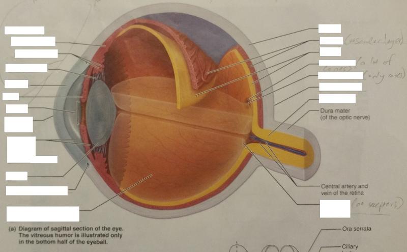
- The outer layer of the eyeball is a tough, white, opaque membrane called the sclera (the white of the eye). The slight bulge in the sclera at the front of the eye is a clear, thin, dome-shaped tissue called the cornea.
- The middle layer is the choroid. ...
- The inner layer is the retina, which lines the back two-thirds of the eyeball. ...
Full Answer
What are the basic parts of the eye?
The outer layer is made up of the following:
- Conjunctiva
- Cornea
- Sclera
What are the parts and functions of the eye?
The internal components of the eye include:
- Lens
- Retina
- Aqueous humour
- Optic nerve
- Vitreous humour
What are the major structures of the eye?
There are two kinds of vision cells:
- The rods (light-dark vision, active in evening or darkness)
- The cones (accountable for color vision) Three different kinds of cone cells are essential for color vision:
- Pins for red-visibility (about 46% of all pins)
- Cones for green vision (about 46% of all cones)
What is the posterior segment of the eye?
The posterior segment or posterior cavity is the back two-thirds of the eye that includes the anterior hyaloid membrane and all of the optical structures behind it: the vitreous humor, retina, choroid, and optic nerve. What causes stringy mucus in the eyes? Stringy, white mucus may represent allergic conjunctivitis .
What are the 2 segments of the eye?
The different structures in the eye can be broadly divided into the anterior (front of the eye) and posterior (back of the eye) segments.
What is a segment in the eye?
The anterior segment refers to the front-most region of the eye, and includes the cornea, iris, and lens. Typically, the phrase “anterior segment surgery” refers to surgery performed on the iris and lens (either natural lens, or synthetic intraocular lens placed during cataract surgery).
How many segments does the eye have?
The inside of the eye is divided into three sections called chambers. Anterior chamber: The anterior chamber is the front part of the eye between the cornea and the iris. The iris controls the amount of light that enters the eye by opening and closing the pupil. The iris uses muscles to change the size of the pupil.
What is anterior segment and posterior segment of eye?
2.1. The anterior segment includes the cornea, iris, ciliary body and lens as well as the spaces of the anterior and posterior chambers filled with aqueous humor. The posterior segment includes the retina, choroid and optic nerve head as well as the vitreous compartment filled with vitreous humor.
What is cornea and anterior segment?
A cornea and external disease specialist is an ophthalmologist who focuses on the cornea (the transparent front structure of the eye), conjunctiva (clear mucous membrane covering the white part of the eye) and the anterior segment (front structures of the eye including the iris and lens).
What is the difference between anterior segment and anterior chamber?
The anterior segment is the front third of the eye that includes the structures in front of the vitreous humour: thecornea, iris, ciliary body, and lens. Within the anterior segment are two fluid-filled spaces: the anterior chamber between the posterior surface of the cornea (i.e. the corneal endothelium) and the iris.
What are the segments of the eye and its contents?
The anterior segment contains the cornea, iris, ciliary body, and lens while the posterior segment consists of the vitreous humor, retina, choroid, and optic nerve ( Figure 1).
What are the 3 layers of eye?
The wall of the eyeouter layer – made up of the sclera and cornea (called the fibrous tunic)middle layer – made up of the uvea (called the vascular tunic)inner layer – made up of the retina (called the neural tunic)
Where is the posterior segment of the eye?
The posterior segment of the eye comprises the back two-thirds of the eye, including the vitreous humor, the retina, the choroid and the optic nerve.
What is posterior of the eye?
The posterior segment or posterior cavity is the back two-thirds of the eye that includes the anterior hyaloid membrane and all of the optical structures behind it: the vitreous humor, retina, choroid, and optic nerve.
What does the posterior segment do?
Posterior chamber is an important structure involved in production and circulation of aqueous humor. Aqueous humor produced by the epithelium of the ciliary body is secreted into the posterior chamber, from which it flows through the pupil to enter the anterior chamber.
What is the function of the anterior segment?
The front part of the eye is called the anterior segment. Its main function is to focus the rays of light that penetrate the eye on the retina. It encompasses the most superficial and transparent part of the eye, the cornea and the crystalline lens, our eye's natural lens that helps us to focus objects.
What is meant by posterior segment of the eye?
The posterior segment of the eye comprises the back two-thirds of the eye, including the vitreous humor, the retina, the choroid and the optic nerve.
What does the posterior segment of the eye do?
At the back of the eye, the retina acts like film in a camera. It is made up of millions of light-sensitive neurons, known as rods and cones. The rods are responsible for vision at low light levels, while the cones are responsible for colour vision.
What does the posterior segment do?
Posterior chamber is an important structure involved in production and circulation of aqueous humor. Aqueous humor produced by the epithelium of the ciliary body is secreted into the posterior chamber, from which it flows through the pupil to enter the anterior chamber.
Is glaucoma a posterior segment disease?
Posterior segment eye disease (PSED) epidemiologically is commonly defined as diseases of the retina, choroid and optic nerve and primarily includes: glaucoma, age-related macular degeneration (AMD) and diabetic retinopathy (DR).
Briefly explain the structure of the human eye.
The human eye is a roughly spherical organ, responsible for perceiving visual stimuli. It is enclosed within the eye sockets in the skull and is an...
What are the external structures of the eye?
The external structures of the eye include: Sclera Conjunctiva Cornea Iris Pupil
What is the function of conjunctiva?
Conjunctiva lubricates the front surface of the eye. It also protects the eyes from debris, dust and infection-causing microorganisms.
What is the function of the iris? How many layers does it have?
The iris regulates the amount of light entering the eyes by controlling the diameter and size of the pupil. The iris consists of two layers: Strom...
What are the internal components of the eye?
The internal components of the eye include: Lens Retina Aqueous humour Optic nerve Vitreous humour
What is the role of the cornea and lens in the eye?
By helping to focus light as it enters the eye, the cornea and the lens both play important roles in giving us clear vision. In fact, 70% of the eye's focusing power comes from the cornea and 30% from the lens.
What is the muscle that controls the movement of the eyeball?
This is a strong layer of tissue that covers nearly the entire surface of the eyeball. This illustration shows eye muscles , which control eye movement.
What part of the retina is responsible for the transmission of light?
The retina has special cells called photoreceptors. These cells change light into energy that is transmitted to the brain. There are two types of photoreceptors : rods and cones.
Why do cataracts change shape?
The lens changes shape to help the eye focus on objects up close. Small fibers called zonules are attached to the capsule holding the lens, suspending it from the eye wall. The lens is surrounded by the lens capsule, which is left in place when the lens is removed during cataract surgery .
What part of the eye is the orbit?
Eye Anatomy: Parts of the Eye Outside the Eyeball. The eye sits in a protective bony socket called the orbit. Six extraocular muscles in the orbit are attached to the eye. These muscles move the eye up and down, side to side, and rotate the eye. The extraocular muscles are attached to the white part of the eye called the sclera.
What is the function of the pupil?
Directly behind the pupil sits the lens. The lens focuses light toward the back of the eye. The lens changes shape to help the eye focus on objects up close.
What is the drainage angle of the eye?
The eye is always producing aqueous humor. To maintain a constant eye pressure, aqueous humor also drains from the eye in an area called the drainage angle. Behind the anterior chamber is the eye’s iris (the colored part of the eye) and the dark hole in the middle called the pupil. Muscles in the iris dilate (widen) or constrict (narrow) ...
What is the external structure of the eye?
The External Structure of an Eye. Sclera: It is a white visible portion. It is made up of dense connective tissue and protects the inner parts. Conjunctiva: It lines the sclera and is made up of stratified squamous epithelium. It keeps our eyes moist and clear and provides lubrication by secreting mucus and tears.
What is the eye?
The eye is one of the sensory organs of the body. In this article, we shall explore the anatomy of the eye. The structure of the eye is an important topic to understand as it one of the important sensory organs in the human body. It is mainly responsible for vision, differentiation of colour ...
What is the purpose of vitreous humor?
It contains water (99%), collage, proteins, etc. The main function of vitreous humour is to protect the eyes and maintain its spherical shape.
How many colors can the human eye differentiate?
It is mainly responsible for vision, differentiation of colour (the human eye can differentiate approximately 10 – 12 million colours) and maintaining the biological clock of the human body. The human eye can be compared to a camera as both works by gathering, focusing and transmitting the light through the lens for creating an image of an object.
What is the human eye made of?
It is made up of several muscles and tissues that come together to form a roughly spherical structure. From an anatomical perspective, the human eye can be broadly classified into external structure and internal structure.
What is the most complicated sense organ in the human body?
The human eyes are the most complicated sense organs in the human body. From the muscles and tissues to nerves and blood vessels, every part of the human eye is responsible for a certain action. Furthermore, contrary to popular belief, the eye is not perfectly spherical; instead, it is two separate segments fused together.
Which part of the eye is made up of stratified squamous epithelium?
Conjunctiva: It lines the sclera and is made up of stratified squamous epithelium. It keeps our eyes moist and clear and provides lubrication by secreting mucus and tears. Cornea: It is the transparent, anterior or front part of our eye, which covers the pupil and the iris.
How many segments are there in the eyeball?
Segments and Chambers of Eyeball: Eyeball is divided into two segments, the anterior one and the posterior one. Anterior segment: The segmentation inside the eye is based on the position of the lens.
Where are the eyes located?
Eyes are spheroid shape organs fitted into the two orbitals of the skull. There are three major parts in each eye like
What is the gap between the sclera and the orbitals of the skull called?
The gap between the sclera and the orbitals of the skull is filled with adipose tissue. This sclera in the anterior side extends as a transparent epithelium called the cornea. The junction of cornea and sclera is called the limbus.
What is the outermost part of the eye?
It is made of a dense, strong fibrous wall consisting of the sclera that is 5/6 th and the cornea that is anterior 1/6 th of the eyeball.
What is the color of the iris?
This is the extension of the choroid layer towards the anterior side of the eye. This iris is pigmented due to which there appears color in the eyes. The pigment is genetically determined and can be like black, brown, green, blue, etc.
How is light transmitted from a lens?
The light received from the lens is converted into a nerve impulse and carried backward as the optic nerve into the brain for further processing.
What is the eye?
Eye Anatomy and Physiology a Complete Detail. An eye is the sense organ of vision. It perceives the light energy, converts it into electrical energy, thereby helping us to ‘’see‘’.
What is the ciliary body?
Structurally, the ciliary body is a ring of tissue that surrounds the iris and connects it to the choroid. The ciliary body can’t be seen when you look at the eye, because it’s located behind the iris and sclera, which is the white part of the eye.
What are the best medications for glaucoma?
Some of the most common forms of glaucoma medication that affect the ciliary body include: 1 Carbonic anhydrase inhibitors such as Azopt (brinzolamide) and Trusopt (dorzolamide). 2 Beta blockers such as Betoptic (betaxolol) and Timoptic (timolol). 3 Alpha-adrenergic agonists such as Alphagan P (brimonidine) and Iopidine (apraclonidine).
How does glaucoma affect the ciliary body?
How does glaucoma medication affect the ciliary body? Typically, the initial treatment used for glaucoma is medicated eye drops, which are designed to help regulate eye pressure by reducing the production of aqueous fluid by the ciliary body and/or increasing its drainage from the eye .
What happens when too much fluid is produced in the eye?
Each of these functions is essential for the health of the eye — in fact, conditions such as ocular hypertension and glaucoma can occur if too much aqueous fluid is produced by the ciliary body.
What is the structure of the ciliary body?
Structures contained within the ciliary body include: The ciliary muscle, which influences the shape of the lens inside the eye. Contraction of the ciliary muscle makes the lens become more convex, enabling the eye to focus on near objects. The ciliary muscle is connected to the lens by a series of very thin, radially-arranged fibers called ...
What is the muscle that holds the lens in place?
The ciliary muscle is connected to the lens by a series of very thin, radially-arranged fibers called the ciliary zonules (also called the zonular fibers or zonules of Zinn ), which hold the lens in place within the eye. The ciliary processes, which are about 70 ridges in the ciliary body that contain cells involved in the production ...
Why does presbyopia occur?
Presbyopia occurs because the lens of the eye thickens over time and loses its natural flexibility. The ciliary body retains its ability to function, but the lens fails to change shape to enable near objects to come into focus. Fortunately, presbyopia is treatable with eyeglasses, contact lenses or vision surgery.
What is the effect of subconjunctival injection?
Subconjunctival injection obviates the conjunctival epithelial barrier, which is rate-limiting for permeation of water-soluble drugs. Thus, the transscleral route bypasses cornea–conjunctiva barrier. Nevertheless, various dynamic, static, and metabolic barriers limit drug access to the posterior segment. Dynamic barriers include conjunctival blood and lymphatic circulation. Various authors reported rapid drug elimination via these pathways following subconjunctival administration (33–35). As a result, the formulation is drained into systemic circulation thereby lowering ocular bioavailability. Thus, drug elimination from the subconjunctival space becomes a major determinant of the vitreous drug levels following subconjunctival administration. The molecules that escape conjunctival vasculature permeate through sclera and choroid to reach the neural retina and photoreceptor cells. The sclera is not a major barrier as it is more permeable than the cornea. Moreover, permeability across the sclera is independent of lipophilicity unlike corneal and conjunctival layers but depends primarily on the molecular radius (12,36). However, choroid is a significant barrier as high choroidal blood flow can also eliminate a considerable fraction of drug before it can reach the neural retina. Furthermore, blood–retinal barriers limit drug availability to the photoreceptor cells.
What are the advantages of intravitreal injection?
Unlike periocular injections, the intravitreal injection offers distinct advantages as the molecules are directly inserted into the vitreous. However, drug distribution in the vitreous is non-uniform. Small molecules can rapidly distribute through the vitreous, whereas the diffusion of larger molecules is restricted. This distribution also depends on the pathophysiological condition and molecular weight of the administered drug (37). The vitreous also acts as a barrier for retinal gene delivery following an intravitreal injection. Hyaluronan, a negatively charged glycosaminoglycan present in the vitreous, can interact with cationic lipid, polymeric, and liposomal DNA complexes (38). This interaction can lead to severe aggregation and complete immobilization of DNA/cationic liposome complexes (39). Similarly, mobility of nanoparticles in the vitreous depends on their structure and surface charge. Polystyrene nanospheres do not diffuse freely into the vitreous due to their adherence to collagen fibrillar structures (39). Hence, surface modification of nanospheres with hydrophilic PEG chains has been performed. A recent study using human serum albumin nanoparticles also demonstrated that anionic nanoparticles with a zeta potential of −33.3 mV diffused more freely in the vitreous than cationic particles with a zeta potential of 11.7 mV (40).
How does melanin affect ocular drug delivery?
The presence of melanin may alter ocular drug disposition. Interaction with this pigment may alter the availability of free drug at the targeted site. Thereby, melanin binding may significantly lower pharmacological activity (42). In ocular tissues, melanin is present in uvea and RPE. It binds to free radicals and drugs by electrostatic and Van der Waals forces or by simple charge transfer (43). Based on available information, it may be concluded that all basic and lipophilic drugs bind to melanin (44). Even though drug binding to melanin is not necessarily predictive of ocular toxicity, it has significant pharmacological consequences and requires careful consideration in ocular drug delivery. Melanin binding in the iris–ciliary body affects drug concentrations in anterior ocular tissues and drug response (45). A melanin-bound drug is not usually available for receptor binding necessitating the administration of larger doses (46). Likewise, melanin present in choroid and RPE affects the extent of drug uptake into the retina and vitreous following transscleral or systemic drug administration. As a result of melanin binding, permeation lag-time of lipophilic beta-blockers through bovine choroid-RPE is much longer than more hydrophilic beta-blockers (11). Similarly, binding of lipophilic compounds to the bovine choroid-Bruch's membrane was demonstrated to be higher due to the presence of melanin. Consequently, there is a greater resistance to solute permeation across choroid-Bruch's membrane than the sclera, which is devoid of melanin (47).
What are the two main barriers to ocular drug delivery?
Following systemic administration, the blood–aqueous barrier and blood–retinal barrier are the major barriers for anterior segment and posterior segment ocular drug delivery, respectively. Blood–aqueous barrier consists of two discrete cell layers located in the anterior segment of the eye viz. the endothelium of the iris/ciliary blood vessels and the nonpigmented ciliary epithelium. Both cell layers express tight junctional complexes and prevent the entry of solutes into the intraocular environment (6) such as the aqueous humor. Blood–retinal barrier restricts the entry of the therapeutic agents from blood into the posterior segment. It is composed of two types of cells, i.e., retinal capillary endothelial cells and retinal pigment epithelium cells (RPE) known as the inner and outer blood– retinal barrier, respectively. RPE, located between the neural retina and the choroid, is a monolayer of highly specialized cells. RPE aids in biochemical functions by selective transport of molecules between photoreceptors and choriocapillaris. Furthermore, it maintains the visual system by uptake and conversion of retinoids (11). However, tight junctions of the RPE efficiently restrict intercellular permeation. Following oral or intravenous dosing, drugs can easily enter into the choroid due to its high vasculature compared to retinal capillaries. The choriocapillaris are fenestrated resulting in rapid equilibration of drug molecules present in the bloodstream with the extravascular space of the choroid. However, outer blood–retinal barrier (RPE) restricts further entry of drugs from the choroid into the retina. Even though it is ideal to deliver the drug to the retina via systemic administration, it is still a challenge due to the blood–retinal barrier, which strictly regulates drug permeation from blood to the retina. Hence, specific oral or intravenous targeting systems are needed to transport molecules through the choroid into deeper layers of the retina (12).
What is the barrier between the stroma and the aqueous humor?
Endothelium is the innermost monolayer of hexagonal-shaped cells. Even though endothelium is a separating barrier between the stroma and aqueous humor, it helps maintain the aqueous humor and corneal transparency due to its selective carrier-mediated transport and secretory function (6). Furthermore, the corneal endothelial junctions are leaky and facilitate the passage of macromolecules between the aqueous humor and stroma (7). Thus, corneal layers, particularly the epithelium and stroma, are considered as major barriers for ocular drug delivery. It is vital to understand that the permeant should have an amphipathic nature in order to permeate through these layers. A schematic of the corneal layers that a permeant needs to cross is presented by Barar et al. (6).
How to improve ocular bioavailability?
The traditional approach to improve ocular bioavailability is to modify the drug chemically to achieve the desired solubility and lipophilicity. However, a more rational approach would be a transporter-targeted modification of the drug. Transporters are membrane-bound proteins that play an important role in active transport of nutrients across biological membranes. The presence of transporters has been reported on various ocular tissues. However, in the present article, we have focused on the transporters that are localized in the epithelia of the cornea, conjunctiva, and retina. These transporters may be amenable to bind and transport specific-targeted ligands attached to drug moieties.
What are the layers of the cornea?
In addition, various layers of the cornea, conjunctiva, and sclera play an important role in drug permeation. The cornea, the anterior most layer of the eye, is a mechanical barrier which limits the entry of exogenous substances into the eye and protects the ocular tissues. It can be mainly divided into the epithelium, stroma, and endothelium. Each layer offers a different polarity and a potential rate-limiting structure for drug permeation. The corneal epithelium is lipoidal in nature which contains 90% of the total cells in the cornea and poses a significant resistance for permeation of topically administered hydrophilic drugs. Furthermore, superficial corneal epithelial cells are joined to one another by desmosomes and are surrounded by ribbon-like tight junctional complexes (zonula occludens) (4,5). Presence of these tight junctional complexes retards paracellular drug permeation from the tear film into intercellular spaces of the epithelium as well as inner layers of the cornea.