Special visceral afferent fibers (SVA) are the afferent fibers that develop in association with the gastrointestinal tract. They carry the special senses of smell (olfaction) and taste (gustation). The cranial nerves containing SVA fibers are the olfactory nerve (I), the facial nerve (VII), the glossopharyngeal nerve (IX), and the vagus nerve (X).
What are general visceral efferent fibers?
General visceral efferent fibers. The term general efferent fibers (GVE or visceral efferent or autonomic efferent) refers to the efferent neurons of the autonomic nervous system that provide motor innervation to smooth muscle, cardiac muscle, and glands (contrast with SVE fibers) through postganglionic varicosities.
Are visceral afferent fibers part of the sympathetic or parasympathetic system?
Although general visceral afferent fibers are part of the ANS, they are not classified as part of the sympathetic or parasympathetic system. However, these visceral sensory nerves often colocalize within sympathetic and parasympathetic nerves.[1] GVA fibers carry sensory impulses from internal organs to the central nervous system (CNS).
Which cranial nerves carry visceral efferent fibers?
Special visceral afferent fibers carry impulses from the olfactory and gustatory senses, and cranial nerves I (olfactory), VII, IX, and X (gustatory) contain these fibers. 89. What are general somatic efferent nerves? Which cranial nerves carry them? General somatic efferent fibers carry motor impulses to somatic skeletal muscles.
How does viscerovisceral reflex work?
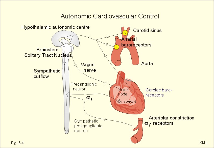
Viscerovisceral Reflexes. The visceral afferent fibers from these course in the glossopharyngeal and vagus nerves to the brain stem, causing a reflex slowing of the heart rate via visceral efferent fibers in the vagus nerve and peripheral vasodilation via inhibition of sympathetic efferent fibers.
What are somatic and visceral fibers?
Somatic efferent fibres innervate voluntary muscles that derive from the myotomes of the embryo. Visceral motor fibres are divided into special visceral efferents, which innervate striped muscles of branchial origin, and general visceral efferents, which innervate involuntary muscles and secreting glands.
What is the function of visceral afferent fibers?
The visceral afferents conduct messages from the organs serving the internal economy of the body; such impulses result in reflex control of these organs (e.g., the rate of the heartbeat and activities of the digestive system).
What is an example of visceral sensory?
Conscious sensations arising from the viscera, in addition to pain, include organ filling, bloating and distension, dyspnea, and nausea, whereas non-visceral afferent activity gives rise to sensations such as touch, pinch, heat, cutting, crush, and vibration.
What is visceral function?
The visceral (or autonomic) motor system controls involuntary functions mediated by the activity of smooth muscle fibers, cardiac muscle fibers, and glands.
What is the function of visceral nerves?
The autonomic nervous system (ANS) is also known as the efferent (motor) visceral nervous system. It controls the autonomic functions of the body such as heart rate, respiration, digestive secretions, sexual arousal, flight to fight responses, reflex actions, such as coughing, vomiting, swallowing, etc.
What is the difference between sensory somatic and visceral fibers?
Somatic sensory input comes from the receptors of the eyes, ears, nose, tongue, and skin. These organs transmit information we associate with the five senses. Visceral sensory input comes from (surprise!) the viscera, or internal organs.
What are visceral neurons?
A peripheral nerve that contains axons of the autonomic nervous system, either transmitting afferent signals from mucous membranes, glands, and vessels (visceral sensory nerves) or transmitting efferent signals to smooth muscles and glands (visceral motor nerves).
What is the difference between somatic and visceral nerves?
Somatic reflex is the nerve circuit of the somatic nervous system. It is responsible for the contraction of skeletal muscles. On the other hand, the visceral reflex is the nerve circuit of the autonomic nervous system. It is responsible for the contraction of smooth muscles and organs inside the body.
What is visceral afferent?
Definition. Visceral Afferents are neurons that sense events occurring within internal organs, the membranes that cover them or their attachments to the body wall.
What are visceral afferent neurons?
Visceral afferent nerves relay sensory information about visceral volume, pressure, contents, or nocioceptive stimuli to spinal centers, where automatic responses are interpreted and reflex responses are generated.
What's the difference between somatic and visceral sensory neurons?
Somatic sensory input comes from the receptors of the eyes, ears, nose, tongue, and skin. These organs transmit information we associate with the five senses. Visceral sensory input comes from (surprise!) the viscera, or internal organs.
What is special visceral efferent?
The term special visceral efferents (SVE) refers to nerve fibers that innervate the voluntary striated muscles of the larynx and pharynx and the muscles of facial expression and mastication.
Where are GVA fibers located?
An organ, or part of an organ, in the pelvis is said to be "above the pelvic pain line" if it is in contact with the peritoneum, except in the case of the large intestine, where the pelvic pain line is said to be located in the middle of the sigmoid colon. GVA fibers from structures above the pain line follow the course of the sympathetic efferent fibers, and GVA fibers from structures below the pain line follow the course of the parasympathetic efferents. Pain from the latter fibers is less likely to be consciously experienced.
How do GVA fibers create pain?
GVA fibers create referred pain by activating general somatic afferent fibers where the two meet in the posterior grey column .
Where do afferent fibers travel?
This means that a signal traveling in an afferent fiber will begin at sensory receptors in the afferent fiber's target organ, travel up to the ganglion where the sympathetic efferent fiber synapses, continue back along a splanchnic nerve ...
Which fibers follow the course of the sympathetic efferents?
GVA fibers from structures above the pain line follow the course of the sympathetic efferent fibers, and GVA fibers from structures below the pain line follow the course of the parasympathetic efferents. Pain from the latter fibers is less likely to be consciously experienced.
Is the visceral nervous system a part of the autonomic nervous system?
They are considered to be part of the visceral nervous system, which is closely related to the autonomic nervous system, but 'visceral nervous system' and 'autonomic nervous system' are not direct synonyms and care should be taken when using these terms. Unlike the efferent fibers of the autonomic nervous system, ...
Which fibers convey visceral information?
General visceral afferent fibers convey visceral information such as distention of organs and chemical conditions from the blood vessels, heart, lungs, digestive system, and other organ systems and glands into the central nervous system via both spinal and cranial (glossopharyngeal and vagus) nerves.
What are the components of the cranial nerve?
Like the vagus nerve, cranial nerve IX has five components, three of which are sensory and two of which are motor . The GSA component is a minor one, which innervates a small region of skin behind the ear. The GSA fibers of the glossopharyngeal nerve have cell bodies located in the superior (jugular) ganglion of IX, and they traverse the spinal tract of V to terminate in the spinal nucleus of V with similar GSA afferents from cranial nerves X, VII, and V. The GVA component of IX has a much more restricted distribution than the GVA component of X. Glossopharyngeal GVA fibers innervate the mucosal membranes that line the internal surface of the middle ear and cover the posterior third of the tongue, the tonsils, the posterior and upper surfaces of the pharynx, and the Eustachian tube. These GVA fibers have cell bodies in the inferior (petrosal) ganglion of IX. They enter the tractus solitarius and terminate in its nucleus. The latter projects to sites that include nucleus ambiguus (for throat reflexes), the dorsal motor nucleus of X, and the reticular formation. The carotid sinus reflex depends on some additional glossopharyngeal GVA fibers that innervate the carotid sinus (which is located at the bifurcation of the internal and external carotid arteries) and are stimulated by increases in arterial blood pressure. These fibers terminate on solitary nucleus neurons that project to the dorsal motor nucleus of X, which, via its projection to the parasympathetic ganglia of the heart, decreases heart rate and the arterial blood pressure. The SVA component of IX innervates taste buds located on the posterior third of the tongue. These fibers have cell bodies located in the inferior (petrosal) ganglion of IX and project to first-order multipolar neurons in the gustatory nucleus, which, as discussed previously, projects to VPMpc in the dorsal thalamus for relay to gustatory cortical areas.
Which cranial nerves carry efferent fibers?
The following cranial nerves carry general visceral efferent fibers: 1. Cranial nerve III (Edinger-Westphal nucleus): the preganglionic fibers from the Edinger-Westphal nucleus terminate in the ciliary ganglion, and the postganglionic fibers innervate the pupil. 2.
Where do glossopharyngeal fibers reach?
The glossopharyngeal general visceral efferent fibers reach the otic ganglion, which lies below the foramen ovale. Postganglionic fibers from the otic ganglion then provide input to the parotid gland.
What are the facial nerves?
The functional anatomy of the facial nerve is determined by its components, including the special somatic efferent fibers innervating muscles derived from the second brachial arch, namely the muscles of facial expression and the stapedius, stylohyoid, posterior belly of the digastric, buccinator, and platysma. 32,69,242 General visceral efferent fibers include preganglionic parasympathetic secretomotor fibers, which innervate lacrimal and seromucous glands in the nasal cavity and palate via the greater superficial petrosal nerve and sublingual and submandibular glands via the chorda tympani nerve. Preganglionic fibers in the greater superficial petrosal nerve travel to the sphenopalatine and pterygopalatine ganglia to synapse with the postganglionic parasympathetic neurons, and the preganglionic fibers in the chorda tympani travel with the lingual nerve to the submandibular ganglion to connect with the postganglionic parasympathetic neurons. 32,69,106,107,242 Taste from the anterior two thirds of the tongue is transmitted via special somatic afferent nerve fibers through the chorda tympani nerve branch of the facial nerve. Taste sensation from the soft palate and nasopharyngeal mucosa is also carried via special somatic afferent nerve fibers through the greater superficial petrosal nerve with cell bodies in the meatal ganglion. 69,106,242 General somatic afferent fibers provide cutaneous sensation from the external auditory canal and concha and course through the posterior auricular branch of the facial nerve, 32,69,106,242 with interconnections with the vagus, trigeminal, and upper cervical cutaneous nerve fibers to the same region. 60,242 The reader is referred to an available review of the anatomy of the sensory components of the facial nerve by Boudreau et al. 29 ( Fig. 50-3) and the functional anatomy as reviewed by Sweeney and Gilden 221 ( Fig. 50-4 ).
What is the name of the cranial nerve that is the most far-reaching?
The Vagus Nerve (CN X) The vagus is by far the most far-reaching of the cranial nerves, indeed its name is derived from the Latin word vago, to wander. The vagus contains both afferent (GVA and special visceral afferent (SVA)) fibres and efferent fibres (GVE and SVE).
Where do preganglionic fibers pass through?
The preganglionic fibers for the submandibular and sublingual salivary glands pass through the geniculate ganglion and then leave the mastoid segment of the facial nerve within the chorda tympani. The chorda tympani travels through the temporal bone within the canal of Huguier and enters the infratemporal fossa via the petrotympanic fissure, where it joins the lingual nerve (CN V). Within the submandibular ganglion, the parasympathetic fibers synapse with the postganglionic cells that innervate the submandibular and sublingual salivary glands.
Where do parasympathetic fibers originate?
Parasympathetic Nucleus. The general visceral efferent fibers originate from the superior salivatory nucleus and the lacrimal nucleus in the dorsal pon s and are carried distally within the nervus intermedius. After joining the motor root of the facial nerve, two main branches carry the parasympathetic fibers to their destination.
What nerves are in the dorsal trunk?
The fibres of the dorsal trunk contain 80% from the right vagus nerve and 20% from the left.