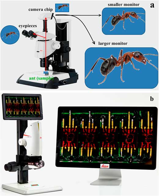
The compound microscope can be used to view a variety of samples some of which include: blood cells cheek cells parasites bacteria algae tissue and thin sections of organs. Compound microscopes are used to view samples that can not be seen with the naked eye.
How does a compound light microscope magnify an image?
Compound microscope – It has two convex lenses. It is called a compound microscope because it compounds the light as it passes through the lenses to magnify. The image of the object being viewed is enlarged because of the lens near the object. An eyepiece, an additional lens, is where real magnification takes place.
What are the parts of a compound light microscope?
What are the parts of a compound light microscope? The three basic, structural components of a compound microscope are the head, base and arm. Head/Body houses the optical parts in the upper part of the microscope. Base of the microscope supports the microscope and houses the illuminator. Arm connects to the base and supports the microscope head.
What are the parts and functions of a microscope?
Microscope Parts: Microscope Parts Functions: Base: Supports the microscope: Arm: Used to carry the microscope: Stage: Platform where the slide with the specimen is placed: Stage Clips: Holds the slide in place on the stage: Eyepiece (containing ocular lens) Magnifies the image for the viewer: Revolving nose piece
What is the magnification of a compound microscope?
Typically, a compound microscope is used for viewing samples at high magnification (40 – 1000x), which is achieved by the combined effect of two sets of lenses: the ocular lens (in the eyepiece) and the objective lenses (close to the sample).

What can you observe with a compound light microscope?
Compound microscopes are used to view small samples that can not be identified with the naked eye. These samples are typically placed on a slide under the microscope. When using a stereo microscope, there is more room under the microscope for larger samples such as rocks or flowers and slides are not required.
What are the 4 types of objectives seen in a compound light microscope?
A compound light microscope often contains four objective lenses: the scanning lens (4X), the low‐power lens (10X), the high‐power lens (40 X), and the oil‐immersion lens (100 X).
Can compound light microscopes view living things?
Compound microscopes are light illuminated. The image seen with this type of microscope is two dimensional. This microscope is the most commonly used. You can view individual cells, even living ones.
What is a light microscope used for?
Light microscopy is used to make small structures and samples visible by providing a magnified image of how they interact with visible light, e.g., their absorption, reflection and scattering.
How many objective lenses are there in a compound microscope?
Objective lens- There is usually 3 or 4 objective lens in the microscope that has different powers. Also, they can magnify objects to a good resolution.
What are the parts of a compound microscope?
Parts of Compound MicroscopeFoot or base. It is a U-shaped structure and supports the entire weight of the compound microscope.Pillar. It is a vertical projection. ... Arm. The entire microscope is handled by a strong and curved structure known as the arm.Stage. ... Inclination joint. ... Clips. ... Diaphragm. ... Nose piece.More items...
What are the parts of compound microscope and their functions?
Monocular or Binocular Head: Structural support that holds & connects the eyepieces to the objective lenses. Arm: Supports the microscope head and attaches it to the base. Nosepiece: Holds the objective lenses & attaches them to the microscope head. This part rotates to change which objective lens is active.
What are the types of microscope?
These five types of microscopes are:Simple microscope.Compound microscope.Electron microscope.Stereomicroscope.Scanning probe microscope.
What is a compound light microscope?
A compound light microscope is a microscope with more than one lens and its own light source. In this type of microscope, there are ocular lenses in the binocular eyepieces and objective lenses in a rotating nosepiece closer to the specimen. Although sometimes found as monocular with one ocular lens, the compound binocular microscope is more ...
Why do compound light microscopes have multiple lenses?
Moreover, because of their multiple lenses, compound light microscopes are able to reveal a great amount of detail in samples. Even an inexpensive one can reveal an incredible view of the world that would be impossible to explore with the naked eye.
How to find the total magnification of a compound light microscope?
In order to ascertain the total magnification when viewing an image with a compound light microscope, take the power of the objective lens which is at 4x, 10x or 40x and multiply it by the power of the eyepiece which is typically 10x.
Which microscope is more commonly used today?
Although sometimes found as monocular with one ocular lens, the compound binocular microscope is more commonly used today.
How much magnifying power does a microscope have?
Microscopes have come a long way since then—today's strongest compound microscopes have magnifying powers of 1,000 to 2,000X.
What is fluorescence microscope?
Fluorescence Microscope - study the most used microscope in medical/biological fields which uses high powered light waves to provide unique image viewing options.
What is oil immersion microscope?
Oil Immersion Microscopy - when used properly increases the refractive index of a sample/specimen. With only a few disadvantages, slides prepared with oil immersion techniques work best under higher magnification where oils increase refraction despite short focal lengths.
Why is the microscope important?
Why the microscope is important for studying biology. Cells are building blocks of life. No only small organisms like protozoa but also large organisms like an elephant, they are all made of cells. However, the size of cells is pretty small, ranging from 1-100 µm (micrometer), which is one-millionth of a meter.
What microscope is used to see the organelles of a cell?
To see the cell organelles, you will need to get a higher magnification (usually with a 40x-100x objective lens). In addition, the electron microscope is required to resolve the structure of mitochondria, bacteria, viruses, and large protein complexes.
How big can the naked eye see?
The human naked eye can see an object as small as 0.1 millimeters (100 µm). Therefore, the human naked eye can barely see cells (most of them <100 µm). Thus, we need the amplifying power of the microscope to see cells and even the structure and organelles inside of cells. Below is a size and length scale in biology, including eggs, cells, ...
What is the purpose of a transmission electron microscope?
The transmission electron microscope is required to see the internal structure of the mitochondria, including the inner membrane, outer membrane, intermembrane space, matrix, and cristae. The structure we saw from the transmission electron microscope is more like an illustration image.
Is the mitochondria visible?
Yes. It is visible. You can see a vague image of mitochondria with very little detail, even with the aid of fluorescence dye. The green color of the image below labeled the mitochondria. Their morphology is tubular, and it is very different from the cartoon illustration, which contains lots of membranes inside.
Can you see viruses with an electron microscope?
You will need an electron microscope to see the viruses. The original electron microscopic image of viruses is black-and-white. The colors are artificially painted post-production for better visualization. We have a detailed post explaining why a light microscope cannot see viruses, what limits the resolution of a light microscope, ...
