
What can stain gram variable? Some bacteria, after staining with the Gram stain, yield a gram - variable pattern: a mix of pink and purple cells are seen. In cultures of Bacillus, Butyrivibrio, and Clostridium
Clostridium
Clostridium is a genus of Gram-positive bacteria. This genus includes several significant human pathogens, including the causative agent of botulism. The genus formerly included an important cause of diarrhea, Clostridioides difficile, which was separated after 16S rRNA an…
What are the steps in a Gram stain?
Stain Reaction: The four basic steps of the Gram Stain are: 1) Application of the primary stain Crystal Violet (CV) to a heat-fixed smear of bacterial culture. CV dissociates in aqueous solutions into CV+ and Cl – ions. These two ions then penetrate through the cell wall and cell membrane of both Gram-positive and Gram-negative cells.
Is Gram stain positive or negative?
Gram-positive bacteria are bacteria with thick cell walls. In a Gram stain test, these organisms yield a positive result. The test, which involves a chemical dye, stains the bacterium’s cell wall purple. Gram-negative bacteria, on the other hand, don’t hold the dye. They stain pink instead.
Which bacteria will not stain using the Gram staining method?
This waxy barrier also prevents stains from penetrating the cell, which is why the Gram stain does not work with mycobacteria such as Mycobacterium, which are pathogens of humans and animals. For these bacteria, the acid–fast staining technique is used. Figure 6. Acid-fast bacilli in sputum
What is a Gram stain used for?
The Gram stain is a differential method of staining used to assign bacteria to one of two groups (gram-positive and gram-negative) based on the properties of their cell walls. It is also known as Gram staining or Gram's method.
What causes Gram stain variability?
The exposure of heat-fixed bacterial smears to relative humidities of 0, 52 and 98%, following the iodine step in a dry Gram stain procedure, markedly influenced the rate of decolorization upon exposure to 95% ethyl alcohol.
What are examples of Gram variable bacteria?
Bacilli: Escherichia coil, Pseudomonas species, Proteus species, and Klebsiella species. Examples of gram variable organisms include: Actinomyces species.
What organisms are Gram variable rods?
There are five medically important genera of gram-positive rods: Bacillus, Clostridium, Corynebacterium, Listeria, and Gardnerella. Bacillus and Clostridium form spores, whereas Corynebacterium, Listeria, and Gardnerella do not.
What can Gram stain identify?
Definition. A Gram stain is a test used to identify bacteria. It is one of the most common ways to quickly diagnose bacterial infection in the body.
Is E. coli Gram-variable?
E coli is a gram-negative bacillus that grows well on commonly used media. It is lactose-fermenting and beta-hemolytic on blood agar. Most E coli strains are nonpigmented.
Is bacillus a Gram-variable?
If gram-positive (or gram-variable) bacilli contain spores, Clostridium or Bacillus species are likely [T1] [I1d]. Although Clostridium organisms are classically gram-positive bacilli that may or may not contain spores, in our experience they often stain gram-negative in clinical speci- mens.
What are Gram-variable organisms?
Gram-variable organisms are those that cannot be grouped as either negative or positive. The appearance of organisms that stain gram-positive or -negative means that the smear contains organisms. These organisms may be pathogenic or nonpathogenic.
What is Gram variability?
Definition of gram-variable : staining irregularly or inconsistently by Gram's stain.
Do Gram-variable organisms cause disease?
Gram-positive and gram-negative bacteria stain differently because their cell walls are different. They also cause different types of infections, and different types of antibiotics are effective against them. Some Gram-positive bacteria cause disease.
What information can be gathered from looking at a gram stained bacterial smear?
A Gram stain is used, along with a culture of the sample from an infected site, to identify the cause of a bacterial infection. The Gram stain provides preliminary results on whether bacteria are present and the general type, such as the shape and whether they are Gram-positive or Gram-negative.
What does a positive Gram stain indicate?
If your test result reveals a positive Gram stain, it means that bacteria were present in your sample. If your result is positive, it usually includes information about what kind of organism was present on the sample slide, including: Type of bacteria: Gram-positive or gram-negative.
Why is Gram staining used to identify bacteria?
Gram staining differentiates bacteria by the chemical and physical properties of their cell walls. Gram-positive cells have a thick layer of peptidoglycan in the cell wall that retains the primary stain, crystal violet.
Is Bacillus subtilis a gram-variable?
Bacillus subtilis is a Gram-positive, rod-shaped bacterium that forms heat-resistant, dormant spores.
What is gram-variable?
Definition of gram-variable : staining irregularly or inconsistently by Gram's stain.
What are gram indeterminate bacteria?
Indeterminate Gram staining is observed in bacteria that are neither Gram-positive nor Gram-negative, also known as atypical bacteria. Gram-indeterminate bacteria have no response to gram staining. Examples include Gram-variable and acid fast bacteria.
What are the 5 Gram-positive bacteria?
Gram-positive bacteria can be staphylococci, streptococci, pneumococci, Bacillus anthracis, and Corynebacterium diptheriae.
What is Gram staining?
The Gram staining is one of the most crucial staining techniques in microbiology. It gets its name from the Danish bacteriologist Hans Christian Gram who first introduced it in 1882, mainly to identify organisms causing pneumonia.[1] .
What is the primary color of gram stain?
Often the first test performed, gram staining involves the use of crystal violet or methylene blue as the primary color.[2] . The term for organisms that retain the primary color and appear purple-brown under a microscope is Gram-positive organisms.
What is the term for organisms that retain the primary color and appear purple-brown under a microscope?
The term for organisms that retain the primary color and appear purple-brown under a microscope is Gram-positive organisms. The organisms that do not take up primary stain appear red under a microscope and are Gram-negative organisms. The Gram staining is one of the most crucial staining techniques in microbiology.
When was Gram staining first used?
Last Update: February 23, 2021. Introduction. The Gram staining is one of the most crucial staining techniques in microbiology. It gets its name from the Danish bacteriologist Hans Christian Gram who first introduced it in 1882, mainly to identify organisms causing pneumonia.[1] . Often the first test performed, ...
How long to counterstain a smear?
The smear is counterstained with basic fuchsin solution for 40 to 60 seconds. The fuchsin solution is washed off with water, and excess water is blotted with the bibulous paper. The slide can also be air-dried after shaking off excess water. 3.
Which cocci are Gram positive?
Gram-positive cocci in clusters: Usually characteristic of Staphy lococcusspecies such as S. aureus.
How thick should a smear be?
The smear should be one cell thick with no overlapping of cells
What is the most common gram stain?
A Gram stain of mixed Staphylococcus aureus ( S. aureus ATCC 25923, gram-positive cocci, in purple) and Escherichia coli ( E. coli ATCC 11775, gram-negative bacilli, in red), the most common Gram stain reference bacteria. Gram stain or Gram staining, also called Gram's method, is a method of staining used to distinguish ...
How does gram staining work?
Gram staining differentiates bacteria by the chemical and physical properties of their cell walls. Gram-positive cells have a thick layer of peptidoglycan in the cell wall that retains the primary stain, crystal violet. Gram-negative cells have a thinner peptidoglycan layer that allows the crystal violet to wash out on addition of ethanol. They are stained pink or red by the counterstain, commonly safranin or fuchsine. Lugol's iodine solution is always added after addition of crystal violet to strengthen the bonds of the stain with the cell membrane. Gram staining is almost always the first step in the preliminary identification of a bacterial organism. While Gram staining is a valuable diagnostic tool in both clinical and research settings, not all bacteria can be definitively classified by this technique. This gives rise to gram-variable and gram-indeterminate groups.
Why is Lugol's iodine added to the gram stain?
Lugol's iodine solution is always added after addition of crystal violet to strengthen the bonds of the stain with the cell membrane. Gram staining is almost always the first step in the preliminary identification of a bacterial organism.
What is the size of a Gram stain of Candida albicans?
Gram stain of Candida albicans from a vaginal swab. The small oval chlamydospores are 2–4 µm in diameter.
What did Gram do to make bacteria more visible?
Gram devised his technique not for the purpose of distinguishing one type of bacterium from another but to make bacteria more visible in stained sections of lung tissue. He published his method in 1884, and included in his short report the observation that the typhus bacillus did not retain the stain.
What is the difference between a purple and a pink gram positive?
Purple-stained gram-positive (left) and pink-stained gram-negative (right) Gram-positive bacteria have a thick mesh-like cell wall made of peptidoglycan (50–90% of cell envelope), and as a result are stained purple by crystal violet, whereas gram-negative bacteria have a thinner layer (10% of cell envelope), so do not retain ...
Which phyla have monoderms?
Gram-positive bacteria generally have a single membrane ( monoderm) surrounded by a thick peptidoglycan. This rule is followed by two phyla: Firmicutes (except for the classes Mollicutes and Negativicutes) and the Actinobacteria. In contrast, members of the Chloroflexi (green non-sulfur bacteria) are monoderms but possess a thin or absent (class Dehalococcoidetes) peptidoglycan and can stain negative, positive or indeterminate; members of the Deinococcus–Thermus group stain positive but are diderms with a thick peptidoglycan.
What is Gram staining?
Gram staining is an empirical method of differentiating bacterial species into two large groups (Gram-positive and Gram-negative) based on the chemical and physical properties of their cell walls. The method is named after its inventor, the Danish scientist Hans Christian Gram (1853-1938), who developed the technique in 1884 (Gram 1884). The importance of this determination to correct identification of bacteria cannot be overstated as all phenotypic methods begin with this assay.
What color is a Gram negative?
Gram’s method involves staining the sample cells dark blue, decolorizing those cells with a thin cell wall by rinsing the sample, then counterstaining with a red dye. The cells with a thick cell wall appear blue (gram positive) as crystal violet is retained within the cells, and so the red dye cannot be seen. Those cells with a thin cell wall, and therefore decolorized, appear red (gram negative).
How to decolorize a cell?
Decolorize the sample by applying 95% ethanol or a mixture of acetone and alcohol. This can be done in a steady stream, or a series of washes. The important aspect is to ensure that all the color has come out that will do so easily. This step washes away unbound crystal violet, leaving Gram-positive organisms stained purple with Gram-negative organisms colorless. The decolorization of the cells is the most “operator-dependent” step of the process and the one that is most likely to be performed incorrectly.
What is the purpose of decolorizing a cell wall?
The decolorizing mixture dehydrates cell wall, and serves as a solvent to rinse out the dye-iodine complex. In Gram-negative bacteria it also dissolves the outer membrane of the gram-negative cell wall aiding in the release of the dye. It is the thickness of the cell wall that characterizes the response of the cells to the staining procedure.
How much crystal violet is good for decolorization?
The standard 0.3% solution is good, if decolorization does not generally exceed 10 seconds.
Which peptidoglycan layer is agglutinin?
Fluorescence-labeled wheat germ agglutinin binds specifically to N-acetylglucosamine in the outer peptidoglycan layer of gram-positive bacteria. The peptidoglycan layer of gram-negative bacteria is covered by a membrane and is not labeled by the lectin.
Is it a prudent practice to always include a positive and negative control on the staining procedure?
It is a prudent practice to always include a positive and negative control on the staining procedure to confirm the accuracy of the results ( Murray et al 1994) and to perform proficiency testing on the ability of the technicians to correctly interpret the stains (Andserson, et al. 2005).
What are the factors that can influence gram stain results?
The many variables that can affect this stain are age of the culture, amount of decolorizer used, the time of decolorization, the type of organism (acid-fast bacteria and spores do not stain well), thickness of the smear, and the general care of the stainer.
Which step is the most crucial or most likely to cause poor results in Gram staining?
The thickness of the smear used in the Gram stain will affect the result of the stain. The step that is most crucial in effecting the outcome of the stain is the decolorizing step.
What is the outcome of a well executed Gram stain quizlet?
What is the Gram Stain method? Hans: What is the outcome of a well-executed Gram stain? Gram- positive appear purple, and Gram-negative appear pink. Hans: Why is the bacterial cell well of such great importance?
What happens to iodine and crystal violet in Gram stain?
During the process of staining, the iodine and crystal violet would form a complex within the heat fixed cell, and in organisms that are gram-negative, this complex would be readily washed out by agents, such as alcohol or acetone, but would appear red as they retain only the counterstain.
What is the characteristics of Gram-negative bacteria?
Gram negative bacteria are characterized by the presence of the periplasmic space, which is a single layer of peptidoglycan sandwiched between the cytoplasmic membrane and the outer membrane. Peptidoglycan, also known as murein, is a polymer that consists of a carbohydrate backbone and amino acids.
Is E coli gram variable?
Gram stain is used to identify the organism causing a disease or pathological condition. … Gram-negative bacilli include Escherichia coli, Pseudomonas species, Proteus species, and Klebsiella species. Gram- variable organisms include Actinomyces species.
What does gram variability mean?
Gram-variable organisms are those that cannot be grouped as either negative or positive. The appearance of organisms that stain gram-positive or -negative means that the smear contains organisms. These organisms may be pathogenic or nonpathogenic.
Why is Mycobacterium gram variable?
tuberculosis has an unusual, waxy coating on its cell surface primarily due to the presence of mycolic acid. This coating makes the cells impervious to Gram staining, and as a result, M. tuberculosis can appear either Gram-negative or Gram-positive.
What bacteria are gram-negative rods?
Commonly isolated Gram-negative organisms include Pseudomonas, Klebsiella, Proteus, Salmonella, Providencia, Escherichia, Morganella, Aeromonas, and Citrobacter. Occasionally, Gram-positive organisms (e.g., Streptococcus, Corynebacteria) are the primary organisms, or are found concurrently with Gram-negative bacteria.
Are Streptococcus rods?
Streptococcus bacteria subdivide into Strep. pyogenes (Group A), Strep. agalactiae (Group B), enterococci (Group D), Strep viridans, and Strep pneumonia. Gram-positive bacilli (rods) subdivide according to their ability to produce spores.
Where are gram-positive rods found?
When gram-positive bacteria are shaped like rods, they’re known as bacilli. Most of these bacteria are typically found on the skin, but some can cause serious medical conditions.
What is the Gram stain?from asm.org
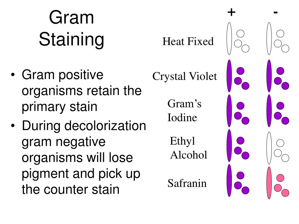
The Gram stain, the most widely used staining procedure in bacteriology, is a complex and differential staining procedure. Through a series of staining and decolorization steps, organisms in the Domain Bacteria are differentiated according to cell wall composition. Gram-positive bacteria have cell walls that contain thick layers of peptidoglycan (90% of cell wall). These stain purple. Gram-negative bacteria have walls with thin layers of peptidoglycan (10% of wall), and high lipid content. These stain pink. This staining procedure is not used for Archeae or Eukaryotes as both lack peptidoglycan. The performance of the Gram Stain on any sample requires four basic steps that include applying a primary stain (crystal violet) to a heat-fixed smear, followed by the addition of a mordant (Gram’s Iodine), rapid decolorization with alcohol, acetone, or a mixture of alcohol and acetone and lastly, counterstaining with safranin.
What are the morphological shapes of Gram positive bacteria?from microbeonline.com
Some of the typical morphological shapes of the Gram-positive bacilli are summarized in the table below: In Gram stain, these bacteria appear as V-in Y-shaped arrangements or in clumps that resemble Chinese letters. Due to the presence of spherical and terminal spore.
What are the problems with multiresistant Gram negative infections?from ncbi.nlm.nih.gov
One of the chief difficulties in the treatment of multiresistant gram-negative infections is the excessive use of antibiotics, not only those acquired by the community but also in hospitals. As the MDRs became epidemic, it is observable that overuse of these antimicrobials was one of the causes that urgently requires addressing. Thus, measures aimed at changing the use of these drugs, with educational campaigns, as well as the fight against self-medication with practices such as monitoring of drug consumption and their registration in pharmacies are necessary to change the habit of the population and also health professionals.
How to prevent multiresistant GNB?from ncbi.nlm.nih.gov
Thus, simple measures such as regular hand hygiene, adequate sterilization of medical equipment, isolation of patients suspected or diagnosed with MDR microorganisms - especially those submitted to invasive procedures - with caution regarding the entry of external persons. All individuals should abide by infectious disease preventive rules established in the healthcare setting. Another significant action is the laboratory worker who isolates MDR pathogens to immediately inform the epidemiological surveillance the staff of the health establishment so that prevention and control measures are enacted promptly.
What is the best method to identify bacterial strains resistant to multiple drugs?from ncbi.nlm.nih.gov
Phenotypic methods such as Modified Hodge (MHT) and Combined Diffusion Disc (CDT) using EDTA are a good alternative.[27] The MHT is a test based on the inactivation of carbapenems by bacterial strains containing the enzymes carbapenemases, enabling a susceptible strain to extend its growth to a disc containing the antimicrobial along the inoculum of the strain tested. This test is recommended for strains with high minimum inhibitory concentration (MIC) or reduced zones of inhibition in the disk.[28] The CDT comprises a test disc-diffusion with carbapenem antibiotics, where carbapenem discs are placed with and without EDTA, and their connection predicts whether the organism produces or does not carbapenemase. [29]
Why do drum sticks stain?from microbeonline.com
Drum stick or tennis racket appearance. Due to the presence of spherical and terminal spore. Nocardia. Branching filaments. In Gram stain, these bacteria stain irregularly and appear beaded. Gram-positive bacilli are responsible for “classical” diseases such as anthrax, diphtheria, and listeriosis and also for newer syndromes, ...
What is the name of the Gram positive bacteria that produces antibiotics?from microbeonline.com
Stre ptomyces: Streptomyces is a Gram-positive bacteria which shape resembles filamentous fungi. Streptomyces is known for its ability to produce bioactive secondary metabolites such as antifungals, antivirals, and mainly antibiotics. Today, nearly 80% of the antibiotics are sourced from the genus Streptomyces such as penicillin, streptomycin, ...
Overview
In microbiology and bacteriology, Gram stain or Gram staining, also called Gram's method, is a method of staining used to classify bacterial species into two large groups: gram-positive bacteria and gram-negative bacteria. The name comes from the Danish bacteriologist Hans Christian Gram, who developed the technique in 1884.
History
The method is named after its inventor, the Danish scientist Hans Christian Gram (1853–1938), who developed the technique while working with Carl Friedländer in the morgue of the city hospital in Berlin in 1884. Gram devised his technique not for the purpose of distinguishing one type of bacterium from another but to make bacteria more visible in stained sections of lung tissue. He published his method in 1884, and included in his short report the observation that the typhus ba…
Uses
Gram staining is a bacteriological laboratory technique used to differentiate bacterial species into two large groups (gram-positive and gram-negative) based on the physical properties of their cell walls. Gram staining is not used to classify archaea, formerly archaeabacteria, since these microorganisms yield widely varying responses that do not follow their phylogenetic groups.
Staining mechanism
Gram-positive bacteria have a thick mesh-like cell wall made of peptidoglycan (50–90% of cell envelope), and as a result are stained purple by crystal violet, whereas gram-negative bacteria have a thinner layer (10% of cell envelope), so do not retain the purple stain and are counter-stained pink by safranin. There are four basic steps of the Gram stain:
Examples
Gram-positive bacteria generally have a single membrane (monoderm) surrounded by a thick peptidoglycan. This rule is followed by two phyla: Bacillota (except for the classes Mollicutes and Negativicutes) and the Actinomycetota. In contrast, members of the Chloroflexota (green non-sulfur bacteria) are monoderms but possess a thin or absent (class Dehalococcoidetes) peptidogl…
Orthographic note
The term Gram staining is derived from the surname of Hans Christian Gram; the eponym (Gram) is therefore capitalized but not the common noun (stain) as is usual for scientific terms. The initial letters of gram-positive and gram-negative, which are eponymous adjectives, can be either capital G or lowercase g, depending on what style guide (if any) governs the document being written. Lowercase style is used by the US Centers for Disease Control and Prevention and other style reg…
See also
• Bacterial cell structure
• Ziehl–Neelsen stain
External links
• Gram staining technique video