What is the normal duration of a P wave?
Normal P Wave Size. Duration <120ms (3mm) Amplitude <2.5mm. The P wave is directed inferiorly and therefore should be positive in leads I and II. It is often biphasic in lead V1.
What are P and S waves also called?
The behavior of Earthquake Waves
- The ‘P’ waves or primary waves (longitudinal nature)
- Secondary waves or ‘S’ waves (transverse in nature)
- Surface waves or ‘L’ waves are long.
What is the difference from P waves and S waves?
P waves travel faster than S waves, and are the first waves recorded by a seismograph in the event of a disturbance. P waves travel at speeds between 1 and 14 km per second, while S waves travel significantly slower, between 1 and 8 km per second. The S waves are the second wave to reach a seismic station measuring a disturbance.
What do P waves and QRS waves represent?
There are three main components to an ECG: the P wave, which represents the depolarization of the atria; the QRS complex, which represents the depolarization of the ventricles; and the T wave, which represents the repolarization of the ventricles.

Why is it called the P wave?
His labeling of the primitive tracing was then mixed: A and B, the first letters of the alphabet, were used to indicate ventricular events, and P, from near the middle of the alphabet, was used to indicate atrial events.
What is called P wave?
P waves, or Primary waves, are the first waves to arrive at a seismograph. P waves are the fastest seismic waves and can move through solid, liquid, or gas. They leave behind a trail of compressions and rarefactions on the medium they move through. P waves are also called pressure waves for this reason.
What does the P wave do?
The first wave (p wave) represents atrial depolarisation. When the valves between the atria and ventricles open, 70% of the blood in the atria falls through with the aid of gravity, but mainly due to suction caused by the ventricles as they expand.
What is difference between P and S waves?
Primary (P) and secondary (S) waves are two types of waves caused by earthquakes. They are defined based on when they arrive and are felt on the surface. P waves, or primary waves, arrive first while S waves, or secondary waves, arrive second.
What does P and S in P and S wave mean?
In P or compressional waves, the vibration of the rock is in the direction of propagation. P waves travel fastest and are the first to arrive from the earthquake. In S or shear waves, rock oscillates perpendicular to the direction of wave propagation.
How do you read P waves on ECG?
1:207:16ECG for Beginners. Understanding the waves of ECG, P wave ... - YouTubeYouTubeStart of suggested clipEnd of suggested clipWhere should we look at our P waves. Well normal P waves should be upright in leads one two andMoreWhere should we look at our P waves. Well normal P waves should be upright in leads one two and upside-down or inverted and lead AVR.
Is the P wave positive or negative?
The Normal P Wave Thus, P waves are positive in lead II and usually in leads I, aVL, and aVF, reflecting the leftward and inferior direction of activation during sinus rhythm. This corresponds to a mean frontal plane P wave axis of approximately 60 degrees.
What are P waves and S waves called?
There are two types of seismic waves, primary waves and secondary waves. Primary waves, also known as P waves or pressure waves, are longitudinal compression waves similar to the motion of a slinky (SF Fig. 7.1 A). Secondary waves, or S waves, are slower than P waves.
What is normal P wave in ECG?
P wave amplitude rarely exceeds two and a half small squares (0.25 mV). The duration of the P wave should not exceed three small squares (0.12 s).
Which best describes P waves?
P-waves or primary waves are compressional waves. The direction of its propagation is parallel to the motion of the particles within the material. As a compressional wave, it can travel through solids, liquids, and gas.
What is P wave movement type?
compressionalSeismic P waves are also called compressional or longitudinal waves, they compress and expand (oscillate) the ground back and forth in the direction of travel, like sound waves that move back and forth as the waves travel from source to receiver. P wave is the fastest wave.
What is an earthquake?
An earthquake is the trembling or shaking of the Earth when multiple tectonic plates suddenly slip past each other.
What are seismic waves?
The waves or bursts of energy that propagate through the Earth and instigate earthquakes are called seismic waves.
What are the two types of seismic waves?
Body waves and surface waves are the two types of seismic waves.
What are the two types of body waves?
P waves and S waves are the two types of body waves.
What are P waves?
P waves are the first waves that are detected by a seismograph. They are the fastest seismic waves and can travel through gases, liquids, or solids.
What are the P waves?
P waves. P waves, or Primary waves, are the first waves to arrive at a seismograph. P waves are the fastest seismic waves and can move through solid, liquid, or gas. They leave behind a trail of compressions and rarefactions on the medium they move through. P waves are also called pressure waves for this reason.
How to understand P waves?
To understand P waves, we have to first look into the basics of seismology and seismic waves. The waves of energy that travel through the earth and cause earthquakes and related phenomena are seismic waves. There are two types of seismic waves : 1 Body waves 2 Surface waves
What are the two types of seismic waves?
There are two types of seismic waves : Body waves. Surface waves. Body waves are the waves that can travel through the layers of the earth. They are the fastest waves and as a result, the first waves that seismographs can record. Body waves can move through all states of matter including rocks and molten lava.
Can shear waves move through solids?
They are compression waves. They are shear waves. Can move through solids and liquids. Can only move through solids. Shake the medium in the direction in which they are propagating. Shake the medium in the direction perpendicular to which they are moving.
Is the outer core liquid or solid?
It is after studying the trajectory of S waves through the layers of earth, scientists were able to conclude that the earth’s outer core is liquid . Following is the table explaining concepts related to waves:
What is the P wave in ECG?
The P wave and PR segment is an integral part of an electrocardiogram (ECG). It represents the electrical depolarization of the atria of the heart. It is typically a small positive deflection from the isoelectric baseline that occurs just before the QRS complex. It can sometimes have abnormalities in morphology or timing that can be indicative of significant clinical pathology.[1] An understanding of the normal and abnormal P wave morphology is, therefore, a crucial part of ECG interpretation. This article will review the basics of P wave interpretation, including characteristics of both the normal P wave and its pathologic abnormalities.
What is a second degree AV block?
Another abnormality in the PR interval is a second-degree AV block. There are two types of second-degree AV blocks: If there is a prolonged PR segment with increased lengthening of the PR segment with each consecutive beat occasionally followed by no QRS complex, it is known as a second degree Mobitz type I block, also known as Wenckebach block. In the other type of second-degree block known as Mobitz type II block, the PR segment can be normal or prolonged but remains constant with every beat, and occasionally, a sudden unexpected drop of the QRS complex is present. In Mobitz type II, there is significant conduction disease in this His-Purkinje system that is irreversible and requires pacemaker placement compared to Mobitz type I blocks. [4]
What is the PR interval?
The PR interval represents the time between atrial depolarization and ventricular depolarization. Abnormalities in the timing of the PR segment can indicate pathology.
What is the role of ECG interpretation?
ECG interpretation and identification of pathology on electrocardiograms should be an integral part of monitoring any patient placed on telemetry or who presents with signs and symptoms of abnormal cardiac function. The healthcare team, including physicians, nurses, and technicians, should be able to quickly recognize common abnormal ECG patterns and intervene or notify a healthcare member, such as a cardiologist, that can treat the pathology found on the electrocardiogram rhythm.
Why is it important to alert clinicians of an abnormal rhythm or electrical conduction defect on ECG?
Early identification by nursing staff to alert clinicians of an abnormal rhythm or electrical conduction defect on ECG can lead to rapid management and decreased morbidity and mortality of patients. It is in the patients' best interest for all healthcare team members to have the ability and confidence to point out P wave abnormalities and PR segment prolongations or heart blocks.
What is the Creative Commons 4.0 license?
This book is distributed under the terms of the Creative Commons Attribution 4.0 International License (http://creativecommons.org/licenses/by/4.0/), which permits use, duplication, adaptation, distribution, and reproduction in any medium or format, as long as you give appropriate credit to the original author(s) and the source, a link is provided to the Creative Commons license, and any changes made are indicated.
What is NCBI bookshelf?
NCBI Bookshelf. A service of the National Library of Medicine, National Institutes of Health.
What is MAT in cardiology?
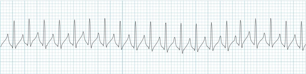
Multifocal atrial tachycardia (MAT) - an irregularly irregular narrow complex tachycardia with at least three different P wave morphologies and variable PP intervals, with an isoelectric baseline.
What is bifid P wave?
Bifid P waves are also referred to as P mitrale. Their presence indicates dyssynchrony between right and left atrial depolarisation; this may be normal, or suggestive of left atrial enlargement.
What does it mean when there are no P waves?
A lack of visible P waves preceding QRS complexes suggests a lack of sinus beats; this may occur with sinus dysfunction or in the presence of fibrillation or flutter waves. The P wave may also be hidden within the QRS complex.
What is QRS complex?
The QRS complex represents the depolarization (activation) of the ventricles. It is always referred to as the “QRS complex” although it may not always display all three waves. Since the electrical vector generated by the left ventricle is many times larger than the vector generated by the right ventricle, the QRS complex is actually a reflection of left ventricular depolarization. QRS duration is the time interval from the onset to the end of the QRS complex. A short QRS complex is desirable as it proves that the ventricles are depolarized rapidly, which in turn implies that the conduction system functions properly. Wide (also referred to as broad) QRS complexes indicate that ventricular depolarization is slow, which may be due to dysfunction in the conduction system.
How many vectors are generated by depolarization of the ventricles?
Depolarization of the ventricles generates three large vectors, which explains why the QRS complex is composed of three waves. It is fundamental to understand the genesis of these waves and although it has been discussed previously a brief rehearsal is warranted. Figure 7 illustrates the vectors in the horizontal plane. Study Figure 7 carefully, as it illustrates how the P-wave and QRS complex are generated by the electrical vectors.
What is ECG interpretation?
ECG interpretation includes an assessment of the morphology (appearance) of the waves and intervals on the ECG curve. Therefore, ECG interpretation requires a structured assessment of the waves and intervals. Before discussing each component in detail, a brief overview of the waves and intervals is given.
Why are R waves high?
It is important to assess the amplitude of the R-waves. High amplitudes may be due to ventricular enlargement or hypertrophy. To determine whether the amplitudes are enlarged, the following references are at hand:
What is ST segment?
The ST segment corresponds to the plateau phase (phase 2) of the action potential. The ST segment must always be studied carefully since it is altered in a wide range of conditions. Many of these conditions cause rather characteristic ST segment changes. The ST segment is of particular interest in the setting of acute myocardial ischemia because ischemia causes deviation of the ST segment ( ST segment deviation ). There are two types of ST segment deviations. ST segment depression implies that the ST segment is displaced, such that it is below the level of the PR segment. ST segment elevation implies that the ST segment is displaced, such that it is above the level of the PR segment. The magnitude of depression/elevation is measured as the height difference (in millimeters) between the J point and the PR segment. The J point is the point where the ST segment starts. If the baseline (PR segment) is difficult to discern, the TP interval may be used as the reference level.
Which side of the ventricular septum is depolarized?
The ventricular septum receives Purkinje fibers from the left bundle branch and therefore depolarization proceeds from its left side towards its right side . The vector is directed forward and to the right. The ventricular septum is relatively small, which is why V1 displays a small positive wave (r-wave) and V5 displays a small negative wave (q-wave). Thus, it is the same electrical vector that results in an r-wave in V1 and q-wave in V5.
Why is Q wave important?
The Q-wave. It is crucial to differentiate normal from pathological Q-waves, particularly because pathological Q-waves are rather firm evidence of previous myocardial infarction. However, there are numerous other causes of Q-waves, both normal and pathological and it is important to differentiate these.