Full Answer
What are the characteristics of Plasmodium ovale?
Plasmodium Ovale. Ring form - Of P. ovale is characterized by one or two large chromatin dots as well as a sturdy/thick cytoplasm. As they mature, the Schuffner's dots may develop. Trophozoites - Like in the ring forms, the cytoplasm of trophozoites is sturdy with a few chromatin dots.
How is Plasmodium falciparum diagnosed?
When looking at a blood smear under a microscope, Plasmodium falciparum can be identified by the presence of a high proportion of infected red blood cells and crescent-shaped gametocytes. Plasmodium malariae can be identified by its three unique characteristics:
What are the morphological characteristics of Plasmodium falciparum malaria?
It's responsible for severe malaria (malignant malaria) which is characterized by irregular paroxysms and high fever and may cause death if not treated. The morphological characteristics of the parasite vary depending on the diagnostic form/stage. The ring form of P. falciparum is found inside the red cells.
How do you see malaria parasites under a microscope?
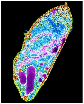
Once in the body, the malaria parasite infects the red cells where it thrives. When a positive slide is viewed under the microscope, it's possible to see the parasite inside the red cells (intracellular) as well as outside the cells (extracellular). Microscopy of malaria parasites (Protozoa)
See more
How do you identify Plasmodium under a microscope?
Malaria parasites can be identified by examining under the microscope a drop of the patient's blood, spread out as a “blood smear” on a microscope slide. Prior to examination, the specimen is stained (most often with the Giemsa stain) to give the parasites a distinctive appearance.
Can you see malaria under a microscope?
Microscopic examination remains the gold standard for laboratory confirmation of malaria in the malaria-endemic world, as well as in the United States.
What is the shape of Plasmodium?
Gametocytes of Plasmodium falciparum are crescent- or sausage-shaped, and are usually about 1.5 times the diameter of an RBC in length.
What are the characteristics of a Plasmodium?
Characteristics of Plasmodium They characteristically show the presence of apical complex. Typically, pigment is produced in erythrocyte during developmental stages of parasite. These pigments are visualized by light microscopy. Each microgamont produces 8 microgametes on exflagellation.
How is malaria parasite different from under a microscope?
Light microscopy of thick and thin stained blood smears remains the standard method for diagnosing malaria. It involves collection of a blood smear, its staining with Romanowsky stains and examination of the Red Blood Cells for intracellular malarial parasites.
What does malaria look like on a blood smear?
Examination of Giemsa-stained peripheral blood smear is the standard test for the diagnosis of malarial infection. Classic ring-shaped/headphone-shaped trophozoites are seen in case of Plasmodium falciparum infection.
Where is Plasmodium found?
Plasmodium falciparum predominates in Africa south of the Sahara, one reason why malaria is so severe in that area.
Is Plasmodium Gram positive or negative?
Plasmodium are not bacteria and as such do not have a uniform gram stain pattern, for example P. vivax is known to display a gram positive or gram negative stain depending on the conditions.
What is the difference between Plasmodium and Plasmodium?
Plasmodium is a sporozoan parasite that causes malaria. Whereas the plasmodium is a wall-less mass of multinucleated protoplasm of plasmodial slime molds.
How big is a Plasmodium?
1 to 20 micronsDepending on the developmental stage and species, malaria parasites can be spherical, ring shaped, elongated, or crescent shaped, and can range in size from 1 to 20 microns in diameter (1 micron equals 1 millionth of a meter or approximately 125,000 of an inch).
Is Plasmodium a bacteria or virus?
The Plasmodium parasite that causes malaria is neither a virus nor a bacteria – it is a single-celled parasite that multiplies in red blood cells of humans as well as in the mosquito intestine.
How does Plasmodium enter the body?
Malaria infection begins when an infected female Anopheles mosquito bites a person, injecting Plasmodium parasites, in the form of sporozoites, into the bloodstream. The sporozoites pass quickly into the human liver. The sporozoites multiply asexually in the liver cells over the next 7 to 10 days, causing no symptoms.
Can you have malaria and still test negative?
If your results were negative, but you still have malaria symptoms, you may need retesting. The number of malaria parasites can vary at times. So your provider may order blood smears every 12-24 hours over a period of two to three days. It's important to find out whether you have malaria so you can get treated quickly.
What tests are done for malaria?
Polymerase chain reaction (PCR). This test detects parasite nucleic acids and identifies the species of malaria parasite. Complete blood count (CBC). This checks for anemia or evidence of other possible infections.
How do you detect malaria parasite in thick film?
A thick blood smear is a drop of blood on a glass slide. Thick blood smears are most useful for detecting the presence of parasites, because they examine a larger sample of blood. (Often there are few parasites in the blood at the time the test is done.)
How is typhoid and malaria tested?
Blood smear examination, Rapid malaria antigen test, blood culture, and Widal test were done for the diagnosis of Malaria and typhoid respectively.
How to identify malaria parasites?
Malaria parasites can be identified by examining under the microscope a drop of the patient’s blood, spread out as a “blood smear” on a microscope slide. Prior to examination, the specimen is stained (most often with the Giemsa stain) to give the parasites a distinctive appearance.
How to detect parasitic nucleic acids?
Parasite nucleic acids are detected using polymerase chain reaction (PCR). Although this technique may be slightly more sensitive than smear microscopy, it is of limited utility for the diagnosis of acutely ill patients in the standard healthcare setting. PCR results are often not available quickly enough to be of value in establishing the diagnosis of malaria infection.
How long does it take to get malaria test results?
Such immunologic (“immunochromatographic”) tests most often use a dipstick or cassette format, and provide results in 2-15 minutes. These “Rapid Diagnostic Tests” (RDTs) offer a useful alternative to microscopy in situations where reliable microscopic diagnosis is not available. Malaria RDTs are currently used in some clinical settings and programs. The World Health Organization is conducting comparative performance evaluations of many of the RDTs which are commercially available worldwide based on a panel of parasites derived from a global network of collection sites. Results of this testing is available at: http://www.wpro.who.int/sites/rdt/home.htm#N#External#N#file_external#N#.
Why is malaria recognized?
Malaria must be recognized promptly in order to treat the patient in time and to prevent further spread of infection in the community via local mosquitoes.
What is the endpoint of in vitro testing?
In vitro tests: The parasites are grown in culture in the presence of increasing concentrations of drugs; the drug concentration that inhibits parasite growth is used as endpoint.
Can a laboratory test show malaria?
However, for a definitive diagnosis to be made, laboratory tests must demonstrate the malaria parasites or their components. A patient with fever who had recently traveled to a malaria-endemic country is being evaluated in the emergency room. Diagnosis of malaria can be difficult:
Can malaria be confirmed by a laboratory test?
Clinical findings should always be confirmed by a laboratory test for malaria.
What color is the GIEMSA stain?
When staining the smear, the nucleic parts of the parasite which is acidic will appear purple while the background will appear to be blue in color given that it is acidophilic.
How to make a thin smear?
Take a glass slide and collect two drops from the finger tip at the center of the glass slide. Using another slide with a smooth edge, spread the blood drops on the slide to create a thin smear. (You can collect another blood drop using another clean slide for a thick smear)
What is a Parasite?
Essentially, a parasite is a living organism that lives in (or on) another living organism (host) for survival. In most cases, the parasite is unable to live independently and thus requires a host that provides favorable conditions for growth and multiplication.
Where do parasites enter the body?
In endoparasitism, the parasites may enter the body through such routes as the mouth, nose, skin and even through the anus. Different endoparasites will move and live in different parts of the body.
What are the most common ectoparasites?
Fluke. Parasitic flatworms such as schistosomes. Ectoparasites - Ectoparasites are also multicellular organisms. Unlike helminths, ectoparasites live on the host and can be found on the skin or scalp. Some of the most common ectoparasites include: Fleas. Lice.
What are some examples of protozoa?
These include: Protozoa - Protozoa are single-celled organisms that live and thrive within the host. A good example of protozoa is plasmodium malariae, which can be found within the red blood cells of the host (the destruction of red cells by the parasites causes anemia).
How to get malaria out of your finger?
Using a clean, spirit swab, wipe the tip of the third finger and allow it to dry (of the patient with malaria) Using the needle/lancet, prick the cleaned finger tip and allow a drop or two to ooze out. Take a glass slide and collect two drops from the finger tip at the center of the glass slide.