
What type of epithelium lines the epithelial ducts of the lungs?
These ducts are lined by stratified squamous epithelium near the opening and the lumens are frequently filled with desquamated cells. Deeper in the connective tissue, the ducts acquire a stratified columnar appearance that is really a cuboidal duct cell sitting on a myoepithelial cell as in the sweat gland.
What is the structure of the sweat glands?
In general, sweat glands tend to comprise a secretory unit and a duct through which sweat or secretory product is passed. Sweat glands are situated in the dermis and are surrounded by adipose tissue. At the base of each sweat gland there is a structure known as the secretory coil.
How many layers of epithelium are present in ducts?
Many ducts appear to be composed of 2 layers of cuboidal epithelium. The inner layer are the actual ductal epithelial cells whereas the outer layer of cells is, in fact, a layer of myoepithelial cells.
What type of epithelium is found in the stomach?
Simple cuboidal epithelium: This type of epithelium is typically found in glandular (secreting) tissue and kidney tubules. Simple columnar epithelium: This type of epithelium is often specialized for absorption and usually has apical cilia or microvilli. These cells line your stomach and intestines.
What type of epithelium makes up the sweat gland ducts?
cuboidal epitheliumThe ducts are lined by stratified (2 layers) cuboidal epithelium. Long thin myoepithelial cells are arranged helically around the periphery between the secretory cells and their basement membrane.
Do sweat glands have epithelial tissue?
Sweat gland epithelial cells were found to detach from gland coils and were polygonal in shape. The cells continued to divide for up to 2 weeks and formed a circular monolayer surrounding the tissues.
Where is the duct of sweat glands?
Their ducts do not open onto the skin surface; this is because these glands originate from the stratum germinativum of the epidermis. Therefore, down-growth does not produce a duct open to the skin surface. Instead, the ducts open into hair follicles, and sweat is released through the hair opening in the skin.
What layer of epidermis are sweat glands located?
dermisYour dermis contains collagen and elastin, which help make your dermis thick and supportive of your skin's overall structure. All of your connective tissues, nerve endings, sweat glands, oil glands and hair follicles exist in your dermis.
Where is stratified squamous epithelium found?
The outer layer of your skin (the epidermis) is made of stratified squamous epithelial cells.
Where do you find stratified cuboidal epithelium?
This type of tissue can be observed in sweat glands, mammary glands, circumanal glands, and salivary glands. They protect areas such as the ducts of sweat glands, mammary glands, and salivary glands. They are also observed in the linings of urethra.
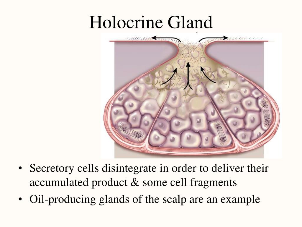
What type of epithelium is in sebaceous gland?
Sebaceous glands are composed of clusters of pale-staining, highly vacuolated epithelial cells (sebocytes) that are located adjacent to follicles. Sebaceous glands are responsible for producing sebum, a lipid-containing compound with moisturizing and antimicrobial properties.
What structure is a type of sweat gland?
Eccrine sweat glands occur over most of the body and open directly onto the skin's surface. Apocrine glands open into the hair follicle, leading to the surface of the skin. Apocrine glands develop in areas with many hair follicles, such as on the scalp, armpits and groin.
What glands are known as sweat glands quizlet?
Sweat glands, also known as sudoriferous or sudoriparous glands, from Latin sudor, meaning "sweat", are small tubular structures of the skin that produce sweat. Sweat glands are a type of exocrine gland, which are glands that produce and secrete substances onto an epithelial surface by way of a duct.
Which tissue within skin is responsible for producing sweat?
Connection: The hypodermis connects your dermis layer to your muscles and bones. Insulation: The hypodermis insulates your body to protect you from the cold and produces sweat to regulate your body temperature, protecting you from the heat.
What is sweat glands in skin?
Sweat glands, also known as sudoriferous or sudoriparous glands, from Latin sudor 'sweat', are small tubular structures of the skin that produce sweat. Sweat glands are a type of exocrine gland, which are glands that produce and secrete substances onto an epithelial surface by way of a duct.
What are sweat glands made of?
Basic structure. In general, sweat glands tend to comprise a secretory unit which is located either in the deep dermis or in the subcutaneous tissue, and a duct which continues from the secretory unit towards the body surface, through which sweat or secretory product is passed.
Which membrane contains epithelial tissue?
Mucous membranesMucous membranes are epithelial membranes that consist of epithelial tissue that is attached to an underlying loose connective tissue. These membranes, sometimes called mucosae, line the body cavities that open to the outside.
What tissues form glands?
Epithelial tissues are widespread throughout the body. They form the covering of all body surfaces, line body cavities and hollow organs, and are the major tissue in glands.
Which of the following functions is not associated with epithelial tissue?
All of the following functions are associated with epithelial tissue except: -generating electrical signals.
Which type of epithelial tissue is found lining the heart blood vessels and lymphatic vessels?
A simple squamous epithelium, called "endothelium," lines blood vessels, lymphatic vessels, and the chambers of the heart. When sections through endothelial cells are viewed with the light microscope, the cytoplasm cannot be seen, because the flattened cell is so thin.
What is the epithelium?
The epithelium is a type of body tissue that forms the covering on all internal and external surfaces of your body, lines body cavities and hollow organs and is the major tissue in glands . Epithelial tissue has a variety of functions depending on where it’s located in your body, including protection, secretion and absorption.
What are epithelial cells?
Epithelial tissue is made up of epithelial cells. The cells can be different shapes and can be arranged in a single layer or multiple layers depending on where they are in your body and what kind of functions they have.
What is the difference between epithelium, endothelium and mesothelium?
Epithelium, endothelium and mesothelium are three types of epithelial cell layers that line your internal organs, body cavities and form the outer layer of your skin.
What are the different kinds of epithelial cell tests?
Since epithelial cells exist in several important parts of your body, several types of tests examine epithelial cells to check for certain medical conditions. In medicine, pathology is the laboratory examination of cells in samples of body tissue or fluids for diagnostic purposes. A scientist called a pathologist examines the cells.
Which membrane is attached to cells of a pseudostratified epithelium?
B.)All cells of a pseudostratified epithelium are attached to the basement membrane.
Where are epithelia found?
This "category" of epithelia is found in areas exposed to abrasion and friction.
What tissue allows the bladder to stretch when filled with urine and then shrink back to its original shape when the bladder is?
Transitional tissue allows the urinary bladder to stretch when filled with urine and then shrink back to its original shape when the bladder is emptied.
What is the term for the cells that acquire nutrients and other vital substances through diffusion from the underlying connective tissue layer?
The cells of the epithelium acquire nutrients and other vital substances through diffusion from the underlying connective tissue layer. *Avascular means that there are no blood vessels present in epithelial tissue.
Where are goblet cells found?
lining the respiratory tract and in the simple columnar epithelium found in the stomach and small intestine. What is the function of goblet cells?
Where are the nuclei located in a tissue?
The nuclei located within the cells are fairly evenly dispersed in the middle of the cell or closer to the basal surface. How would you classify this tissue?
Is epithelial tissue avascular?
Epithelial tissues are avascular. How do the cells of this tissue obtain nutrients and other vital substances?
Which epithelium lines the urinary tract?
Correct answer 4. The urinary tract - epithelium shown is transitional epithelium (note the DOME-shaped cells), which lines the urinary tract. (skin would be lined by stratified squamous keratinizing epithelium
What is the epithelium of the skin?
This epithelium is found at the surface of the skin and is known as the epidermis. As protection against desiccation, it undergoes a process known as cornification or keratinization. As cells move toward the surface, they differentiate and eventually die, leaving an outermost layer of dead cells filled with keratin View Image. The absence of nuclei in this layer shows that it is devoid of live cells. In some slides, the keratinized region is gray, but occasionally it has been penetrated in places by red stain. Note the differences in morphology of the cells as they move toward the surface. You will learn the names of these layers when we study the skin. In the lower strata, look for the layer of spinous cells (the spines look like little lines between cells, and can be difficult to see) View Image; the spines are sites where desmosomes attach the cells to one another.
What is the difference between the inner and outer layers of epithelial cells?
The inner layer are the actual ductal epithelial cells whereas the outer layer of cells is, in fact, a layer of myoepithelial cells. In slide 258 (active gland), you can see that the amount of the glandular tissues has increased, while that of the connective tissue has decreased.
What are the structures of the mammary gland?
Be able to identify the histological components of the mammary gland, specifically the structures associated with the nipple and the areola, the overall organization into lobes and lobules, as well as secretory alveoli (acini), lactiferous ducts and sinuses and the intralobular and interlobular connective tissue.
What is the squamous epithelium?
Simple squamous epithelial cells are flattened, i.e., wider than they are tall. A simple squamous epithelium, called "endothelium," lines blood vessels, lymphatic vessels, and the chambers of the heart. When sections through endothelial cells are viewed with the light microscope, the cytoplasm cannot be seen, because the flattened cell is so thin. Thus, endothelium is generally identified on the basis of the structure and position of nuclei alone; that is, the nuclei are also often flattened and elongated, and are found lining the lumen of the vessel. Observe the endothelial lining of blood and lymph vessels in the mesentery in slide 30 View Image. Sometimes the blood vessels contain red blood cells and can be identified that way. Otherwise, look for tubular or circular profiles at low power and examine the endothelial lining of these vessels at high power. Note that the endothelium may be damaged during processing such that it separates from the vessel wall or it may slough off entirely and not be visible at all. In areas where you can find an endothelium, note that the nuclei do not always look flattened in vessels that have contracted. Another excellent place to look for endothelial cells is in the many small vessels in the wall of the intestine shown in slide 29 --look for the vessels in the submucosal layer (the lightest staining area in the wall of the intestine) View Image.
Why is the orientation of the tissue confusing?
The orientation of the tissue can be confusing because of connective tissue projections that push up into the epithelium. Unlike keratinizing epithelium, nuclei are still present in most surface cells (although they are often difficult to see in sectioned tissue.) 2. Stratified squamous keratinizing epithelium.
How are epithelia organized?
In epithelia, cells are organized in sheets, either a single layer thick (simple epithelia) or made up of multiple layers (stratified epithelia). Be able to identify the classes of epithelia underlined in the text below, and give some thought to why these different classes of epithelia have such different morphologies. The glass slide sets occasionally contain different stains or even different slides of the same tissue in place of the slide that you have in your set. When these differences occur, the lab guide will usually refer to a particular slides as being in either even or odd-numbered slide collections. Please try to look at the alternates by borrowing from your lab mates. Certain stains are much more instructive than others and different (alternate) tissues often help to explain functional changes.
