What is articular cartilage and why is it so important?
- Joint pain, especially when you put weight on the joint (and even while at rest)
- A grinding or clicking sensation when moving a joint
- Swelling and tenderness in and around the joint
- The joint giving out, locking, or catching
What function does the articular cartilage serve?
- Layer 1—Collagen fibers have a random orientation.
- Layer 2—Collagen fibers are in bundles. The bundles intersect at various angles. ...
- Layer 3—Collagen fibers have circular orientation. The external and internal fibers merge with fibers of the transverse acetabular ligament. ...
What kind of tissue is articular cartilage made of?
One of the most studied tissues is cartilage, a complex and avascular tissue that displays a limited self-repair capacity after injuries. Herein, the development of alginate-based hydrogels and scaffolds containing different microstructure is presented and the printability of alginate by 3D bioprinting is studied.
Why is articular cartilage necessary for long bones?
Why it is important to have articular cartilage at the ends of long bones? As the primary buffer between bones, this pliable, rubbery tissue known as cartilage is responsible for supporting your weight when you stand, bend, run, and so on. Long bone. Hyaline cartilage provides support and flexibility to different parts of the body.
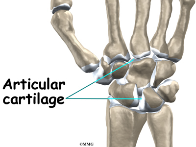
Where is articular cartilage located in the body?
In a joint, hyaline cartilage is referred to as articular cartilage. This is because the cartilage covers bones' surfaces where they articulate, or meet to form the joint. For example, at the knee joint, the top of the tibia, the bottom of the femur, and the back of the kneecap are covered with articular cartilage.
What does articular mean?
or relating to a jointMedical Definition of articular : of or relating to a joint.
What is articular cartilage quizlet?
what is articular cartilage? a layer of fibrous connective tissue covering the articular surface of bones in synovial joints.
What does articular mean in bones?
The articular surfaces of bones. adjective. 1. 4. Of or relating to a joint or joints.
What is articular joint?
Introduction. Joints, also known as articulations, are a form of connection between bones. They provide stability to the skeletal system as well as allowing for specialized movement.
Which is a function of articular cartilage quizlet?
What is Articular Cartilage? Hyaline cartilage covering bone ends at moveable joints. What is the purpose of the outer layer (fibrous capsule) of the Articular Capsule? It strengthens the joint so that the bones don't pull apart.
What is the function of articular cartilage within a joint?
Articular cartilage is a highly specialized connective tissue of diarthrodial joints. Its principal function is to provide a smooth, lubricated surface for articulation and to facilitate the transmission of loads with a low frictional coefficient.
What type of cartilage is articular cartilage?
Hyaline Cartilage Example : Connection between ribs and sternum, nasal cartilage and articular cartilage (which covers opposing bone surfaces in many joints).
What is another word for articular?
In this page you can discover 15 synonyms, antonyms, idiomatic expressions, and related words for articular, like: articulary, osteochondral, trabecular, interosseous, chondral, cartilage, soft-tissue, osseous, medial, serosal and calcaneum.
What is an articular disease?
Inflammatory articular diseases encompass a spectrum of conditions that range from acute forms of septic or sterile arthritis to chronic, often polyarticular conditions. They may involve inflammation of synovial tissue of joints and other tissues, such as muscles and tendons, mucosal and epithelial tissues, and organs.
What is an articular process?
The articular processes or zygapophyses (Greek ζυγον = "yoke" (because it links two vertebrae) + απο = "away" + φυσις = "process") of a vertebra are projections of the vertebra that serve the purpose of fitting with an adjacent vertebra.
What does non articular mean?
Medical Definition of nonarticular : affecting or involving soft tissues (as muscles and connective tissues) rather than joints nonarticular rheumatic disorders.
Which type of cartilage is more flexible, hyaline or diarthrodial?
diarthrodial cartilage articular cartilage. elastic cartilage cartilage that is more opaque, flexible, and elastic than hyaline cartilage, and is further distinguished by its yellow color. The ground substance is penetrated in all directions by frequently branching fibers that give all of the reactions for elastin.
Which cartilage is on the right side of the aortic?
aortic cartilage the second costal cartilage on the right side. arthrodial cartilage ( articular cartilage) that lining the articular surfaces of synovial joints. arytenoid c's two pyramid-shaped cartilages of the larynx. connecting cartilage that connecting the surfaces of an immovable joint.
What is Reichert's cartilage?
Reichert's cartilage the dorsal cartilage of the second branchial arch. reticular cartilage elastic cartilage. semilunar cartilage one of the two interarticular cartilages of the knee joint. temporary cartilage cartilage that is normally destined to be replaced by bone.
What is floating cartilage?
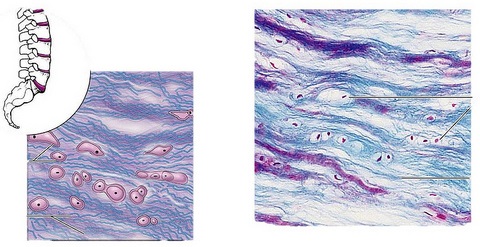
floating cartilage a detached portion of semilunar cartilage in the knee joint. hyaline cartilage flexible, somewhat elastic, semitransparent cartilage with an opalescent bluish tint, composed of a basophilic fibril-containing substance with cavities in which the chondrocytes occur. Hyaline cartilage.
What is the most important part of the skeleton?
a specialized, fibrous connective tissue present in adults, and forming most of the temporary skeleton in the embryo, providing a model in which most of the bones develop, and constituting an important part of the organism's growth mechanism; the three most important types are hyaline cartilage, elastic cartilage , and fibrocartilage.
What is articular cartilage made of?
Developing articular cartilage is thin, matrix-poor, and made of small flat cells. The prospective articular cartilage in newborn mice is thin and dense, consisting of 6–8 layers of small cells with little matrix (Rhee et al., 2005 ). The substantial morphological appearance of articular cartilage is observed in 2–4-week-old mice. During this time, the tissue undergoes a remarkable increase in thickness, with round chondrocytes oriented randomly and separated by considerable matrix ( Decker et al., 2017 ). From 4 weeks of age the articular cartilage displays a zonal anisotropic organization and an abundant and unique extracellular matrix ( Hunziker et al., 2007) ( Fig. 1 ). The superficial zone consists of flat, tightly bound cells that sustain frictionless joint motion by producing hyaluronan and lubricin. The intermediate/medial zone is made of round chondrocytes oriented randomly and separated by appreciable matrix. The deep zone is thick and made of very large, round chondrocytes aligned in vertical columns perpendicular to the articular surface and separated by abundant inter-columnar resilient matrix ( Julkunen et al., 2009; Decker et al., 2015 ). It has been hypothesized that the articular cartilage grows and thickens by a mechanism of apposition: proliferation of cells within the superficial zone leads to lateral expansion of the articular surface, and rapid proliferation of cells in the deeper zone, leads to vertical expansion of the cartilage ( Hunziker et al., 2007; Archer et al., 1994; Hayes et al., 2001; Dowthwaite et al., 2004 ). However, in contrast to this hypothesis, Decker et al. recently demonstrated using inducible ROSA-CE/R26-Confetti mice that localized proliferation plays a role in the early stages of postnatal articular cartilage growth, whereas the main mechanism of articular cartilage thickening is attributable to the increase in cell volume of resident cells and local alignment of non-daughter cells to form vertical stacks ( Decker et al., 2017 ).
How does articular cartilage get its nutrition?
Articular cartilage thus receives its nutrition by diffusion from the synovial fluid at all ages. In the time period prior to skeletal maturity, the cartilage of the lowermost part of the articular cartilage and the outermost part of the epiphyseal cartilage forms a miniplate, which undergoes the endochondral sequence.
What is the purpose of articular cartilage?
Articular cartilage (AC) covers the ends of bones forming synovial joints and facilitates the transmission of loads across articular surfaces whilst permitting almost friction-free movement and minimizing pressure on the underlying subchondral bone (Bhosale and Richardson, 2008 ). Chondrocytes are the active cells in AC, being responsible for secreting the tissue’s ECM which contains a network of collagen (predominantly type II) and noncollagenous proteins within a viscous water-like substance ( Lafont, 2010; Poole et al., 2001 ). The ECM is also composed of large proteoglycans with the core protein, aggrecan, predominating ( Fox et al., 2009 ). Articular cartilage is avascular, and so intermittent loading is important for altering intraarticular hydrostatic pressure and facilitating oxygen and nutrient diffusion from the synovial fluid to the AC ( Bhosale and Richardson, 2008; Carter et al., 1987; Fermor et al., 2007; Vanwanseele et al., 2002b ), thereby maintaining chondrocyte health and AC morphology and function ( Milner et al., 2012; Vanwanseele et al., 2002b ).
What is the avascular nature of articular cartilage?
The avascular nature of articular cartilage predisposes the individual to progressive symptoms and degeneration owing to the extremely slow and frequent inability to heal. Nonoperative rehabilitation and palliative care are frequently unsuccessful, and further treatment is required to alleviate symptoms.
What are the inflammatory cells in the synovium?
Other inflammatory cells in the synovium include macrophages, lymphocytes, and mast cells. Macrophages produce MMP-1, MMP-9, TIMP-1, and TIMP-2. uPA and cathepsins B, L, and D also are secreted from activated macrophages.
Is articular cartilage avascular?
The articular cartilage is always avascular throughout the entire period of its development and in adult life . In the human and in the other relatively large species in which cartilage canals form, they are never present within the articular cartilage regions.
Which cells are responsible for secreting collagen?
Chondrocytes are the active cells in AC, being responsible for secreting the tissue’s ECM which contains a network of collagen (predominantly type II) and noncollagenous proteins within a viscous water-like substance ( Lafont, 2010; Poole et al., 2001 ).
Learn about this topic in these articles
Articular cartilage (cartilage that covers the articulating part of a bone) is of the type called hyaline (glasslike) because thin sections of it are translucent, even transparent. Unlike bone, it is easily cut by a sharp knife. It is deformable but elastic, and…
major reference
Articular cartilage (cartilage that covers the articulating part of a bone) is of the type called hyaline (glasslike) because thin sections of it are translucent, even transparent. Unlike bone, it is easily cut by a sharp knife. It is deformable but elastic, and…