The temporal bones are situated at the sides and base of the skull. CT scans use X-ray technology and advanced computer analysis to create detailed pictures of the body. This cross-sectional scanning method allows the radiologist to look at different levels or slices of the temples or sides of the skull bone using a rotating X-ray beam.
How to read a temporal bone CT?
Temporal bone radiology
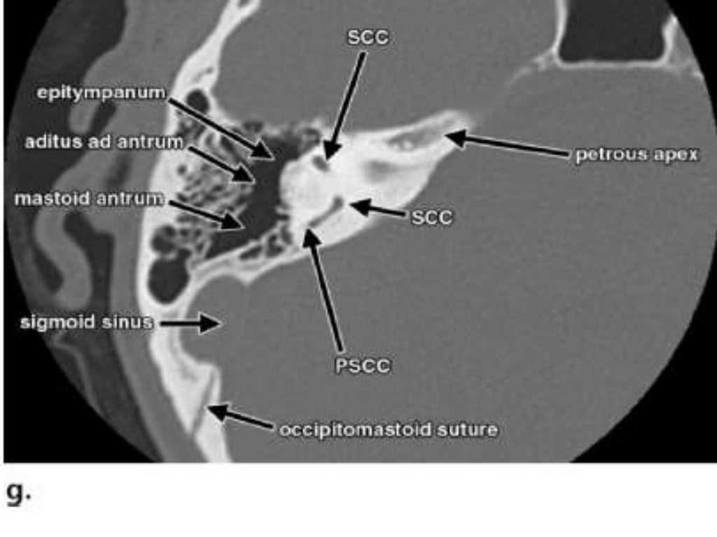
- HRCT Temporal bone anatomy
- Planes of scanning Axial 30 Degrees to anthropological base line Parallel to lateral SCC. Best displays inner & middle ear. ...
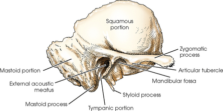
- Temporal Bone 1.Squamous 2.Petrous 3.Mastoid 4.Tympanic 5. ...
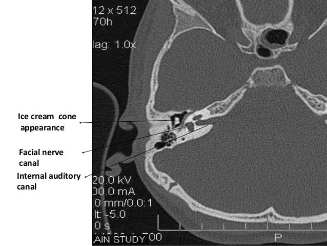
- EAR IS DIVIDED INTO 3 PARTS • EXTERNAL EAR • MIDDLE EAR • INTERNAL EAR 5 EAC Tympanic cavity Bony & membranous labyrinth
What is difference between CT scan and MRI scan?
- When compared to a CT scan, MRI produces more detailed images of soft tissues and behind bones. ...
- CT scan is less expensive and costs half the price of MRI. ...
- The time required for the procedure depends on whether or not you need a contrast. ...
How often are CT scans?
- Radiation exposure during a single chest X-ray (0.014 mSv) is equivalent to 3 days of natural radiation
- An abdomen X-ray (0.7 mSv) is equivalent to 4 months of natural background radiation
- A CT head scan (2 mSv) is equal to 1 year of natural radiation exposure 2
Is a bone scan better than a MRI scan?
Speak with your doctor about your personal concerns, as they can help determine what your best course of action is. Research has shown that MRI’s are able to detect bone cancer better than traditional bone scans can. MRI can show the physical abscess and abnormal appearance of bones infected by harmful diseases, specifically osteomyelitis.
How do you scan a temporal bone in CT?
2:2112:24Temporal Bone Anatomy on CT Imaging - MRI Online - YouTubeYouTubeStart of suggested clipEnd of suggested clipScan. So the tympanic membrane is identified as the medial. Most portion of the external auditoryMoreScan. So the tympanic membrane is identified as the medial. Most portion of the external auditory canal so the pathology is going to be in the cartilaginous.
Is a CT scan of temporal bone safe?
General comments about temporal bone CTs: Note that most temporal bone CT scans are "high radiation" procedures because enough Xray energy must be used to "see" into a very hard bone (temporal bone). All Xrays increase cancer risk. Accordingly, CT scans should not be done to "screen" for SCD.
What is temporal bone diseases?
Paget disease of the temporal bone is often associated with a mixed hearing loss. There is typically an air-bone gap in the low frequencies with a downsloping, high-frequency sensorineural hearing loss. The pathogenesis for the conductive and sensorineural hearing loss is not known.
How long does a CT scan of temporal bone take?
The entire process should take around 30 minutes.
Why do I need a CT scan on my ear?
CT Scans. If the results of hearing tests indicate that conductive hearing loss might be causing your symptoms, doctors may recommend a CT scan to visualize the middle ear in sharp detail.
What causes temporal bone pain?
stress, tooth grinding, direct trauma to the Temporalis muscle, excessive gum chewing. In rare cases a condition called Coronoid Process Hyperplasia may be the cause of Temporal Tendinitis. The diagnosis of Temporal Tendinitis by palpation of the tendon as it inserts into the coronoid process.
Why is it called temporal bone?
Its exact etymology is unknown. It is thought to be from the Old French temporal meaning "earthly," which is directly from the Latin tempus meaning "time, proper time or season." Temporal bones are situated on the sides of the skull, where grey hairs usually appear early on.
Is the temporal bone a facial bone?
The temporal bone: Anatomy and function. The temporal bone consists of a pair of bones that help make up the skull. Many cranial nerves and blood vessels pass through the temporal bone. Injuries to this bone can cause a loss of function in the facial muscles, as well as hearing loss and heavy bleeding.
What is temporal bone CT?
Temporal bone CT is a limited kind of head CT that focuses on the lower part of the skull and the surrounding soft tissues , and is often used in patients with hearing loss, chronic ear infections, and middle and inner ear diseases.
Why do we need a CT scan of the brain?
CT scans of the head/brain can provide more detailed information about brain tissue and brain structures than standard X-rays of the head. Therefore, this can provide more information related to injuries and brain diseases, and can be used to evaluate patients with headaches, stroke symptoms, masses, or cancers elsewhere in the body. ...
What is CT scan of temporal bones?
Your doctor has requested a computed tomography scan (CT or CAT) of your temporal bones. The temporal bones are situated at the sides and base of the skull. CT scans use X-ray technology and advanced computer analysis to create detailed pictures of the body. This cross-sectional scanning method allows the radiologist to look at different levels or slices of the temples or sides of the skull bone using a rotating X-ray beam. The radiologist is able to check each slice for injury or bony abnormalities. Our team of expert physicians, nurses and technologists is led by Barry Pressman, MD, chief of Neuroradiology and Head and Neck Radiology.
How long does it take for a CT scan to be read?
The results will be sent to your physician, usually within 48 hours. Your physician will discuss these results with you and explain what they mean in relation to your health.
How to do a head scan?
Before Arriving for Your Procedure 1 Please leave your jewelry and valuables at home and wear comfortable clothing. Because this is a scan of the head, anything like dentures, false teeth or implants, earrings, or hairpins can interfere with the scan and should be left at home. 2 If your doctor gave you an order, please bring it with you. 3 Although we do not anticipate any delay in your scheduled appointment, we recommend that you bring a book, a magazine or a music player to help pass any time you may have to wait.
What is CT and MRI used for?
CT and MRI are primarily used for imaging of the temporal bone. We first present the standard technique and protocols most often used, then review the special considerations for both modalities. A brief overview of the roles of plain radiographs, ultrasound (US), positron emission tomography (PET), and PET/CT is given at the end of this section.
What is CT venography?
CT arteriography (CTA) or CT venography (CTV) of the temporal bone may be used to evaluate for tinnitus. At our institution, the standard CT protocol for temporal bone imaging is employed, but the injection rate is increased to 3 to 4 cc per second for CTA. A power injector is employed if a 22-gauge IV or larger is available.
What is the head phantom?
The head phantom is a cylinder with a diameter of 16 cm and a height of 15 cm. The effective dose E is used to assess the radiation detriment from partial-body as opposed to whole-body irradiation (e.g., irradiation of only the head or only the abdomen).
What is a plain film radiograph?
Plain film radiographs have limited application to imaging of the temporal bone. A plain radiograph in the Stenvers projection, however, may be used for intraoperative or postoperative confirmation of position of a cochlear implant lead. The patient’s head is placed in a 45-degree obliquity contralateral to the implanted ear that places the axis of the implanted temporal bone parallel to the film and then in a 15-degree Townes projection. For example, if the left ear has been implanted, the radiographer would turn the head 45 degrees to the right, with the film behind the head of the patient, and shoot a single radiograph with the beam tilted 15 degrees inferiorly toward the patient ( Fig. 1.4 ). Familiarity with this projection and with the proper position of a cochlear implant is one of the few instances in current imaging where plain film radiography is critical, as the intraoperative assessment is often made while the patient is still anesthetized on the operating room table.
Where is the temporal bone located?
Temporal bone. The temporal bone is situated on the sides and the base of the cranium and lateral to the temporal lobe of the cerebrum. The temporal bone is one of the most important calvarial and skull base bones.
What are the parts of the temporal bone?
Gross anatomy. The temporal bone is divided into several main parts/portions 1-3: In addition, there are several bony projections: The temporal bone can also be divided into otologic zones:
Which otologic zone is the mastoid process?
mastoid process (considered a component of the mastoid part or synonymous with it) The temporal bone can also be divided into otologic zones: the medial third of the external auditory canal (part of the external ear) middle ear (tympanic cavity) inner ear. internal auditory canal.