What are filling and contour defects in the bladder?
Filling defects in the bladder must be differentiated from contour defects. A filling defect is an area of incomplete opacification on computed tomography (CT), excretory urography, or cystography and denotes something lying free within the bladder lumen. Examples include calculi, blood clots, and foreign bodies.
What is a filling defect?
A filling defect is a general term used to refer to any abnormality on an imaging study which disrupts the normal opacification (filling) of a cavity or lumen.
What is normal bladder filling and storage rate?
Characteristics of normal bladder filling and storage include (1) filling rates between 20 and 100 mL/h, (2) urethral pressures greater than bladder pressures, (3) low bladder pressures (0 to 20 cm H2 O), (4) a bladder capacity of 400 to 500 mL, and (5) maintenance of continence and awareness of bladder fullness when the bladder is full.
What is a filling defect in a barium swallow?
A filling defect would be a dark spot somewhere in the esophagus amongst the white of the swallowed barium. The filling defect just means that something is preventing the white barium from covering that area. Usually something that occupies space. Another example would be a CT done to look at the bladder.

What does filling defect mean?
A filling defect is a general term used to refer to any abnormality on an imaging study which disrupts the normal opacification (filling) of a cavity or lumen.
What does focal filling defect mean?
An imaging term of art referring to a reduction in filling of a focal, typically rounded hollow viscus (e.g., urinary bladder, gallbladder, colon) by radiocontrast due the presence of a mass that projects into the lumen.
What happens during bladder filling?
Bladder filling activates afferent nerve fibers in the bladder wall, which results in stimulation of sympathetic efferent activity in the hypogastric nerve, which leads to contraction of the smooth muscles in the bladder base and proximal urethra as well as relaxation of the detrusor from activation of beta-adrenergic ...
Can a hole in the bladder be fixed?
Bladder fistula is most often treated with surgery to remove the damaged part of the bladder. Healthy tissue is moved between the bladder and the other organ to block the opening.
What causes filling defect?
Differential Diagnoses Acute and chronic PE are the most common causes of filling defect in the pulmonary artery. CT findings in acute PE include intraluminal filling defects, acute angles of the filling defect with the vessel wall, total cutoff of vascular enhancement and enlargement of an occluded vessel [15].
How do you know if you have a pulmonary embolism on CT?
CT pulmonary angiography ― also called CT pulmonary embolism study ― creates 3D images that can detect abnormalities such as pulmonary embolism within the arteries in your lungs. In some cases, contrast material is given intravenously during the CT scan to outline the pulmonary arteries.
What are the two stages of filling the bladder?
Physiology. The micturition cycle involves two phases: bladder filling/urine storage and bladder emptying.
How long does it take to recover from a cystoscopy?
You may feel the need to urinate more often, and your urine may be pink. These symptoms should get better in 1 or 2 days. You will probably be able to go back to work or most of your usual activities in 1 or 2 days. This care sheet gives you a general idea about how long it will take for you to recover.
How painful is a cystoscopy for a woman?
People often worry that a cystoscopy will be painful, but it does not usually hurt. Tell your doctor or nurse if you feel any pain during it. It can be a bit uncomfortable and you may feel like you need to pee during the procedure, but this will only last a few minutes.
How do they repair a hole in the bladder?
With an intraperitoneal injury, there are often other intraabdominal injuries; therefore, the bladder injury is repaired during laparotomy. It is repaired in a 2-layer fashion with absorbable suture. The bladder is then drained via a transurethral catheter or suprapubic catheter.
What is it called when you poop and pee at the same time?
Bladder or bowel incontinence means there is a problem holding in urine or stool. You may have unwanted passage of urine or stool that you can't control.
How long does it take for a hole in the bladder to heal?
Extraperitoneal Rupture The urine and blood drain into a collection bag. It usually takes at least 10 days for the bladder to heal.
How long does it take for bladder to fill?
A healthy bladder can hold about 2 cups of urine before it's considered full....Pee table.AgeAverage bladder sizeTime to fill bladderChild (4–12 years)7–14 ounces2–4 hoursAdult16–24 ounces8–9 hours (2 ounces per hour)2 more rows•Jul 30, 2019
How painful is urethral dilation?
After dilation, your urethra may be sore at first. It may burn when you urinate. You may feel the need to urinate more often, and you may have some blood in your urine. These symptoms should get better in 1 or 2 days.
How do you fill a bladder?
If you do have to force yourself, here are 10 strategies that may work:Run the water. Turn on the faucet in your sink. ... Rinse your perineum. ... Hold your hands in warm or cold water. ... Go for a walk. ... Sniff peppermint oil. ... Bend forward. ... Try the Valsalva maneuver. ... Try the subrapubic tap.More items...
How do doctors stretch your bladder?
A doctor may use a procedure called bladder stretching, or hydrodistention, to treat your bladder pain, if only for a short time. Bladder stretching occurs when a doctor stretches your bladder by filling it with fluid. You will be given a local or general anesthesia link to help you tolerate the bladder stretching.
What is the bladder response to filling?
The normal adult bladder response to filling at a physiologic rate is an almost imperceptible change in intravesical pressure. During at least the initial stages of bladder filling, after unfolding of the bladder wall from its collapsed state, this very high compliance (Δ volume/Δ pressure) of the bladder primarily results from its elastic and viscoelastic properties. Elasticity allows the constituents of the bladder wall to stretch to a certain degree without any increase in tension. Viscoelasticity allows stretch to induce a rise in tension, followed by a decay (i.e., stress relaxation) when the filling (i.e., stretch stimulus) slows or stops. Brading and colleagues 6 think there is continuous contractile activity in the smooth muscle cells to adjust their length during filling but without (normally) synchronous activity that would increase intravesical pressure, would impede filling, and could cause urinary leakage. Clinically and urodynamically, the bladder seems relaxed. The urothelium also expands but must preserve its barrier function while doing so. There may be an active component to the storage properties of the bladder. The mucosa and lamina propria are normally the most compliant layers of the bladder. Coplen and associates 7 have hypothesized that the smooth muscle layer may have a chronic effect on compliance in the midportion of the cystometric filling curve through a complex interaction between muscle and extracellular matrix. This layer may acutely affect compliance in response to neurologic input as well.
How does the bladder maintain continence?
Continence is maintained through the action of the urinary sphincters. The internal sphincter, or the bladder neck, is richly innervated with α -adrenergic receptors. During bladder filling this structure remains closed through constant sympathetic discharge via the hypogastric plexus. The external sphincter, composed of striated muscle, also maintains a resting tone to maintain continence. It is believed that the fibers of the external sphincter are primarily of the slow twitch variety and thus can maintain tension for long periods of time. With rapid increases in intra-abdominal pressure, fast twitch fibers are recruited to contract and further increase the urethral resistance to avoid urinary leakage.
What is bladder accommodation?
Bladder accommodation during filling is a primarily passive phenomenon. It is dependent on the elastic and viscoelastic properties of the bladder wall and the lack of parasympathetic excitatory input. An increase in outlet resistance occurs via the striated sphincter somatic guarding reflex. In at least some species a sympathetic reflex also contributes to storage by (1) increasing outlet resistance by increasing tension on the smooth sphincter, (2) inhibiting bladder contractility through an inhibitory effect on parasympathetic ganglia, and (3) causing a decrease in tension of bladder body smooth muscle. Continence is maintained during increases in intra-abdominal pressure by the intrinsic competence of the bladder outlet and the pressure transmission ratio to this area with respect to the intravesical contents. A further increase in striated sphincter activity, on a reflex basis, is also contributory.
Why do children fire the sphincter?
The child has learned to fire the external sphincter to prevent an episode of incontinence.
Does the smooth sphincter contribute to bladder filling?
Although it seems logical and compatible with neuropharmacologic, neurophysiologic, and neuromorphologic data to assume that the muscular component of the smooth sphincter also contributes to the change in urethral response during bladder filling, it is extremely difficult to prove this experimentally or clinically. The direct and circumstantial evidence in favor of such a hypothesis has been summarized by Wein and Barrett, 1 Elbadawi, 11 and Brading. 12 The passive properties of the urethral wall deserve mention because they undoubtedly play a large role in the maintenance of continence. 6,13 Urethral wall tension develops within the outer layers of the urethra; however, urethral pressure is a product of the active characteristics of smooth and striated muscle and of the passive characteristics of the elastic, collagenous, and vascular components of the urethral wall, because this tension must be exerted on a soft or plastic inner layer capable of being compressed to a closed configuration—the “filler material” representing the submucosal portion of the urethra. The softer and more plastic this area is, the less pressure required by the tension-producing area to produce continence. Whatever the compressive forces, the lumen of the urethra must be capable of being obliterated by a watertight seal. This mucosal seal mechanism explains why a thin-walled rubber tube requires less pressure to close an open end when the inner layer is coated with a fine layer of grease than when it is not, and the latter case is much like scarred or atrophic urethral mucosa.
Does bladder filling cause urethral pressure to increase?
There is a gradual increase in urethral pressure during bladder filling, contributed to by at least the striated sphincteric element and perhaps by the smooth sphincteric element as well. The rise in urethral pressure seen during the filling and storage phase of micturition can be correlated with an increase in efferent pudendal nerve impulse frequency and in electromyographic activity of the periurethral striated musculature. This constitutes the efferent limb of a spinal somatic reflex, the so-called guarding reflex, which results in a gradual increase in striated sphincter activity.
Does the nervous system affect bladder filling?
Does the nervous system affect the normal bladder response to filling? At a certain level of bladder filling, spinal sympathetic reflexes facilitatory to bladder filling and storage are evoked in animals, a concept developed over the years by de Groat and associates, 3 who have also cited indirect evidence to support such a role in humans. This inhibitory effect is likely mediated primarily by sympathetic modulation of cholinergic ganglionic transmission. Through this reflex mechanism, two other possibilities exist for promoting filling and storage. One is neurally mediated stimulation of the predominantly α-adrenergic receptors in the area of the smooth sphincter, the net result of which would be to cause an increase in resistance in that area. The second is neurally mediated stimulation of the predominantly β-adrenergic receptors (inhibitory) in the bladder body smooth musculature, which would cause a decrease in bladder wall tension. McGuire and colleagues 10 also cited evidence for direct inhibition of detrusor motor neurons in the sacral spinal cord during bladder filling that results from increased afferent pudendal nerve activity generated by receptors in the striated sphincter. Good evidence exists to support a tonic inhibitory effect of other neurotransmitters on the micturition reflex at various levels of the neural axis. Bladder filling and consequent wall distention may also release autocrine-like factors (e.g., nitric oxide, prostaglandins, peptides) that influence contractility.
What is filling defect?
A filling defect is an area of incomplete opacification on computed tomography (CT), excretory urography, or cystography and denotes something lying free within the bladder lumen. Examples include calculi, blood clots, and foreign bodies.
Why does my bladder have gas in the lumen but not in the wall?
Gas within the lumen but not in the bladder wall is usually the result of catheterization or instrumentation in which air is introduced during the procedure. It may also be caused by a fistula between the bladder and the colon, small bowel, or vagina. Only rarely is such a finding caused by cystitis alone.
What is focal thickening on a cystogram?
If no localized extravesical mass is seen, focal thickening may indicate a bladder tumor. On CT, the attenuation of the edematous bladder mucosa is only slightly less than that of soft tissue and presents as a contour defect of the bladder wall. CT is unlikely to distinguish edema from other causes of bladder wall thickening, including infiltrating bladder tumor.
What causes focal mucosal edema?
Focal mucosal edema may also result from bladder irritation by extravesical conditions such as acute appendicitis, Crohn disease, or sigmoid diverticulitis. Another cause of focal edema is an indwelling catheter, whose tip may irritate the bladder wall, typically near the dome.
Why is there a higher incidence of tumors in the bladder lumen than in the bladder lumen?
There is a tendency for such tumors to spread outside the bladder more rapidly than bladder lumen tumors because the walls of false diverticula consist only of urothelium protruding between muscle bundles and are not surrounded by detrusor muscle.
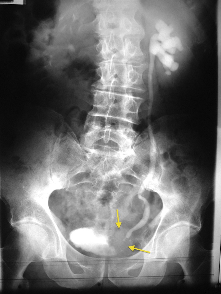
What are the foreign bodies in the bladder?
Foreign bodies within the bladder are most commonly inserted by the patient. Among other items, pens, pencils, matches, wire, tubing, and string can be seen within the bladder lumen. Foreign bodies may also be iatrogenic (e.g., broken or shorn catheter fragments) or may result from penetrating trauma (e.g., bullets).
Where is the urothelial neoplasm in the diverticulum?
A small soft-tissue mass is seen in the middle of a large diverticulum along the right posterolateral aspect of the bladder. An area of high attenuation located more posteriorly represents calcification within the tumor.