What is a giant cell in medical terms?
Giant cell. A giant cell ( multinucleated giant cell, multinucleate giant cell) is a mass formed by the union of several distinct cells (usually histiocytes ), often forming a granuloma. It can arise in response to an infection, such as from tuberculosis, herpes, or HIV, or foreign body.
What is a giant cell made of?
Giant cell. Giant cell, also called Langhans giant cell, large cell characterized by an arc of nuclei toward the outer membrane. The cell is formed by the fusion of epithelioid cells, which are derived from immune cells called macrophages. Once fused, these cells share the same cytoplasm, and their nuclei become arranged in an arc near...
What are the key points about giant cell tumors?
Key points about giant cell tumors. A giant cell tumor is a rare, aggressive non-cancerous tumor. It usually develops near a joint at the end of the bone. Most occur in the long bones of the legs and arms. Giant cell tumors most often occur in young adults when skeletal bone growth is complete. The exact cause of giant cell tumors remains unknown.
What is multinucleated giant cell?
A giant cell ( multinucleated giant cell, multinucleate giant cell) is a mass formed by the union of several distinct cells (usually histiocytes ), often forming a granuloma. It can arise in response to an infection, such as from tuberculosis, herpes, or HIV, or foreign body.

What is the function of giant cells?
Foreign body giant cells (FBGC) most commonly are observed at the tissue/material interface of implanted medical devices, prostheses, and biomaterials (**2). In this context, adherent macrophages and foreign body giant cells constitute the foreign body reaction (Figure 1).
What are giant cells called?
giant cell, also called Langhans giant cell, large cell characterized by an arc of nuclei toward the outer membrane. The cell is formed by the fusion of epithelioid cells, which are derived from immune cells called macrophages.
What is the cause of giant cell?
The cause of giant cell tumors is unknown. The tumors occur spontaneously. They are not known to be caused by trauma, environmental factors, or diet. Giant cell tumors of bone are not inherited.
What are giant cells in bone?
Giant Cell Tumours (GCT) are benign (non-cancerous) tumours that develop in the bone. They mostly occur in the long bones found in the arms and legs. GCT often affects people between the ages of 20 and 45 years old.
Are osteoclasts giant cells?
One must consider the fact that, in general, giant cells are larger than osteoclasts, i.e. contain hundreds of nuclei, while osteoclasts usually contain only a few, except in diseases such as Paget's disease, and in response to bone implants on which osteoclast-like cells/giant cells differentiate.
What is giant cell reaction?
Any reparative tissue reaction with multinucleated epithelioid histiocytes, that may be due to exogenous material–eg, sutures, or endogenous material–eg, the contents of a ruptured epidermal inclusion cyst, chalazion, or fat.
How long can you live with giant cell tumor?
The data on long-term prognosis of metastatic GCT (mGCT) are mainly limited to small retrospective studies. The largest of them, recently published by Yang et al., found a 94.4% 5-year survival rate based on data from 42 patients (21).
Can a giant cell tumor be cancerous?
Most giant cell tumors occur at the ends of the long bones of the arms and legs, near a joint (such as the knee, wrist, hip, or shoulder). Most are benign (not cancer) but some are malignant (cancer).
What foods should I avoid with giant cell arteritis?
Pain is a big part of living with giant cell arteritis (GCA), a type of vasculitis affecting the temporal, cranial, and other carotid system arteries. You'll often feel pain in your head, scalp, jaw, and neck....Avoid or limit anything that can contribute to inflammation, including:sweets.fried foods.processed foods.
Is a bone cyst a tumor?
A unicameral, or simple, bone cyst is a common, benign (noncancerous) bone tumor that primarily occurs in children and adolescents.
Is giant cell tumor fatal?
Giant Cell tumors (GCT) are benign tumors with potential for aggressive behavior and capacity to metastasize. Although rarely lethal, benign bone tumors may be associated with a substantial disturbance of the local bony architecture that can be particularly troublesome in peri-articular locations.
What percentage of giant cell tumor is malignant?
The most recent data from the 4 large GCTB patient series showed that the frequency of malignancy was 1.1% to 11.3% (Table 1).
What are macrophages?
Listen to pronunciation. (MA-kroh-fayj) A type of white blood cell that surrounds and kills microorganisms, removes dead cells, and stimulates the action of other immune system cells. Enlarge.
How many types of giant cells are there?
Types include foreign-body giant cell, Langhans giant cell, Touton giant cells, Giant-cell arteritis, and Reed–Sternberg cell.
What are rare giant cells?
A giant cell tumor is a rare, aggressive non-cancerous tumor. It usually develops near a joint at the end of the bone. Most occur in the long bones of the legs and arms. Giant cell tumors most often occur in young adults when skeletal bone growth is complete. The exact cause of giant cell tumors remains unknown.
What is multinucleated cells?
Multinucleated giant cells (MNGCs) are a special class of giant cell formed by the fusion of monocytes/macrophages abundantly found in human tissues.
What is the semifluid substance of a cell that is external to the nuclear membrane and internal to the
cytoplasm. Cytoplasm, the semifluid substance of a cell that is external to the nuclear membrane and internal to the cellular membrane, sometimes described as the nonnuclear content of protoplasm. In eukaryotes (i.e., cells having a nucleus), the cytoplasm contains all of the organelles.
How are cells formed?
The cell is formed by the fusion of epithelioid cells, which are derived from immune cells called macrophages. Once fused, these cells share the same cytoplasm, and their nuclei become arranged in an arc near the outer edge of the cell.
What are the cells that make up platelets called?
The megakaryocytes, the normal bone-marrow cells thought to be the source of the blood platelets, are also called giant cells. giant cell. Multinucleated giant cells (micrograph with hematoxylin and eosin stain). Nephron. Britannica Quiz. Diseases, Disorders, and More: A Medical Quiz. What condition is caused by the deposition of salts of uric acid?
What is the nucleus?
Nucleus, in biology, a specialized structure occurring in most cells (except bacteria and blue-green algae) and separated from the rest of the cell by a double layer, the nuclear membrane.
What is a cell in biology?
cell, in biology, the basic membrane-bound unit that contains the fundamental molecules of life and of which all living things are composed. A single cell is often a complete organism in itself, such as a bacterium or yeast. Other cells acquire specialized functions as they mature. These cells cooperate with…. nucleus.
What is an encyclopedia editor?
Encyclopaedia Britannica's editors oversee subject areas in which they have extensive knowledge, whether from years of experience gained by working on that content or via study for an advanced degree. ...
Where are Langhans giant cells found?
Langhans giant cells typically form at the centre of granulomas (aggregates of macrophages) and are found in the tubercle, or primary focus of infection, in tuberculosis, in lesions of syphilis, leprosy, and sarcoidosis, and in fungal infections.
What is the most important differential diagnostic consideration of giant cell carcinoma?
The most important differential diagnostic consideration of giant cell carcinoma is sarcomatoid carcinoma of the prostate with or without heterologous elements, which occasionally exhibits neoplastic giant cells. Sarcomatoid carcinoma is composed of spindle cells with large, pleomorphic, hyperchromatic nuclei.
What is the growth pattern of giant cell angiofibroma?
Microscopically, giant cell angiofibroma shows noninfiltrative growth of patternless round to spindle cells in a stroma composed of collagen fibers or occasionally myxoid material with irregular pseudovascular spaces. The giant cells are interspersed among the spindle cells and partially line the walls of the pseudovascular spaces. These cells are multinucleated floret-type giant cells with nuclei located at the periphery of the cell. Immunohistochemically, the spindle and giant cells are positive for estrogen, progesterone, vimentin, and CD34, confirming the relationship to solitary fibrous tumor.
What is a giant cell fibroblastoma?
(Left) Giant cell fibroblastoma (GCFB) is a benign but locally aggressive fibroblastic neoplasm that most frequently arises on the trunk, usually in children. As shown in this image, GCFB is a superficial lesion and arises in the dermis &/or subcutis.
Which tumors exhibit giant cells?
Other tumors that exhibit giant cells, such as giant cell carcinoma of the bladder, urothelial carcinoma with osteoclast-type giant cells or trophoblastic differentiation, and leiomyosarcoma with frank nuclear anaplasia, should also be considered.
How many nuclei are there in a giant cell tumor?
Giant cells in giant cell tumor usually have > 12 nuclei. •. Mononuclear cells in GCRG are spindle-shaped and not oval/round as in giant cell tumor of bone. ○. Nuclei of mononuclear cells in giant cell tumor of bone are identical to nuclei in osteoclast-type giant cells. –.
Where are osteoclasts found in PFHT?
(Left) Osteoclast-like multinucleated giant cells are often seen within histiocytoid nodules in PFHT; however, they are absent in a subset of cases. Note also that the cells at the periphery of the nests may be spindled and appear to encircle the plumper epithelioid cells .
Where is giant cell angiofibroma found?
Since then, several case reports have described this tumor at various extraorbital sites, including the submandibular region, the parascapular area, and the posterior mediastinum. The condition is also reported to occur in the oral cavity. Currently, the giant cell angiofibroma is thought to be a variant of the solitary fibrous tumor. The condition presents as a slow-growing nodule or mass with normal overlying mucosa. It behaves in a benign fashion with only rare local recurrences and no tendency to metastasize.
What is the goal of giant cell cancer?
Your tolerance for specific medications, procedures, or therapies. Expectations for the course of the disease. Your opinion or preference. The goal for treatment of a giant cell tumor is to remove the tumor and prevent bone damage.
How do you know if you have a giant cell tumor?
Symptoms may include: A visible mass. Bone fracture. Fluid buildup in the joint nearest the affected bone. Limited movement in the nearest joint. Swelling. Pain at the nearest joint.
How to treat a tumor that can't be removed?
Bone reconstruction. Physical therapy to regain strength and mobility. Surgery to remove the tumor and any damaged bone. Tumors that can’t be removed surgically can often be controlled and sometimes destroyed with radiation therapy .
What is radionuclide scan?
Radionuclide bone scans. A nuclear imaging test used to detect bone diseases and tumors, and to determine the cause of bone pain or inflammation. X-rays. A diagnostic test that uses invisible electromagnetic energy beams to make images of internal tissues, bones, and organs on film.
What tests are done to determine if a person has cancer?
A test in which tissue samples are removed from the body and examined under a microscope to determine if cancer or other abnormal cells are present. Radionuclide bone scans.
Where do giant cell tumors occur?
A giant cell tumor is a rare, aggressive non-cancerous tumor. It usually develops near a joint at the end of the bone. Most occur in the long bones of the legs and arms. Giant cell tumors most often occur in young adults when skeletal bone growth is complete. The exact cause of giant cell tumors remains unknown.
Where does a giant cell tumor of the bone develop?
It generally happens in adults between ages 20 and 40 when skeletal bone growth is complete. It usually develops near a joint at the end of the bone. The location of a giant cell tumor is often in the knee, but can also involve the bones of the arms and the legs.
Overview
Giant cell arteritis is an inflammation of the lining of your arteries. Most often, it affects the arteries in your head, especially those in your temples. For this reason, giant cell arteritis is sometimes called temporal arteritis.
Symptoms
Giant cell arteritis causes inflammation of certain arteries, especially those near the temples.
Causes
With giant cell arteritis, the lining of arteries becomes inflamed, causing them to swell. This swelling narrows your blood vessels, reducing the amount of blood — and, therefore, oxygen and vital nutrients — that reaches your body's tissues.
Risk factors
Several factors can increase your risk of developing giant cell arteritis, including:
Complications
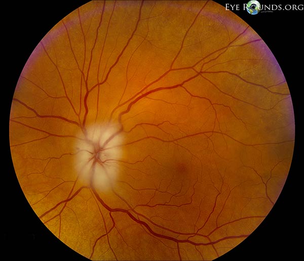
Blindness. Diminished blood flow to your eyes can cause sudden, painless vision loss in one or, rarely, both eyes. Loss of vision is usually permanent.
The Mayo Clinic experience and patient stories
Our patients tell us that the quality of their interactions, our attention to detail and the efficiency of their visits mean health care like they've never experienced. See the stories of satisfied Mayo Clinic patients.
What is the name of the condition that causes pain and swelling in the blood vessels?
Giant cell arteritis (or GCA) is a medical condition that can cause pain and swelling in blood vessels. Blood vessels are tubes that carry blood around the body. GCA affects arteries, which are the largest of the three types of blood vessels. Arteries take blood with oxygen in from the heart to different parts of the body.
What is the area of the head that is affected by GCA?
Arteries take blood with oxygen in from the heart to different parts of the body. GCA commonly affects arteries in the head and neck. This condition can cause pain and tenderness in the soft part at the side of the head in between your eyes and ears, known as the temples. When the condition affects this part of the head it can be called temporal ...
What is GCA in medical terms?
GCA is one of a group of conditions called vasculitis. The word vasculitis means inflammation in blood vessels. There are different types of vasculitis, because different blood vessels can be affected.
How to treat GCA?
Steroid tablets. While there’s currently no cure for GCA, treatment with steroid tablets is very effective and usually starts to work within a few days. Prednisolone is the most commonly used steroid tablet. Steroid tablets slow down the activity of the immune system, and reduce inflammation in blood vessels.
What is temporal arteritis?
When the condition affects this part of the head it can be called temporal arteritis. GCA can also affect other large arteries and their branches that take blood elsewhere around the body. This condition is treatable, usually with steroid tablets.
How do you know if you have GCA?
Most people will have some, but not all of these. The most common symptoms of GCA are: headaches, often with severe pain and tenderness over the temples and the scalp – it may be painful to brush your hair or to shave.
How long does it take to get rid of GCA?
To treat GCA, you’ll usually be given between 40 mg and 60 mg of steroid tablets every day to begin with. This dose is usually continued for three to four weeks. If you’re well after that, and your blood tests show that your condition has improved, your doctor will start reducing the dose.
What is the most common form of vasculitis?
Giant cell arteritis (GCA) is the most common form of vasculitis that occurs in adults. Almost all patients who develop giant cell arteritis are over the age of 50. GCA commonly causes headaches, joint pain, facial pain, fever, and difficulties with vision, and sometimes permanent visual loss in one or both eyes.
How long does it take for a biopsy to be positive?
Patients dramatically improve within 24 to 72 hours of beginning therapy, and the ESR usually normalizes within 1 month.
How much of the fever is caused by giant cell arteritis?
Although giant cell arteritis accounts for only 2% of all fever of unknown origin, it accounts for 16% of fever of unknown origin in patients over age 65 years and is often associated with rigors and sweats.
How long does it take for prednisone to work?
Most patients improve rapidly and dramatically on this dose, with improvement of most symptoms in 1–3 days. Unfortunately, if blindness has occurred as a symptom it is usually irreversible, which only emphasizes the importance of early detection and treatment. Almost all patients experience side effects from prednisone.
Can giant cell arteritis cause headaches?
Giant cell arteritis can begin suddenly or gradually with nonspecific symptoms such as malaise, weight loss, depression, and fatigue or with the classic symptoms of headache, scalp tenderness, jaw claudication, visual changes, or polymyalgia rheumatica. Polymyalgia rheumatica which can occur with or without giant cell arteritis, ...
What is the sed rate of erythrocytes?
The sed rate measures how fast a patient’s red blood cells settle when placed in a small tube. In inflammatory conditions, red blood cells settle more quickly than in non–inflammatory states.
What are the symptoms of a stroke?
Other symptoms can include tenderness of scalp (it hurts to comb the hair), cough, throat pain, tongue pain, weight loss, depression, stroke, or pain in the arms during exercise . Some patients have many of these symptoms; others have only a few.
What is the best test to confirm a diagnosis?
A small sample of tissue is taken and test. This is usually needed to confirm diagnosis. Radionuclide bone scans. This is a nuclear imaging test. It can show any degenerative or arthritic changes in the joints, find bone diseases and tumors, and find the cause of bone pain or inflammation.
How do you know if you have a giant cell tumor?
Symptoms may include: A visible bump. Bone fracture. Fluid buildup in the joint nearest the affected bone. Limited movement in the nearest joint. Swelling. Pain at the nearest joint.
Where do giant cell tumors grow?
A giant cell tumor is a rare, aggressive non-cancerous tumor. It usually grows near a joint at the end of the bone. Most occur in the long bones of the legs and arms. Giant cell tumors most often occur in young adults when skeletal bone growth is complete. The exact cause of giant cell tumors remains unknown.
What is the best test to rule out infection?
This test helps to rule out any infection or fractures. X-rays. This is a test that uses a small amount of radiation to make images of tissues, bones, and organs on film. CT scan. This is a test that uses a series of X-rays and a computer to make detailed images of the tissues in the body. MRI.
Can a giant cell tumor come back?
The goal for treatment of a giant cell tumor is often to remove the tumor and prevent damage to the affected bone. Tumors that can’t be removed surgically can often be controlled and sometimes destroyed with radiation therapy. Giant cell tumors can come back.
Can a tumor come back after surgery?
Amputation, in severe cases. Tumors that can’t be removed with surgery can often be controlled and sometimes destroyed with radiation therapy. Giant cell tumors can come back. Follow-up with your healthcare provider may be needed for several years.
