
A lumbar MRI specifically examines the lumbar section of your spine — the region where back problems commonly originate. The lumbosacral spine is made up of the five lumbar vertebral bones (L1 thru L5), the sacrum (the bony “shield” at the bottom of your spine), and the coccyx (tailbone).
What is the average cost of a lumbar spine MRI?
The price of a lumbar MRI is influenced by several factors such as the facility/hospital you choose, whether or not any insurance is available, the geographical location, and whether or not a contrast substance will be used. However, you should plan on spending anywhere between $550 and more than $3,200 without insurance for a lumbar spine MRI.
How long does a MRI of the lumbar spine take?
The test usually takes 30 to 60 minutes but can take as long as 2 hours. Does your whole body go in for a lumbar MRI? An MRI can be performed on any part of your body. A lumbar MRI specifically examines the lumbar section of your spine — the region where back problems commonly originate. Do you go in feet first for a lumbar MRI?
What does a lumbar sacral MRI show?
MRI LS scan with a contrast dye is especially used in the following conditions:
- To differentiate causes of lower back pain and lumbar radiculopathy
- To distinguish recurrent disc herniation after operation of spine
- To show nerve roots in viral or inflammatory conditions
- To detect tumours
What does this MRI for lumbar spine indicate?
The lumbar MRI will help them plan the procedure before making an incision. An MRI scan provides a different kind of image from other imaging tests like X-rays, ultrasound, or CT scans. An MRI of the lumbar spine shows the bones, disks, spinal cord, and the spaces between the vertebral bones where nerves pass through.
How long does a MRI spine lumbar and sacral take?
The test usually takes 30 to 60 minutes but can take as long as 2 hours.
What does an MRI of the lumbar spine show?
A lumbar spine MRI can detect a variety of conditions in the lower back, including problems with the bones (vertebrae), soft tissues (such as the spinal cord), nerves, and disks.
What will an MRI of the sacrum show?
Used to evaluate pain in the region of the tailbone or low back pain not attributed to disorders of the lumbar spine. MRI can screen for causes of both chronic pain, as well as evaluate for fractures after episodes of trauma.
How long does it take for a lumbar spine MRI?
The test most often lasts 30 to 60 minutes, but may take longer.
Can a lumbar MRI show nerve damage?
An MRI may be able help identify structural lesions that may be pressing against the nerve so the problem can be corrected before permanent nerve damage occurs. Nerve damage can usually be diagnosed based on a neurological examination and can be correlated by MRI scan findings.
Does a lumbar MRI show inflammation?
Some of the inconsistencies that a lumbar spine MRI may show include compression or inflammation of the spinal cord and adjacent nerves, degeneration of joints such as vertebral facet joints, disc herniation, infection of the discs, spinal cord and vertebrae, trauma to the tissues, and tumors.
What are symptoms of sacrum damage?
SymptomsIntense pain in the pelvis or hip area, as well as lower back.Pain near the buttocks.Intensifying pain during physical activities or exercises.Tender areas in the lower back region.More items...
Where do you feel sacrum pain?
Sacroiliac joint pain is most commonly felt in the low back and buttock but can also be referred into the thigh and leg. If numbness and tingling or weakness is present, an alternative diagnosis should be considered.
What does pain in the sacrum feel like?
What are the symptoms? The signs and symptoms of SI pain start in the lower back and buttock, and may radiate to the lower hip, groin or upper thigh. While the pain is usually one sided, it can occur on both sides. Patients may also experience numbness or tingling in the leg or a feeling of weakness in the leg.
How do you prepare for a lumbar spine MRI?
Preparation Instructions: Please do not wear jewelry, hairpins, or any metal objects. Glasses, dentures, hearing aids, and watches will be removed at the time of the examination; you will be provided a locker. IF YOUR MRI REQUIRES CONTRAST: do not eat or drink anything 4 hours prior to exam.
What position are you in for a lumbar MRI?
In some cases, you may need to wait up to an hour for the dye to work its way through your bloodstream and into your spine. The MRI technician will have you lie on the bench, either on your back, side, or stomach. You may receive a pillow or blanket if you have trouble lying on the bench.
What should you not do before an MRI?
What to Avoid Before an MRIDon't Get Any New Piercings. When you go in for your MRI, you'll need to remove any body piercings or earrings. ... Don't Disregard Doctor's Instructions. ... Don't Disrupt Your Schedule.
What are the symptoms of L4 L5 nerve damage?
Tingling, numbness (pins and needles), and an aching or burning sensation in the leg and on top of the foot are widespread. In severe cases, an L4-L5 slipped disc leads to weakness in the legs or feet. Some may even have an inability to walk, leading to an inability to stand.
Can you see sciatica on lumbar MRI?
An MRI of the lumbar spine will show many causes of low back pain and sciatica, including disc herniations, facet arthritis, and lumbar spinal stenosis. Digital x-rays and CT scans may also be used to diagnose the cause of sciatica.
Can a herniated disc heal on its own?
Herniated disks get better on their own over time or with nonsurgical treatment for 9 out of 10 people. If other treatments don't relieve your symptoms, your healthcare provider may recommend surgery.
Which is worse bulging or herniated disk?
Compared with a bulging disk, a herniated disk is more likely to cause pain because it generally protrudes farther and is more likely to irritate nerve roots. The irritation can be from compression of the nerve or, much more commonly, the herniation causes a painful inflammation of the nerve root.
What is a lumbosacral spine MRI?
What is a Lumbosacral (LS) spine MRI? Magnetic resonance imaging lumbosacral spine (MRI LS Spine) is a test that produces detailed images of the lumbar area (lower part of the spine), sacrum (base of the spine) and the coccyx (the tailbone). The test helps to identify or look for any problems in these areas of the spine.
What do the results of a lumbosacral (LS) spine MRI mean?
The MRI images will be interpreted by the radiologist, who will send a detailed report to your physician. Then, the physician will discuss the results with you. Abnormal results may be due to any of the following conditions:
How should I prepare for a lumbosacral (LS) spine MRI?
Generally, you are not supposed to eat or drink for four to six hours before the scheduled time of the procedure. If you have a fear of closed spaces (claustrophobia), you may be given a sedative or anaesthesia that will help you feel sleepy or less anxious.
How will a lumbosacral (LS) spine MRI feel like?
MRI is a painless procedure. You might feel the table of the MRI scanner to be hard/cold. In such instances, you can ask for a pillow or blanket. The scanner may make loud noises during the procedure. The radiography staff will provide you with earplugs to help reduce the noise.
What are the risks and benefits of a lumbosacral (LS) spine MRI?
The test involves a safe scanning procedure. Some complications that may occur are as follows:
What is contrast dye used for in MRI?
MRI LS scan with a contrast dye is especially used in the following conditions: To differentiate causes of lower back pain and lumbar radiculopathy. To distinguish recurrent disc herniation after operation of spine. To show nerve roots in viral or inflammatory conditions. To detect tumours.
What is the disorder of the lower back?
Osteoporosis resulting in fracture of the lower back. Spinal cord injury / abscess. Syringomyelia (a disorder characterised by a fluid-filled pocket formed within the spinal cord) Ankylosing spondylitis (inflammation of the joints that primarily affects the spine)
What is a lumbar MRI?
A lumbar MRI is a powerful diagnostic tool that doctors may use to: check spinal alignments. detect abnormalities of vertebrae or the spinal cord. evaluate any inflammation of the spinal cord or nerves. check for tumors on or around the spinal cord. monitor damage to the spine after an injury.
Why do doctors do lumbar MRIs?
Medical professionals perform lumbar MRIs for a variety of reasons. If someone is experiencing pain in their lower back, a doctor may recommend a lumbar MRI scan to help diagnose the source of the pain. A doctor may also order a lumbar MRI for a person who is about to undergo back surgery. In this case, the surgical team uses the results ...
How does an MRI work?
The MRI machine uses a strong magnetic field that aligns and stimulates particles called protons in the body, for cing them to spin out of alignment. When the technician halts the magnetic field, the protons begin to spin in their usual way. As they do this, they give off energy that the MRI machine detects.
What is magnetic resonance imaging?
Magnetic resonance imaging is a noninvasive diagnostic tool that uses radio waves and a magnetic field to produce detailed images of the inside of a body. Doctors can use them to examine a person’s lower spine, or lumbar region, and the surrounding tissues. The exam itself is very safe because it does not use ionizing radiation, ...
What are the symptoms of a lumbar MRI?
A doctor may order a lumbar MRI if a person has any of the following symptoms: sudden back pain that occurs alongside fever. injury or trauma to the lower spine. severe and persistent lower back pain. multiple sclerosis. leg pain that suggests a lumbar disc herniation. bowel or bladder incontinence.
How long does it take to get a lumbar MRI?
According to one MRI provider, the scanning phase of a lumbar scan takes around 20 to 35 minutes. The MRI machine looks like a giant doughnut.
Why do I need an MRI for my back?
A lumbar MRI can check spinal alignments and tumors and explore different causes of back pain. According to the American Academy of Family Physicians, low back pain is the fifth most common reason for outpatient visits to hospitals, clinics, or other healthcare facilities.
What is the MRI of the lumbar spine?
An MRI scan of this area is used to accurately depict soft tissue in and around the lumbosacral spine. Measurements mainly focus on a change in signal intensities and less on absolute distances or angles. Various pathologies affect the configuration of the soft tissue, which can lead to clinical symptoms such as neural compression syndromes, most commonly due to degeneration, congenital disorders, and benign or malignant neoplasms. Structures visible on MRI include neural structures, meninges, cerebrospinal fluid, blood vessels, fat, ligaments, bone and joints, discs, muscles, and organs. Knowledge of normal anatomy and the systematic assessment of these structures will help to generate the most accurate and clinically relevant scan interpretation. In addition, it is important to have a clinical question in mind before requesting and assessing a lumbosacral MRI, since, for example, degenerative abnormalities are common and asymptomatic in the majority of people. More importantly, among patients without clinical signs pointing toward a specific condition, imaging does not improve patient outcomes and may lead to worse outcomes, unnecessary harm, and increased costs.
What are the structures that are visible on an MRI?
Structures visible on MRI include neural structures, meninges, cerebrospinal fluid, blood vessels, fat, ligaments, bone and joints, discs, muscles, and organs. Knowledge of normal anatomy and the systematic assessment of these structures will help to generate the most accurate and clinically relevant scan interpretation.
How many lumbar vertebrae are there?
The most common number of lumbar vertebrae is five. In up to 20% of the general population lumbosacral transitional vertebrae (LSTV) are seen, of which the sacralization of L5 is the most common (~ 17%) and the lumbarization of S1 is rarer (~ 2%).
What does deviation of vertebrae from the midsagittal plane indicate?
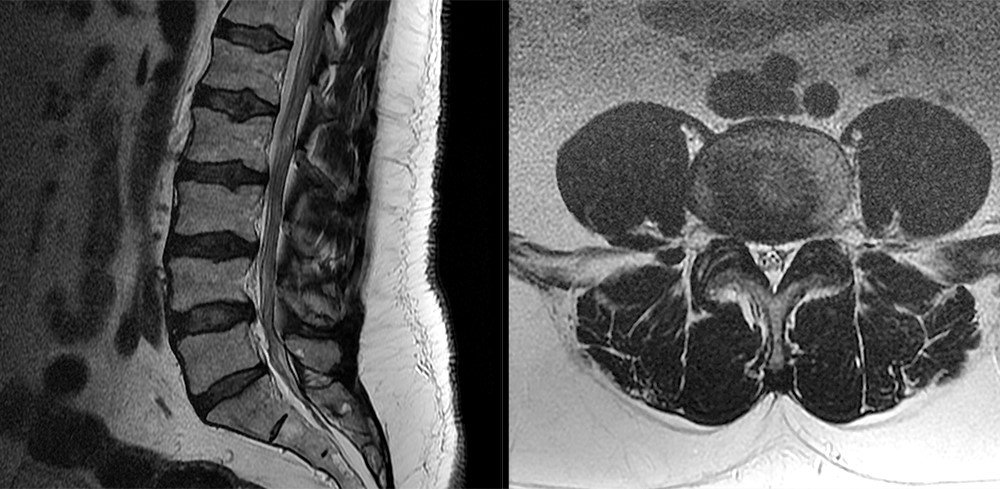
Deviation of vertebrae from the midsagittal plane may indicate scoliosis; however, as previously stated, MRI is not the imaging modality of choice to categorize the scoliosis, to measure angles and to make a treatment plan. Fig. 1. T2-weighted images of a normal lumbosacral spine. The most common number of lumbar vertebrae is five.
What is ABCDE in MRI?
ABCDE-Method for Structured Assessment of the Lumbar Spine on MRI.
Is a lumbosacral MRI a clinical question?
In addition, it is important to have a clinical question in mind before requesting and assessing a lumbosacral MRI, since, for example, degenerative abnormalities are common and asymptomatic in the majority of people.
Is degeneration in the lumbar spine a sign of symptoms?
As previously stated, the presence of degeneration in a lumbar spine is a common finding and by no means an indicator of symptoms. The etiology of these common degenerative changes is multifactorial: mechanical, traumatic, nutritional, inflammatory, and genetic factors are involved.
How many vertebrae are in the lumbar spine?
Before deciphering the abnormalities listed in the lumbar MRI report, it’s important to understand the normal anatomy. The lumbar spine consists of bones (usually five vertebral bodies) stacked on top of each other and separated by five discs. The vertebral bodies are usually labeled 1 through 5, and the discs are named by the bones above and below.
What are the problems with the lumbar spine?
Problems in the Lumbar Spine. As we get older, changes occur naturally in the spine. The discs tend to lose their water content (desiccate). The annulus of the disc may bulge, protrude or herniate. The bones may develop bone spurs (osteophytes). The ligamentum flavum may enlarge (hypertrophy).
What is the L5-S1 disc?
Therefore the L4 and L5 vertebral bodies are separated by the L4-5 disc. Just below the lumbar spine is the sacrum so the bottom disc is called L5-S1. This normal configuration is reported in the MRI report as both sagittal and axial depictions. The sagittal view is a profile picture. The axial view is a cross-section.
Why does my lower back hurt?
Low back pain is one of the most common diseases in the United States. Low back and leg pain can be caused by a spine problem such as a bone spur or disc herniation, but can also be caused by hip problems, pelvic problems, kidney problems, muscle problems or unknown reasons.
Which nerves go to the groin?
The back side of the spine is more bone called the lamina and spinous process. The nerves go to specific regions of the leg. The L1 and L2 nerves tend to go to the groin region. The L3 nerve to the front of the thigh. The L4 nerve to the shin and instep. The L5 nerve to the top of the foot and big toe.
What nerve is the L5 nerve?
The L5 nerve to the top of the foot and big toe. The S1 nerve to the outside and bottom of the foot. The disc normally is composed of 2 parts. These are microscopic and cannot exactly be differentiated on an MRI. The central softer part of the disc is the nucleus and the out layer is the annulus.
Where does the spinal cord end?
The spinal cord itself ends around the T12-L1 level , but the nerves continue down the lumbar spine as the cauda equina. At each disc level, a nerve exits the spine and goes to a specific region of the leg. The back side of the spine is more bone called the lamina and spinous process.
Why do we use MRI of the sacral spine?
As you can see, the apparatus for MRI of the sacral spine and other human organs, as well as the examination methodology, are designed to ensure maximum patient safety, eliminate panic among them and , if possible, ensure their comfort.
What is sacral MRI?
MRI of the sacral or sacro-coccygeal spine involves the identification of pathological changes in the coccyx, and in the sacrum and sacroiliac joints, where there are multiple blood vessels and nerve roots that provide innervation to the pelvic region and lower extremities.
How many vertebrae are in the lumbar region?
lumbar, consisting of 5 separate vertebrae, sacral, in which there are also 5 vertebrae, the size of which decreases as they approach the coccyx (in adolescence, the sacral vertebrae fuse into one bone), coccygeal, which may include from 4 to 5 small vertebrae (they also fuse together) Of the 3 parts of the lower spine in adults, ...
How to diagnose a hernia of the spine?
It is possible to diagnose hernia of the spine if there are visible changes in the intervertebral disks in the picture: their displacement and protrusion, unequal height over the entire disk or reduction of the height of one of the intervertebral disks, rupture of the disc sheath (fibrous ring), narrowing of the spinal canal at the displacement of the disk.
What is a suspected intervertebral hernia?
Suspected intervertebral hernia or protrusion of vertebrae in the lumbosacral region as a result of damage to the fibrous ring.
Which part of the spine is mobile?
Of the 3 parts of the lower spine in adults, only the lumbar is mobile. In children and adolescents, the sacral region also has some mobility, whose vertebrae grow together only in adolescence. The tailbone is considered a rudimentary body, inherited from tail ancestors and eventually lost its relevance.
What is the purpose of magnetic resonance examination of the sacral section?
In order to understand under what diseases and symptoms a doctor may suggest a magnetic resonance examination of the sacral section, it would be helpful to understand the structure of the lower part of the spine. At the same time, it is not at all necessary to delve into the evidence-based framework, but rather recall the information from the school course of anatomy.
