Does the AV node control the heart rate?
The regulation of electrical signals by the AV node ensures that electrical impulses do not move too rapidly, which can result in atrial fibrillation. In atrial fibrillation, atria beat irregularly and very rapidly at rates of between 300 to 600 times per minute. Normal heart rate is between 60 to 80 beats per minute.
Is the AV node responsible for contractions of the heart?
t/f: the sinoatrial node (SA) relays nerve impulses into the AV bundle of the interventricular septum and the antrioventricular (AV) node is responsible for the rhythmic contractions of the heart t/f Arteries are strong, elastic vessels that carry blood to the heart. Nice work!
What is the firing rate of AV node?
AV node is also known as pace setter of the heart. The normal firing rate of AV node is 40-50 times per minute. There are two pathways through the AV node; one is slow conduction pathway while other is fast conduction pathway. SA Node vs. AV Node. SA node stands for the sinoatrial node, whereas AV node stands for the atrioventricular
What does the SA node and AV node do?
Whereas SA node serves as the pacemaker of the heart, AV node serves as the pacesetter of the heart. The autonomic nervous system regulates SA node while AV node is regulated by the SA node.

What is the meaning of AV node?
atrioventricular node. n. A small mass of specialized cardiac muscle fibers, located near the ostium of the coronary sinus and giving rise to the atrioventricular bundle of the conduction system of the heart. A-V node.
Is AV node a pacemaker?
The AV node is a small area of tissue whose job is to send an electrical heartbeat signal from your upper chambers (atria) to your lower chambers (ventricles). After an AV node ablation, a permanent pacemaker placed before or during the procedure will take over the job of transmitting this electrical signal.
What are the 3 functions of the AV node?
Abstract. As well as transmitting the impulse from the atria to the ventricles the atrioventricular node has two other important functions namely: synchronisation of atrial and ventricular contractions by a varying delay; and protection of the ventricles from rapid atrial arrhythmias.
What happens if AV node is damaged?
If your AV node is not working well, you may develop a condition known as heart block. First-degree heart block is when it takes too long for your heartbeat to travel from the top to the bottom of your heart.
Why is AV node important?
The AV node controls the passage of the heart's electrical signal from the atria to the ventricles. After an electrical impulse is generated by the sinus node (located at the top of the right atrium), it spreads across both atria, causing these chambers to beat.
How long does it take to recover from an AV node ablation?
You may have some discomfort and bruising in the groin. Symptoms will typically improve over several days. You should avoid exercise, driving, and heavy lifting for 5 days after the procedure. Please arrange to take 1 week off work for your recovery.
What is another name for AV node?
atrioventricular nodeThe atrioventricular node (also known as the AV node) is a collection of specialized cardiac muscle cells, which are bundled together to form a node within the wall of the interatrial septum.
Can a heart block go away?
Heart block occurs when the electrical signal is slowed down or does not reach the bottom chambers of the heart. Your heart may beat slowly, or it may skip beats. Heart block may resolve on its own, or it may be permanent and require treatment.
Why does AV node delay?
The atrioventricular node delays impulses by approximately 0.09s. This delay in the cardiac pulse is extremely important: It ensures that the atria have ejected their blood into the ventricles first before the ventricles contract.
What is the treatment for AV block?
Permanent pacing is the therapy of choice in patients with symptomatic atrioventricular (AV) block with bradycardia. Temporary transcutaneous or transvenous pacing is required if a slow heart rate (or asystole) caused by AV block requires correction and permanent pacing is not immediately indicated or not available.
What are the early signs of heart blockage?
Symptomsslow or irregular heartbeats, or palpitations.shortness of breath.lightheadedness and fainting.pain or discomfort in the chest.difficulty in doing exercise, due to the lack of blood being pumped around the body.
What heart conditions require a pacemaker?
Pacemakers are used to treat heart rhythm disorders and related conditions such as: Slow heart rhythm (bradycardia) Fainting spells (syncope) Heart failure.
Which node is the pacemaker of the heart?
The sinus nodeThe sinus node is sometimes called the heart's "natural pacemaker." Each time the sinus node generates a new electrical impulse; that impulse spreads out through the heart's upper chambers, called the right atrium and the left atrium (figure 2).
What is an AV pacemaker?
The AV node is a nerve that conducts electrical impulses from the top chambers to the bottom chambers of the heart, controlling heart rate. Patients who undergo an AV node ablation are also implanted with a pacemaker to help maintain a normal heart rate.
Why SA node is called pacemaker?
The sinus node continuously generates electrical impulses, thereby setting the normal rhythm and rate in a healthy heart. Hence, the SA node is referred to as the natural pacemaker of the heart.
Why AV node is called reserve pacemaker?
The explanation for the correct answer : The Sinoatrial node or SA node is referred to as the natural pacemaker of the human heart. SA node transmits impulses originating in the right atrium to the atrioventricular node (AVN). It helps in maintaining the rhythmicity of the heart.
Where is the AV node located?
The atrioventricular (AV) node is a small structure in the heart, located in the Koch triangle,[1] near the coronary sinus on the interatrial septum . In a right-dominant heart, the atrioventricular node is supplied by the right coronary artery. The purpose of this structure is to connect the electrical systems of the atria and the ventricles, providing electrical impedance from the atria and an intrinsic pacemaker in its absence. The intrinsic rate of the AV node is 40 to 60 beats per minute (bpm).
Why is it important to understand the anatomy and function of the AV node?
Understanding the anatomy and function of the AV node is crucial for understanding pathologic conditions that may present in the clinical setting.
What is junctional ectopic tachycardia?
Junctional ectopic tachycardia is a rare condition typically seen in newborns and post-cardiac surgery. [9][10] In JET, there is a short circuit through the AV node, which bypasses the normal gatekeeper function. Consequently, impulses generated by the atria can be conducted to the ventricles without impedance, up to a 1 to 1 ratio. JET can be distinguished from AVNRT using adenosine; in JET, the tachycardia will continue without AV dissociation; however, in AVNRT, adenosine should terminate the arrhythmia.
What is the rate of accelerated junctional rhythm?
In accelerated junctional rhythm, a pathologic AV node generates an electrical impulse at a rate of 60 to 100 bpm; in junctional tachycardia, the accelerate rate is higher than 100 bpm.[7] In both cases, the AV node becomes a generator of ventricular depolarization, providing a narrow QRS complex, and a regular rhythm on EKG. In the setting of heart block, accelerated AV nodal rhythms may be concurrent with other forms of supraventricular tachycardia (SVT).
What is the importance of AV nodal pathology?
Identification of an AV nodal pathology is crucial to the diagnosis and management of potentially fatal syndrome from nonspecific symptoms. Often the presentation of these potentially fatal syndromes is with apparently benign symptoms such as palpitations. In some cases, patients may present with syncope as well. However, upon evaluation, the EKG may only have subtle signs of disease which require astute observation and a high index of suspicion. Thus, an interprofessional team, including physicians, electrophysiological, and telemetry trained nurses, are required in the care of these patients. Each member of the interprofessional team plays an important role in diagnosing these patients in a timely manner. When clinical suspicion is high, cardiac specialists should be consulted for evaluation and consideration of diagnostic or therapeutic procedures. A collaborative interprofessional team can greatly improve outcomes in patients with AV nodal disease and arrhythmias.
How does ANRT work?
In typical AVRT, the impulse conducts through the normal pathway, from the sinoatrial node to the bundle branches; instead of the impulse terminating at the bundle branches, it conducts through the accessory pathway towards the AV node. When the original impulse arrives at the AV node, it reenters the normal conduction pathway, forming a loop and causing additional action potentials. This condition is differentiated from AVNRT by the conduction path. In AVRT, the conduction travels through the bundle branches and reenters the AV node through an accessory pathway. In AVNRT, the loop is contained within the AV node and does not require an accessory pathway outside the AV node. Attached media provides a visual representation.
What is the P wave in AVNRT?
In typical AVNRT, the P wave is conducted through the fast pathway, anterograde towards the ventricles; the signal is then conducted through the slow pathway, retrograde back to the atria. This can be seen on EKG as a P wave after the QRS complex. Multiple atypical AVNRT variations exist, depending on the pathway configurations. Ablation therapy may resolve AVNRT. [8]
What is the AV node?
The atrioventricular node (also known as the AV node) is a collection of specialized cardiac muscle cells, which are bundled together to form a node within the wall of the interatrial septum. It is an important component of the cardiac conduction system and is responsible for transmitting impulses that originate in the sinoatrial (SA) node to the ventricles of the heart. An important feature of the AV node is its ability to slightly delay electrical signals, thus coordinating the contraction firstly of the atria and secondly of the ventricles.
What is the primary function of the AV node?
Although the AV node has the primary function of transmitting impulses into the ventricles of the heart , its nodal cells are also able to produce their own electrical impulses ( self-excitation ). When the SA node or the connected conducting fibers are dysfunctional and electrical signals are not transmitted further, the AV node is able to independently produce impulses in order to maintain the contractions of the ventricles. It is therefore often referred to as the secondary pacemaker of the heart.
What is the atrioventricular node?
The atrioventricular node is the second of several components of the cardiac conduction system. This intrinsic conduction system is responsible for generating impulses for the contractions of the heart and ensuring coordinated blood flow through the chambers of the heart. It consists of the following components:
Where does the atrioventricular node receive its blood supply?
The atrioventricular node receives its arterial blood supply from the atrioventricular nodal artery, which is a large arterial branch in the interventricular septum, arising from the right coronary artery .
Where is the atrioventricular node located?
The atrioventricular node is an oblique, oval-shaped collection of cells located in the wall of the posteroinferior region of the interatrial septum, close to the coronary sinus . It lies within the triangle of atrioventricular node (or Koch’s triangle ), which is the area bordered by the right atrioventricular valve anteriorly, the opening of the coronary sinus basally and the tendon of the inferior pyramidal space (tendon of Todaro) posteriorly.
What are the nodal cells?
The cells of the AV node are specialized cardiac muscle cells (cardiomyocytes), also known as nodal cardiac muscle cells, which are smaller than typical cardiomyocytes and lack intercalated discs. A special feature of nodal cells is the ability of self-excitation, through which they are able to independently produce spontaneous electrical impulses. Nodal cells transmit these impulses onto adjacent/perinodal cardiac myocytes through gap junctions. Important to note is that the electrical signals are delayed slightly within the AV node (by approx. 40 ms), which is due to the lower number of gap junctions present on these cells.
How does an AV block work?
They team up to pump blood through your body. An electrical signal starts out in a spot called the sinoatrial (SA) node. It's known as your heart's natural pacemaker. The current heads down to a group of cells called the atrioventricular (AV) node.
What is the condition where the heart is blocked?
But sometimes this current gets delayed or stopped. The result: a condition called atrioventricular (AV) block or heart block. Certain health conditions, heart defects, and medicines can cause it.
How many beats per minute does your heart beat?
But these signals are less reliable. Your heart may only beat 30-50 beats per minute. Symptoms.
What is the first degree of heart failure?
First-degree. You'll have a delay in electrical signals. Your heartbeat won't get blocked, but it may slow down. This kind is more common in athletes and young people.
What medications slow your heartbeat?
Medication. Certain drugs can slow your heartbeat. This includes blood pressure medicine like beta-blockers and calcium channel blockers.
What is the name of the test that checks the heart?
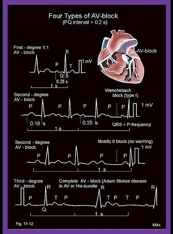
They'll also check your heart with an electrocardiogram. You may hear this called an ECG or EKG. It puts the rate, rhythm, and electrical timing of your heart on a graph. And each type of AV block has its own electrical pattern.
Why does my heart hurt?
Some diseases and infections can hurt your heart, like rheumatic fever, sarcoidosis, lupus, and Lyme disease. Electrolyte imbalance. Your heart may not beat normally if you don't have the right amount of minerals in your blood. AV block may happen if you have too much potassium. Heart surgery.
What is AV node ablation?
AV node ablation. AV (atrioventricular) node ablation is a treatment for an abnormally fast and disorganized heartbeat called atrial fibrillation. It uses heat (radiofrequency) energy to destroy a small amount of tissue between the upper and lower chambers of your heart ( AV node). If you have AV node ablation, ...
What happens after an AV node is destroyed?
Your doctor and nurse will tell you how to prepare and what to bring to the hospital. After an AV node ablation, a pacemaker is necessary for proper heart function.
Why do doctors recommend AV node ablation?
Why it's done. Your doctor may recommend AV node ablation if you have atrial fibrillation that hasn' t improved with medication or other treatments . It's generally considered the last option because it requires the placement of a pacemaker.
When to put pacemaker after AV node ablation?
You may have this device placed several weeks before your ablation to make sure it is working well, or it may be done the day of your ablation.
Where is cardiac ablation done?
Cardiac ablation is done in the hospital. A specialist will insert an IV into your forearm or hand and give you medication to help you relax. During the procedure, you may be awake, lightly sedated or under general anesthesia (fully asleep), depending on your type of arrhythmia and other health conditions.
What is AvnRT in cardiology?
Atrioventricular nodal reentry tachycardia (AVNRT) Atrioventricular nodal reentry tachycardia (AVNRT) is the most common type of supraventricular tachycardia. People with AVNRT have episodes of an abnormally fast heartbeat (more than 100 beats per minute) that often start and end suddenly.
What is the use of sound waves to produce images of your heart's size, structure and motion?
Echocardiogram, which uses sound waves to produce images of your heart's size, structure and motion
What is supraventricular tachycardia?
Treatment. Supraventricular tachycardia is an abnormally fast heartbeat. It occurs when faulty electrical connections in the heart set off a series of early beats in the upper chambers of the heart (atria). Most people with AVNRT don't need medical treatment.
What is the purpose of ECG?
Electrocardiogram (ECG) to measure the electrical activity of your heart and measure the timing and duration of each heartbeat
How does a catheter ablation work?
Catheter ablation. In this procedure, your doctor threads one or more catheters through your blood vessels to your heart. Sensors at the tip of the catheter use heat energy (radiofrequency) or extreme cold (cryoablation) to scar a small area of heart tissue and block the faulty signals that are causing your arrhythmia.
Can AvnRT affect anyone?
AVNRT tends to occur more often in young women, but it can affect anyone.
Do you need medical attention for Avnrt?
Most people with AVNRT don't need medical treatment . However, if you have prolonged or frequent episodes, your doctor may recommend:
What is the AV system?
The AV system consists of the atrioventricular node (AV node) and the His-Purkinje system. These structures conduct the atrial impulse to the ventricles. Impulse conduction through the atrioventricular node is slow. This is explained by the scarcity of gap junctions in the cells of the atrioventricular node. Contractile cells and, in particular, Purkinje fibers, have an abundance of gap junctions which enables rapid impulse conduction. Nevertheless, the slow impulse conduction through the atrioventricular node has a physiological purpose. It causes a delay which gives the atria enough time to empty their blood into the ventricles, before ventricular contraction starts.
Where is the AV block located?
There are, fortunately, some rules of thumb that should be noted. The block in first-degree AV block is mostly located in the atrioventricular node. The block in second-degree AV block Mobitz type 1 is also mostly located in the atrioventricular node. These types of AV block are the most benign.
What is AV block?
Impulse conduction from the atria to the ventricles may be abnormally delayed or even blocked. These conditions are referred to as atrioventricular (AV) blocks, which are subdivided according to the degree of block. First-, second- and third-degree AV block may all be diagnosed using the ECG.
What is the effect of vagal stimulation on the heart?
Vagal stimulation: The Vagus nerve slows heart rate as well as conduction through the AV node. Vagal activity is increased in the following situations: carotid sinus massage (intentional or not), Valsalva maneuver, acute pain and hypersensitive carotid sinus reflex. Vagal fibers unload acetylcholine on AV nodal cells which slows conduction and may even block conduction with ensuing asystole. In the vast majority of cases the asystole is transient.
What is the term for the electrical dissociation of the atria and ventricles?
The atria and the ventricles are electrically dissociated from each other. This condition is referred to as atrioventricular (AV) dissociation. Importantly, for the ventricles to have any electrical (and thus pumping) activity at all, an escape rhythm must arise in an ectopic focus (located distal to the block).
What is second degree AV block?
In second-degree AV block some impulses are completely blocked, which means that not all P-waves are followed by QRS complexes . Second-degree AV-block occurs in the following two variants:
What causes AV block?
The block may be located in the atrioventricular node, His bundle, bundle branches and/or fascicles. A wide range of conditions may cause AV blocks.
Where is the AV node?
Atrioventricular (AV) Node. The atrioventricular node lies on the right side of the partition that divides the atria, near the bottom of the right atrium. When the impulses generated by the SA node reach the AV node, they are delayed for about a tenth of a second.
Where do impulses go in the AV node?
Impulses from the AV node are passed along to atrioventricular bundle fibers. The atrioventricular bundle, also called the bundle of His, is a bundle of cardiac muscle fibers located within the septum of the heart. This fiber bundle extends from the AV node and travels down the septum, which divides the left and right ventricles. The atrioventricular bundle splits into two bundles near the top of the ventricles and each bundle branch continues down the center of the heart to carry impulses to the left and right ventricles.
Which layer of the heart is the Purkinje fiber located in?
These fibers extend from atrioventricular bundle branches to the left and right ventricles. Purkinje fibers rapidly relay cardiac impulses to the myocardium (middle heart layer) of the ventricles causing both ventricles to contract. Myocardium is thickest in heart ventricles allowing ventricles to generate enough power to pump blood to the rest ...
Which two nodes are instrumental in the cardiac cycle?
These two nodes are the sinoatrial (SA) node and the atrioventricular (AV) node. 01.
What is a heart node?
our editorial process. Regina Bailey. Updated July 18, 2019. A heart node is a specialized type of tissue that behaves as both muscle and nervous tissue. When nodal tissue contracts (like muscle tissue), it generates nerve impulses (like nervous tissue) that travel throughout the heart wall. The heart has two nodes that are instrumental in cardiac ...
Which ventricle is the thickest?
Myocardium is thickest in heart ventricles allowing ventricles to generate enough power to pump blood to the rest of the body. The right ventricle forces blood along the pulmonary circuit to the lungs. The left ventricle forces blood along the systemic circuit to the rest of the body. Bailey, Regina.
Where is the SA node located?
Located in the upper wall of the right atrium, it generates nerve impulses that travel throughout the heart wall causing both atria to contract. The SA node is regulated by the autonomic nerves of the peripheral nervous system. Parasympathetic and sympathetic autonomic nerves send signals to the SA node to either accelerate (sympathetic) ...
