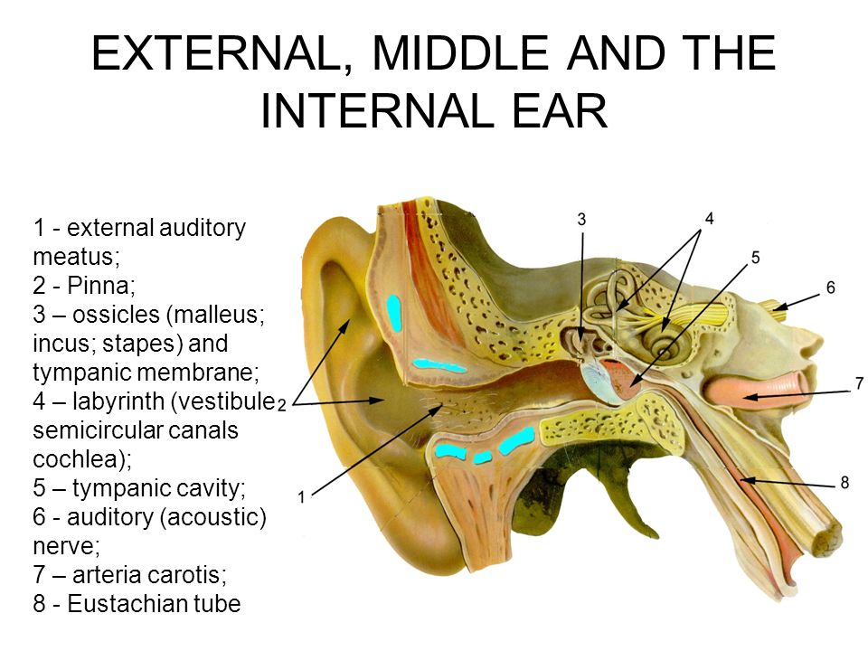
Internal Auditory Meatus
- Neuroradiology of the Temporal Bone and Skull Base. The most common indication for temporal bone imaging is in the evaluation of a suspected CPA or internal auditory canal lesion.
- Head. The vestibular ganglion is found in the trunk of the nerve at, or within, the internal auditory meatus.
- Volume 1
Is the auditory meatus part of the skull?
Anatomical terms of bone. The internal auditory meatus (also meatus acusticus internus, internal acoustic meatus, internal auditory canal, or internal acoustic canal) is a canal within the petrous part of the temporal bone of the skull between the posterior cranial fossa and the inner ear.
What is the fundus of the right internal acoustic meatus?
Diagrammatic view of the fundus of the right internal acoustic meatus. The internal auditory meatus (also meatus acusticus internus, internal acoustic meatus, internal auditory canal, or internal acoustic canal) is a canal within the petrous part of the temporal bone of the skull between the posterior cranial fossa and the inner ear .
What nerve passes through the auditory meatus?
The internal auditory meatus provides a passage through which the vestibulocochlear nerve (CN VIII), the facial nerve (CN VII), and the labyrinthine artery (an internal auditory branch of the basilar artery) can pass from inside the skull to structures of the inner ear and face.
How wide is the internal auditory meatus?
The width of the internal auditory meatus is variable. CT confirmed the abnormally wide internal auditory meatus bilaterally (figure 2). A thorough review of the English-language literature suggests that this is the first report of bilateral SSCD associated with bilateral large internal auditory meatus.
What is a internal auditory meatus mean?
The internal acoustic canal (IAC), also known as the internal auditory canal or meatus (IAM), is a bony canal within the petrous portion of the temporal bone that transmits nerves and vessels from within the posterior cranial fossa to the auditory and vestibular apparatus.
What is a MRI internal auditory meatus both for?
Abstract. Magnetic resonance imaging (MRI) is presently the study of choice for assessment of the internal auditory canal (IAC). MRI provides excellent assessment of the IAC and the bony changes occurring in the canal walls, and it provides excellent demonstration of the content of the canal.
What goes through internal auditory meatus?
The facial nerve enters the internal auditory meatus, passes through the petrous part of the temporal bone, and exits the skull through the stylomastoid foramen. The nerve then enters the parotid gland and breaks up into its five terminal branches: temporal, zygomatic, buccal, mandibular, and cervical.
What does internal auditory canal do?
Unlike the external auditory canal that transmits sound waves, the internal auditory canal carries the the facial and vestibulocochlear nerves that transmit information to the brain.
Why do I need an MRI scan for tinnitus?
An MRI scan may reveal a growth or tumor near the ear or the eighth cranial nerve that could be causing tinnitus. Imaging tests can also help doctors evaluate pulsatile tinnitus. They can show changes in the blood vessels near the ears and determine whether an underlying medical condition is causing symptoms.
Will ear MRI show brain problems?
An MRI scan can detect abscess, as well as meningitis, and infections involving the ears and eyes.
What is the difference between internal and external acoustic meatus?
Unlike the external acoustic meatus that transmits sound waves, the internal acoustic meatus carries the facial and vestibulocochlear nerves that transmit information to the brain.
What is the purpose of the internal acoustic meatus quizlet?
The external acoustic meatus is the opening in the temporal bone that allows sound waves to reach the tympanic membrane. The internal acoustic meatus is the foramen in the temporal bone that allows the vestibulocochlear nerve and the facial nerve to pass into the skull at the base of the brain.
How long does an MRI internal auditory meatus take?
For as non-contrast exam, the exam should take about 25 minutes. For a contrast exam, the exam should take about 45 minutes allowing time for the technologist to start your IV.
Does CT scan show ear problems?
CT scans use electromagnetic radiation to take a series of X-rays of the interior structures of the ear and create a computerized three-dimensional image. CT scans may reveal damage to the bony components of the ear or an abnormal bone growth in the middle ear, a condition called otosclerosis.
Can a CT scan show fluid in ear?
Cultures of the fluid that may show a bacterial infection. A CT scan of the head or mastoids may show that the infection has spread beyond the middle ear.
Can a CT scan detect inner ear problems?
A temporal bone CT scan will therefore show details of external ear canal, middle ear and ossicles, mastoid and inner ear problems.
How long does an MRI internal auditory meatus take?
For as non-contrast exam, the exam should take about 25 minutes. For a contrast exam, the exam should take about 45 minutes allowing time for the technologist to start your IV.
Can MRI results be seen immediately?
This means it's unlikely you'll get the results of your scan immediately. The radiologist will send a report to the doctor who arranged the scan, who will discuss the results with you. It usually takes a week or two for the results of an MRI scan to come through, unless they're needed urgently.
What does MRI IAM both mean?
What is an MRI hand scan? Magnetic Resonance Imaging (MRI) uses a strong magnetic field to produce high quality images in multiple planes or directions. The images are generated using superconducting magnets and pulsed radio waves. An MRI of the IAM or 'internal acoustic meatus' exam- ines the auditory tract.
Where is the internal acoustic meatus located?
temporal boneThe internal auditory canal (IAC), also referred to as the internal acoustic meatus lies in the temporal bone and exists between the inner ear and posterior cranial fossa. It includes the vestibulocochlear nerve (CN VIII), facial nerve (CN VII), the labyrinthine artery, and the vestibular ganglion.
What is the internal auditory meatus?
The internal auditory meatus (IAM) is a canal in the temporal bone that extends from the bony cochlea medially to an opening in the posterior aspect of the petrous portion of the temporal bone. This structure is germane to audiologists because it contains three nerves of interest to audiologists: 1- the auditory nerve, 2- the vestibular nerves, and 3- the facial nerve. These actual compose two of the cranial nerves – number 7 (facial) and 8 (auditory and vestibular).
What is the posterior aspect of the IAM?
In the posterior aspect of the IAM is the vestibular nerves. These can be divided into superior and inferior segments. (Musiek and Baran, 2007; Fatterpekar et al.,1999). One of the best and most common ways to view the IAM and associated nerves in cross section is by a Stenver’s view.
What are the two key disorders of the IAM?
One of course is the vestibular schwannoma (better known as an acoustic tumor) and the vascular loop.
Benign Neoplasms of the Ear and Temporal Bone
Meningiomas occur at a number of sites in the temporal bone, including the internal auditory meatus, the jugular foramen, the geniculate ganglion region, roof of the eustachian tube, and middle ear (cleft). Tumors of the ear and temporal bone are on average <1.5 cm in greatest dimension.
Intracranial tumours in adults
Nyree Griffin MD FRCR, Lee Alexander Grant BA (Oxon) FRCR, in Grainger & Allison's Diagnostic Radiology Essentials, 2013
Syndromes of the Head and Neck
Chris Jo DMD, ... Shahrokh C. Bagheri DMD, MD, in Clinical Review of Oral and Maxillofacial Surgery, 2008
Lower Brainstem and Cranial Nerve Dysfunction
J. Eric Piña-Garza MD, Kaitlin C. James MD, in Fenichel's Clinical Pediatric Neurology (Eighth Edition), 2019
TEMPORAL BONE
Coronal bone CT in the same patient shows the cochlea appears cystic and lacks internal structure . There is a wide communication between the cochlea and vestibule and the internal auditory meatus .
The Head, Neck and Dentition
Pars petrosa ( Figs. 5.30 and 5.33) – the perinatal bone can be recognized at about midfetal life and the descriptions can be found in the main text. Sideing of the perinatal bone depends on identifying the middle ear cavity, which lies laterally and the intracranial surface, which is medial.
Traumatic Facial Nerve Injury
The facial nerve is a mixed nerve that carries fibers from and into three distinct nuclei in the midbrain.
Where is the vestibular ganglion located?
The vestibular ganglion is found in the trunk of the nerve at, or within, the internal auditory meatus. On the more distal side of the vestibular ganglion the nerve divides into a posterior, superior, and inferior branch. The fibers are distributed as follows:
Which nerve pierces the upper surface of petrous temporal bone?
Leaving the geniculate ganglion, the greater petrosal nerve pierces the upper surface of petrous temporal bone, enters the middle cranial fossa, is joined at foramen lacerum by the deep petrosal nerve (sympathetic fibres from internal carotid plexus) to form the nerve of the pterygoid canal, which goes to the pterygopalatine ganglion. Secretomotor fibres are carried to the lacrimal gland by the zygomatic and lacrimal nerves (V), and to mucosal glands of nose and palate by nasal and palatine nerves (V). The nerve to stapedius branches off in the facial canal. The chorda tympani leaves the facial nerve above the stylomastoid foramen, ascends to cross the tympanic membrane and leaves the middle ear via the petrotympanic fissure to join the lingual nerve (V): this carries its taste fibres from the anterior two-thirds of the tongue and its preganglionic secretomotor fibres to the submandibular ganglion for sublingual and submandibular salivary glands. A communicating branch joins the auricular branch of the vagus.
How is the temporal fascia excised?
The temporal fascia is excised along the anterior, superior, and inferior borders of the incision and dissected from the temporal muscle ( Fig. 13.2 A ). All attempts should be made to preserve the integrity of the fascia. The fascia is kept in connection with the sternocleidomastoid muscle, which is then detached from its insertion at the mastoid process to create a large vascularized flap for reconstruction. The temporal muscle is then reflected anteriorly through a subperiosteal dissection ( Fig. 13.2 B).
Where does the facial nerve enter the parotid gland?
The facial nerve enters the internal auditory meatus, passes through the petrous part of the temporal bone, and exits the skull through the stylomastoid foramen. The nerve then enters the parotid gland and breaks up into its five terminal branches: temporal, zygomatic, buccal, mandibular, and cervical. The temporal branch can be found along ...
Where is the auditory canal located?
The opening of the internal auditory canal into the cranial cavity located on the posterior surface of the petrous portion of the temporal bone, or the canal itself.
Is the width of the internal auditory meatus variable?
The width of the internal auditory meatus is variable.