What are microvilli?
Microvilli (singular: microvillus) are microscopic cellular membrane protrusions that increase the surface area for diffusion and minimize any increase in volume, and are involved in a wide variety of functions, including absorption, secretion, cellular adhesion, and mechanotransduction.
What are the function of microvilli?
Microvilli on the surface of epithelial cells such as those lining the intestine increase the cell's surface area and thus facilitate the absorption of ingested food and water molecules.
Where are microvilli found?
the small intestineThey are microscopic protrusions of the cellular membrane. They are present on the surface of the small intestine. They increase the surface area and hence absorption.
What is the function of microvilli and where are they found?
Microvilli are extensions of villi and are made of microfilaments and cytoplasm . The abundance of microvilli is found in the small intestine. Their function here to is to increase surface area in order to increase the amount of nutrients that are absorbed.
What is the structure of microvilli?
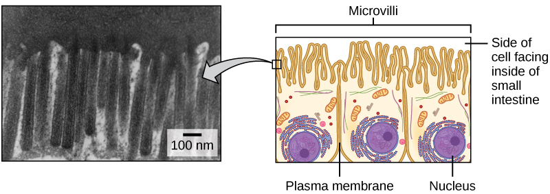
These are small finger like projections, about 1mm in length, and 90nm or so in diameter. The microvilli are shorter and narrower than cilia. They contain bundles of parallel actin filaments held together into a bundle by cross-linking proteins called villin and fimbrin.
What are microvilli made of?
actin filaments(Microvilli is the plural of microvillus.) They contain bundles of parallel actin filaments held together into a bundle by cross-linking proteins called villin and fimbrin. Lateral arms containing myosin I and calmodulin link the actin filament bundle to the plasma membrane.
What are examples of microvilli?
Microvilli are finger-shaped plasma membrane protrusions that are found at the surface of a large variety of cell types but are most numerous and elaborated on simple epithelial, for example intestinal mucosa and the epithelium of the kidney proximal tubule.
What human cells have microvilli?
Microvilli are thin finger-like membrane protrusions that are found on the surface of a wide variety of cell types (8), including intestinal epithelial cells (9), dendritic cells (10), and neurons (11).
What is the role of microvilli in the small intestine?
Every cell lining the small intestine bristles with thousands of tightly packed microvilli that project into the gut lumen, forming a brush border that absorbs nutrients and protects the body from intestinal bacteria.
What's the difference between villi and microvilli?
Microvilli is a part of a cell. Its function is to augment the surface area of the cell. The main function of microvilli includes secretion, absorption, and cellular sticking or adhesion. Villi or intestinal villi, on the other hand, are finger-like projections that are found in the intestinal wall.
How do microvilli absorb nutrients?
In the small intestine, these cells contain microvilli, which are tiny hair-like projections that increase nutrient absorption. These projections increase the surface area of the small intestine allowing more area for nutrients to be absorbed.
Where are microvilli found small intestine?
Microvilli are most often found in the small intestine, on the surface of egg cells, as well as on white blood cells. Thousands of microvilli form a structure called the brush border that is found on the apical surface of some epithelial cells, such as the small intestines.
What is the function of the microvilli in the small intestine?
Every cell lining the small intestine bristles with thousands of tightly packed microvilli that project into the gut lumen, forming a brush border that absorbs nutrients and protects the body from intestinal bacteria.
What is the function of cilia and microvilli?
Cilia are motile and involved in the rhythmic movement of either the whole cell or external objects like microorganisms, dirt, and mucus over the cell surface. In contrast, microvilli are not motile. They enhance the absorption of nutrients of the intestine by increasing the surface area of the intestine.
What are microvilli Class 11?
Microvilli, in the most simple terms, are tiny small microscopic projections that exist in, on, and around cells. - They can exist on their own or in conjunction with villi. - Microvilli function is to increase the surface area of the small intestinal wall for the absorption of the digested food.
What is the function of microvilli in simple columnar epithelium?
SIMPLE COLUMNAR EPITHELIUM: The surface of each villus is covered with simple columnar epithelium . The free surface of these cells has very tiny projections called microvilli , which are specialized for absorption (absorptive cells).
Where do microvilli occur?
They are tiny projections that exist on, in, and around cells. Microvilli occur in various structures and sites in an organism. One such place is the small intestine where the microvilli and villi work to increase the surface area of the intestine, allowing for more absorption of vital nutrients for the organism.
What is the difference between a villi and a microvilli?
It is a similar thing here; the villi would be like the centimeters, while the microvilli would be the millimeters falling in between. The microvilli have their own plasma membrane that covers them.
How to make a microvilli model?
Start by getting a roll of paper towels. Take one paper towel from the roll, cut it in half, and lay it flat.
What is the role of microvilli in egg cells?
They also play a role in egg cells as they help in anchoring the sperm to the egg, thu s allowing for easier fertilization. In white blood cells, the microvilli again act as an anchoring point. They allow the white blood cell that is hurtling through the body to grab and stick to whatever it is going after at the given moment.
What are the folds on the small intestine called?
They can exist on their own or in conjunction with villi. The linings in some mucous membranes, most specifically of the small intestine, there exist tiny folds that project out like numerous fingers. These are called villi. On each of the villi, there are even smaller folds that stick out like fingers called microvilli.
Do microvilli have cytoplasm?
The microvilli have their own plasma membrane that covers them. Inside of them, they have almost no organelles, but they do have cytoplasm (cellular fluid) and some microfilaments (strands of actin, a protein), which help give them their structure. They are essentially bundles of cross-linked actin fibers.
What are microvilli in epithelium?
microvilli. The millions of tiny, hair-fine, finger-like protrusions on the surface cells of EPITHELIUM which greatly increase the effective surface area so as to facilitate absorption. Microvilli occur especially on the secretory and absorptive surfaces, and are formed by extensions of the cell membrane.
Where are the microvilli located in the intestinal tract?
Visualization of mucosal layer of intestinal tract with SEM under higher magnification revealed that the microvilli were densely arranged on the surface of mucosal membrane of anterior intestine and abundant secretory gland cells were be mingled with columnar cells.
What are the hairlike projections above the surface of specialised epithelial cells?
Microscopic hairlike projections above the surface of specialised epithelial cells, which are composed of complex plasma membrane folds surrounding an actin microfilament core. Microvilli greatly increase total cell surface, and therefore also the absorptive capacity of cell.
Does chronic exposure affect microvilli?
Acute exposures did not have much effect, but chronic exposure diminished the absorptive projections on the surface of intestinal cells called microvilli, showed the findings published in the journal NanoImpact.
What are the roles or functions of microvilli?
Microvilli perform numerous functions and most of them include the following:
How do microvilli work?
In the intestines, microvilli work with villi to absorb all the essential nutrients by expanding the intestines’ surface area.
How do microvilli help the body?
The membranes of microvilli are packed with enzymes that help in breaking down complex nutrients into simpler compounds so that they can be easily absorbed by the body. Microvilli play an essential role in fertilization. They make fertilization easy by helping the sperm to anchor to the egg.
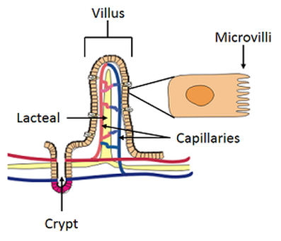
What is the structure of a villus?
In every villus, there are smaller folds called microvilli, which have their own plasma membrane covering them. Inside the microvilli is a cytoplasm or cellular fluid and microfilaments giving microvilli the structure it needs. Image 1:A closer look at the structure of microvilli. Picture Source: britannica.com.
What is the name of the small folds in the villus?
In every villus, there are smaller folds called microvilli, which have their own plasma membrane covering them. Inside the microvilli is a cytoplasm or cellular fluid and microfilaments giving microvilli the structure it needs.
Where are cilia found?
Cilia – They are found in the columnar epithelial cells of the tubular areas of the uterine and respiratory system.
What are tiny projections called?
In the cells, you will find tiny little projections called microvilli. They can be on their own or with villi. In the small intestines, you will find tiny. In the cells, you will find tiny little projections called microvilli. They can be on their own or with villi.
Where are microvilli found?
Microvilli location. Microvilli are found in small intestinal epithelial cells and kidneys. In the intestine, each villus is composed of many cells and each villus has thousands of microvilli on the apical surface forming a brush border. They are also present on the plasma surface of the ovum to help in the anchoring of sperms.
What is the structure of the microvilli?
Microvilli structure increases the surface area to volume ratio. The wall of the small intestine is covered by several mucus membrane folds called plicae circulares. The surface of these folds contains tiny projections called villi, which in turn have microvilli.
Are microvilli anchored by desmosomes?
No, but the desmos ome protein desmopla kin affects the length of microvilli.
What cells are in the small intestine?
They increase the total area for absorption. The small intestine villi are covered by a single layer of simple columnar cells called goblet cells. Goblet cells are scattered among the epithelial cells covering the villi and secrete mucin (which forms mucus). The goblet cells contain many microvilli.
How does microvilli affect the gastrointestinal tract?
Microvilli largely increase the surface area of the cell. It increases the surface of nutrient absorption in the gastrointestinal tract and kidney.
How do microvilli help digestion?
The microvilli help in the digestion and absorption of intestinal contents by increasing the absorbing surface.
Why do microvilli get damaged?
Microvilli in the gut and other lining get damaged due to sloughing of cells, actions of toxins, stresses, etc. Repair of microvilli occurs via intrinsic reparative processes. The entire cell is not regenerated and replaced every time.
What are microvilli?
Microvilli are strands that look like tiny hairs that are attached to the membrane of a cell.
Where are microvilli found?
They are found in the tongue, nose, and even in your ears. It’s the microvilli on your tongue that are the tiniest hairs on the taste buds that send your brain signals.
Why can't you find microvilli in every cell?
You won’t find microvilli on every cell, because they are only found on cells that have a specialized purpose.
What is the function of the microvilli on the surface of white blood cells?
The microvilli on the surface of white blood cells act like bodyguards to protect against dangerous viruses and bacteria.
How many microvilli are there in the small intestine?
Our small intestines have multiple of thousands of microvilli, and there are so many that scientists have given the area that they form the name of “brush border.”
Do microvilli have plasma membranes?
Microvilli have their own plasma membranes covering them .
Where are microvilli located?
Microvilli are small finger-like cytoplasmic projections emanating from the apical surface of the cell into the lumen.
What is the micro villi?
Micro- villi represent the striated border of the intestinal absorptive cells and the brush border of the kidney proximal tubule cells observed by light microscopy.
How many actin filaments are in a microvillus?
1. Each microvillus contains a core fo 25 to 30 actin filament bundle, whose members are cross-linked to each other by a number of actin binding proteins (espin, fascin, villin and fimbrin).
Where are stereocilia found?
Stereocilia are long microvilli found only in the epididymis and on the sensory hair cells of the cochlea (inner ear).
Where do the minus ends of the actin filaments terminate?
The minus ends of the actin filaments terminate in the ter- minal web.
Where are myosin and trophomyosin located?
Tropomyosin and myosin II are located in the terminal web , and, as these two proteins act on the actin filaments and the apical aspect of the cell contracts, the microvilli spread apart, providing more space for molecular transport at the cell apex.
Is microvilli short or sparse?
In less active cells, microvilli may be sparse and short.