How is the pyruvate dehydrogenase complex regulated?
The pyruvate dehydrogenase complex is regulated by covalent modification of the first enzyme, pyruvate dehydrogenase (PDH). Pyruvate dehydrogenase kinase inactivates PDH by phosphorylation with ATP (Fig. 6-5 ).
What is pyruvate dehydrogenase (PDH) deficiency?
Pyruvate dehydrogenase (PDH) deficiency is a congenital degenerative metabolic disease resulting from a mutation of the pyruvate dehydrogenase complex (PDC) located on the X chromosome.
What is the function of E2 in pyruvate dehydrogenase?
Function. Lipoic acid is covalently bound to dihydrolipoamide acetyltransferase (E2), which is the second catalytic component enzyme of PDC. The reaction catalyzed by pyruvate dehydrogenase (E1) is considered to be the rate-limiting step for the pyruvate dehydrogenase complex (PDHc).

How is pyruvate dehydrogenase inactivated?
The pyruvate dehydrogenase complex (PDH complex) of Escherichia coli and its pyruvate dehydrogenase component (E1) are rapidly inactivated by low concentrations of fluoropyruvate in a thiamin pyrophosphate (TPP) dependent process. The inactivation rates for the PDH complex and for its E1 component are similar.
How is pyruvate dehydrogenase kinase inhibited?
The pyruvate dehydrogenase complex is regulated by covalent modification of the first enzyme, pyruvate dehydrogenase (PDH). Pyruvate dehydrogenase kinase inactivates PDH by phosphorylation with ATP (Fig. 6-5). Reactivation is achieved by the action of pyruvate dehydrogenase phosphatase.
Is pyruvate dehydrogenase inhibited by ATP?
ATP, acetyl CoA, and NADH negatively regulate (inhibit) pyruvate dehydrogenase, while ADP and pyruvate activate it.
Which compound inhibits the enzyme pyruvate dehydrogenase?
Regulation of Pyruvate Oxidation Reactivation is achieved by the action of pyruvate dehydrogenase phosphatase. Both of these regulatory enzymes are regulated: PDH kinase is stimulated by NADH and acetyl-CoA. It is inhibited by pyruvate.
Which of the following has inhibitory effect on PDH?
Products of fatty acid oxidation (acetyl-CoA, NADH, and ATP) inhibit PDH and save glucose and amino acids.
Does glucagon inhibit pyruvate dehydrogenase?
It is suggested that glucagon and cyclic AMP inhibit pyruvate dehydrogenase and thus limit the supply of reducing equivalents needed for glucose formation.
Does NADH inhibit PDC?
The PDC is also inhibited by acetyl-CoA and NADH directly.
Does ATP inhibit glycolysis?
In glycolysis, one of the end products is energy in the form of ATP. ATP acts as an inhibitor of phosphofructokinase-1, one of the main rate limiting enzymes in glycolysis.
How is pyruvate dehydrogenase activated?
Pyruvate dehydrogenase kinase is activated by ATP, NADH and acetyl-CoA. It is inhibited by ADP, NAD+, CoA-SH and pyruvate. Each isozyme responds to each of these factors slightly differently. NADH stimulates PDK1 activity by 20% and PDK2 activity by 30%.
What inhibits citric acid cycle?
Reactions steps of the citric acid cycle. The CAC as the pivotal element of energy metabolism is strictly regulated. The cycle is triggered by ADP, inorganic phosphate, and calcium while it is inhibited by NADH and ATP.
What is pyruvate dehydrogenase deficiency?
Collapse Section. Pyruvate dehydrogenase deficiency is characterized by the buildup of a chemical called lactic acid in the body and a variety of neurological problems. Signs and symptoms of this condition usually first appear shortly after birth, and they can vary widely among affected individuals.
Does citrate inhibit glycolysis?
For example, citrate directly inhibits the main regulators of glycolysis, phosphofructokinase-1 (PFK1) and phosphofructokinase-2 (PFK2) [2,3], while it enhances gluconeogenesis by promoting fructose-1,6-biphosphatase (FBPase) [4]. An example of the key role of citrate in normal cells can be found in hepatocytes.
How does Dichloroacetate inhibit pyruvate dehydrogenase kinase?
The pyruvate dehydrogenase (PDH) kinase (PDK) inhibitor dichloroacetate (DCA) activates PDH, the rate-limiting enzyme of aerobic glucose oxidation5. PDH converts glycolysis-produced pyruvate in acetyl-CoA that enters the mitochondria and is consumed in the process of oxidative phosphorylation (OXPHOS).
How is pyruvate dehydrogenase activated?
Pyruvate dehydrogenase kinase is activated by ATP, NADH and acetyl-CoA. It is inhibited by ADP, NAD+, CoA-SH and pyruvate. Each isozyme responds to each of these factors slightly differently. NADH stimulates PDK1 activity by 20% and PDK2 activity by 30%.
How does pyruvate dehydrogenase kinase work?
Pyruvate dehydrogenase kinase (PDK) is a kinase that inactivates the pyruvate dehydrogenase enzyme complex through phosphorylation. By downregulating the activity of this complex, PDK decreases the oxidation of pyruvate in mitochondria and increases the conversion of pyruvate to lactate in the cytosol.
What is the role of pyruvate dehydrogenase kinase?
Pyruvate dehydrogenase kinase (PDK) can regulate the catalytic activity of pyruvate decarboxylation oxidation via the mitochondrial pyruvate dehydrogenase complex, and it further links glycolysis with the tricarboxylic acid cycle and ATP generation.
What is pyruvate dehydrogenase?
Pyruvate dehydrogenase (PDH) deficiency is a congenital degenerative metabolic disease resulting from a mutation of the pyruvate dehydrogenase complex (PDC) located on the X chromosome. While defects have been identified in all 3 enzymes of the complex, the E1-α subunit is predominantly the culprit.
What is the name of the enzyme that converts pyruvate dehydrogenase into ace?
PDC consists of other enzymes, referred to as E2 and E3. Collectively E1-E3 transform pyruvate, NAD +, coenzyme A into acetyl-CoA, CO 2, and NADH. The conversion is crucial because acetyl-CoA may then be used in the citric acid cycle to carry out cellular respiration. To distinguish between this enzyme and the PDC, it is systematically called pyruvate dehydrogenase (acetyl-transferring) .
What enzyme catalyzes the reaction of pyruvate and a lipoamide to give the?
Pyruvate dehydrogenase is an enzyme that catalyzes the reaction of pyruvate and a lipoamide to give the acetylated dihydrolipoamide and carbon dioxide. The conversion requires the coenzyme thiamine pyrophosphate. Pyruvate dehydrogenase is usually encountered as a component, referred to as E1, of the pyruvate dehydrogenase complex (PDC).
What is the name of the enzyme that catalyzes the reaction of pyruvate and a?
Thiamine pyrophosphate (TPP) is shown in grey ball and stick form, two magnesium ions in purple undergoing metal ligation with the TPP, and two potassium ions in orange. Pyruvate dehydrogenase is an enzyme that catalyzes the reaction of pyruvate and a lipoamide to give the acetylated dihydrolipoamide and carbon dioxide.
What is PDBe-KB?
PDBe-KB provides an overview of all the structure information available in the PDB for Human pyruvate dehydrogenase (lipoamide) beta.
What enzyme is responsible for oxidation of pyruvate?
In bacteria, a form of pyruvate dehydrogenase (also called pyruvate oxidase, EC 1.2.2.2) exists that links the oxidation of pyruvate into acetate and carbon dioxide to the reduction of ferrocytochrome. In E. coli this enzyme is encoded by the pox B gene and the protein has a flavin cofactor.
What is the phosphorylation of E1?
Phosphorylation of E1 by pyruvate dehydrogenase kinase (PDK) inactivates E1 and subsequently the entire complex. PDK is inhibited by dichloroacetic acid and pyruvate, resulting in a higher quantity of active, unphosphorylated PDH. Phosphorylation is reversed by pyruvate dehydrogenase phosphatase, which is stimulated by insulin, PEP, and AMP, but competitively inhibited by ATP, NADH, and Acetyl-CoA .
What is the treatment for pyruvate dehydrogenase deficiency?
This can prevent immediate worsening of the disease. [1] [4] Treatment options typically include supplementing cofactors including carnitine, thiamine, and lipoic acid.
Why do people with pyruvate dehydrogenase complex (PDC) deficiency?
This is because many people with pyruvate dehydrogenase complex (PDC) deficiency pass away before they have children. Genetic changes that are new in a person are called de novo. In some cases, people who have a pathogenic variant in PDHA1 inherited the genetic change from their mother.
Why do females have a copy of PDHA1?
However, because females have another copy of the PDHA1 gene that is producing the enzyme of the pyruvate dehydrogenase complex , some females may have less severe symptoms of the disease. [1] In some cases, people who have a pathogenic variant in PDHA1 are the first people in the family with the genetic change. [5] .
How do you know if you have pyruvate dehydrogenase complex?
Signs that may be apparent in pregnancy include poor fetal weight gain and low levels of estriol in the mother’s urine. [4] Some babies with the disease may have brain abnormalities seen on ultrasound. [1] Babies with PDC deficiency may have low scores measuring a baby’s health after birth ( Apgar scores ). A low birth weight is common. Some features that may be characteristic of PDC deficiency include a narrow head, prominent forehead ( frontal bossing ), wide nasal bridge, long philtrum, and flared nostrils. However, these features are not present in all babies with PDC deficiency. [2]
What is PDC deficiency?
Pyruvate dehydrogenase complex (PDC) deficiency is a type of metabolic disease. This means that the body is not able to efficiently break down nutrients in food to be used for energy. Symptoms of PDC deficiency include signs of metabolic dysfunction such as extreme tiredness (lethargy), poor feeding, and rapid breathing ( tachypnea ). Other symptoms may include signs of neurological dysfunction such as developmental delay, periods of uncontrolled movements ( ataxia ), low muscle tone ( hypotonia ), abnormal eye movements, and seizures. Symptoms usually begin in infancy, but signs can first appear at birth or later in childhood. Symptoms may be especially apparent during times of illness, stress, or after meals with high amounts of carbohydrates. [1]
Why is PDC deficiency variable?
The signs and symptoms of PDC deficiency are variable because the amount of enzyme that is available to create energy varies in different people with the disease. [1]
Can pyruvate dehydrogenase deficiency be long term?
Unfortunately, the long-term outlook for people with pyruvate dehydrogenase complex (PDC) deficiency is poor. People who show signs and symptoms early in life may pass away from complications of the disease in the first years of life. If people with the disease survive, they may be affected with complications including intellectual disability. [1] [4] People who have symptoms of PDC deficiency beginning in late childhood may survive longer because they have higher levels of functioning enzymes in the body. However, these individuals can still have complications of the disease. [1]
Which enzymes regulate glycolysis?
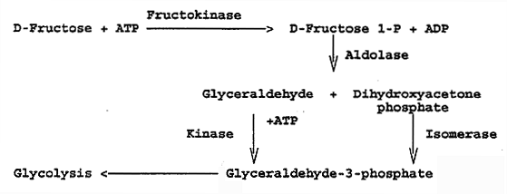
Glycolysis is regulated at the steps catalyzed by hexokinase, PFK-1, and pyruvate kinase.
Which hormone stimulates PDH phosphatase?
PDH phosphatase is stimulated by Ca ++ and insulin.
How are signals transmitted between nucleotide binding sites?
An important mechanism by which signals are transmitted between the nucleotide-binding site and the L2-binding domain may be provided by a long central rod located near the two sites. This rod is composed of two short helices that link the two sites. A central pyruvate/DCA-binding site sits between the two helices ( Figure 3 ). The geometry of the active site is influenced by its nucleotide occupancy, pyruvate, and L2 binding, and, surprisingly, by K + ions. Analysis of human PDK2 with bound DCA indicates that the β -phosphate of ADP chelates K +, together with backbone carbonyl groups of three amino acids, and an oxygen atom of a Ser hydroxyl group, to enhance ADP inhibition. Binding of lipoyl domain shifts the arrangement of the participating amino acid residues, disrupts the K + binding site, and enhances dissociation of ADP from the active site. Binding of K + ions at other sites on the protein stabilizes the L2-binding site and facilitates cross-talk between the nucleotide- and L2-binding sites. Although much less is known about their regulation, ions are also important regulators of PDP1 and PDP2. Both forms are activated by increasing concentrations of free intra-mitochondrial Mg 2+ or Mn 2+ that occur in response to declining levels of ATP. Increases in free mitochondrial Ca 2+ activate PDP1 in tissues with hormonal or exercise-dependent increases in mitochondrial Ca 2+. The ion serves to bridge the interaction of PDP1 with L2 domain of E2, thereby stimulating phosphatase activity. The activities of PDK and PDP2 are regulated in reciprocal fashion by inositol phosphoglycans, compounds that chelate Mn 2+ and Zn 2+ metal ions and that are generated in response to stimulation by insulin.
What can alter PDK expression?
Hormonal regulation and a number of disease states, other than diabetes , can also alter PDK expression significantly. For example, PDK3 and PDK1 are overexpressed in hypoxic cancer cells by the actions of the transcription factor hypoxia-inducible factor-1 (HIF-1).
How does phosphorylation affect E1?
E1 activity can be inhibited by phosphorylation of any of three target Ser residues on its α -subunit. The PDK isoforms show significant variation in their specificity toward individual phosphorylation sites: each can phosphorylate Ser 264 (site 1) and Ser271 (site 2), albeit with different rates, but only PDK1 modifies Ser203 (site 3). These phosphorylation sites are located on two adjacent loops that are close to the active site of E1. Structural studies of human E1 indicate that phosphorylation of site 1, which blocks the majority of E1 activity, disorders two loops adjacent to the E1 active site and disrupts the interaction of ThDP and the E2-bound lipoyl group with their respective binding sites on E1 ( Figure 2 (a) ). Phosphorylation of sites 2 and 3 occurs more slowly and is thought to hinder reactivation of E1, primarily by PDP2.
What enzyme is involved in glycolysis?
Both synthesis from glycolytic precursors and breakdown to either acetyl-CoA or oxaloacetate are tightly regulated. Pyruvate dehydrogenase (EC3.1.3.43) is the enzyme that connects glycolysis to the citric acid cycle. This enzyme complex is inactivated by phosphorylation ( [pyruvate dehydrogenase (lipoamide)] kinase, EC2.7.1.99) of three serines in the E1 subunit and reactivated by removal of these phosphates by
What happens if you have insufficient PDC activity?
Insufficient PDC activity leads to accumulation of pyruvate, which is converted into lactate and H +. This lactic acidosis causes pain and weakness in ischemic skeletal muscles, which are typical symptoms for intermittent claudication. Inhibition of PDHK would increase PDC activity and, consequently: (1) reduce the amount of lactic acid formation and (2) reduce the demand of oxygen for ATP production by shifting metabolism from fatty acid oxidation to glycolysis as primary acetyl-CoA source. Therefore, PDHK inhibition provides a route for therapeutic intervention in diabetes and cardiovascular disorders. 21–28
What is the cause of pyruvate dehydrogenase deficiency?
Pyruvate dehydrogenase deficiency (PCDC) can result from mutations in any of the enzymes or cofactors. Its primary clinical finding is lactic acidosis. Such PCDC mutations, leading to subsequent deficiencies in NAD and FAD production, hinder oxidative phosphorylation processes that are key in aerobic respiration.
Which subunits are bound by pyruvate dehydrogenase?
Main article: Pyruvate dehydrogenase. Initially, pyruvate and thiamine pyrophosphate (TPP or vitamin B 1) are bound by pyruvate dehydrogenase subunits. The thiazolium ring of TPP is in a zwitterionic form, and the anionic C2 carbon performs a nucleophilic attack on the C2 (ketone) carbonyl of pyruvate.
What are the similarities between pyruvate dehydrogenase and enzymes in Gram-positive bacteria?
Similarities of the quaternary structures between pyruvate dehydrogenase and enzymes in gram-positive bacteria point to a shared evolutionary history which is distinctive from the evolutionary history of corresponding enzymes found in gram-negative bacteria.
What is the active site of dihydrolipoyl dehydrogenase?
Dihydrolipoyl dehydrogenase (E3) The dihydrolipoate, still bound to a lysine residue of the complex, then migrates to the dihydrolipoyl dehydrogenase (E3) active site, where it undergoes a flavin -mediated oxidation, identical in chemistry to disulfide isomerase.
What is the E1 subunit of pyruvate dehydrogenase complex?
Coli. The E1 subunit, called the pyruvate dehydrogenase subunit, has a structure that consists of two chains (an “ɑ” and “ꞵ” chain). A magnesium ion forms a 4-coordinate complex with three, polar amino acid residues (Asp, Asn, and Tyr) located on the alpha chain, ...
How many copies of pyruvate dehydrogenase are in a PDC?
In Gram-negative bacteria, e.g. Escherichia coli, PDC consists of a central cubic core made up from 24 molecules of dihydrolipoyl transacetylase (E2). Up to 24 copies of pyruvate dehydrogenase (E1) and 12 molecules of dihydrolipoyl dehydrogenase (E3) bind to the outside of the E2 core.
What is the name of the enzyme that converts pyruvate into acetyl-CoA?
Pyruvate dehydrogenase complex. Pyruvate dehydrogenase complex ( PDC) is a complex of three enzymes that converts pyruvate into acetyl-CoA by a process called py ruvate decarboxylation. Acetyl-CoA may then be used in the citric acid cycle to carry out cellular respiration, and this complex links the glycolysis metabolic pathway to ...
How many CO2 molecules are in pyruvate?
For each molecule of pyruvate, you get one Acetyl CoA, one CO2 molecule, and one NADH.
What is the name of the cystine that oxidizes lipoamide dithiol?
3. Lipoamide dithiol is reoxidized by a cystine of E3 (E2/E3) to give two cysteine residues
What is the transfer of the 2 carbon acetyl group from acetyl-TPP to?
Transfer of the 2 carbon acetyl group from acetyl-TPP to lipoid acid by coenzyme lipoyl transacetylase.
Does pyruvate inhibit PDH kinase?
False. An increase in pyruvate inhibits PDH kinase.

Overview
Regulation
Phosphorylation of E1 by pyruvate dehydrogenase kinase (PDK) inactivates E1 and subsequently the entire complex. PDK is inhibited by dichloroacetic acid and pyruvate, resulting in a higher quantity of active, unphosphorylated PDH. Phosphorylation is reversed by pyruvate dehydrogenase phosphatase, which is stimulated by insulin, PEP, and AMP, but competitively inhibited by ATP, NADH, a…
Mechanism
The thiamine pyrophosphate (TPP) converts to an ylide by deprotonation. The ylide attack the ketone group of pyruvate. The resulting adduct decarboxylates. The resulting 1,3-dipole reductively acetylates lipoamide-E2.
In terms of details, biochemical and structural data for E1 revealed a mechanism of activation of TPP coenzyme by forming the conserved hydroge…
Structure
E1 is a multimeric protein. Mammalian E1s, including human E1, are tetrameric, composed of two α- and two β- subunits. Some bacterial E1s, including E1 from Escherichia coli, are composed of two similar subunits, each being as large as the sum of molecular masses of α- and β- subunits.
Active site
E1 has two catalytic sites, each providing thiamine pyrophosphate (TPP) and magnesium ion as cofactors. The α- subunit binds magnesium ion and pyrophosphate fragment while the β-subunit binds pyrimidine fragment of TPP, forming together a catalytic site at the interface of subunits.
The active site for pyruvate dehydrogenase (image created from PDB: 1NI4) hol…
Pathology
Pyruvate dehydrogenase is targeted by an autoantigen known as anti-mitochondrial antibodies (AMA), which results in progressive destruction of the small bile ducts of the liver, leading to primary biliary cirrhosis. These antibodies appear to recognize oxidized protein that has resulted from inflammatory immune responses. Some of these inflammatory responses could be related to gluten sensitivity as over 50% of the acute liver failure patients in one study exhibited a nonmit…
Related enzymes
In bacteria, a form of pyruvate dehydrogenase (also called pyruvate oxidase, EC 1.2.2.2) exists that links the oxidation of pyruvate into acetate and carbon dioxide to the reduction of ferrocytochrome. In E. coli this enzyme is encoded by the pox B gene and the protein has a flavin cofactor. This enzyme increases the efficiency of growth of E. coli under aerobic conditions.
See also
• Pyruvate dehydrogenase deficiency
Overview
Pyruvate dehydrogenase complex (PDC) is a complex of three enzymes that converts pyruvate into acetyl-CoA by a process called pyruvate decarboxylation. Acetyl-CoA may then be used in the citric acid cycle to carry out cellular respiration, and this complex links the glycolysis metabolic pathway to the citric acid cycle. Pyruvate decarboxylation is also known as the "pyruvate dehydroge…
Regulation
Pyruvate dehydrogenase is inhibited when one or more of the three following ratios are increased: ATP/ADP, NADH/NAD and acetyl-CoA/CoA.
In eukaryotes PDC is tightly regulated by its own specific pyruvate dehydrogenase kinase (PDK) and pyruvate dehydrogenase phosphatase (PDP), deactivating and activating it respectively.
• PDK phosphorylates three specific serine residues on E1 with different affinities. Phosphorylatio…
Structure
The E1 subunit, called the pyruvate dehydrogenase subunit, has a structure that consists of two chains (an “ɑ” and “ꞵ” chain). A magnesium ion forms a 4-coordinate complex with three, polar amino acid residues (Asp, Asn, and Tyr) located on the alpha chain, and the thiamine diphosphate (TPP) cofactor directly involved in decarboxylation of the pyruvate.
Mechanism
Initially, pyruvate and thiamine pyrophosphate (TPP or vitamin B1) are bound by pyruvate dehydrogenase subunits. The thiazolium ring of TPP is in a zwitterionic form, and the anionic C2 carbon performs a nucleophilic attack on the C2 (ketone) carbonyl of pyruvate. The resulting hemithioacetal undergoes decarboxylation to produce an acyl anion equivalent (see cyanohydrin or aldehy…
Structural differences between species
PDC is a large complex composed of multiple copies of 3 or 4 subunits depending on species.
In Gram-negative bacteria, e.g. Escherichia coli, PDC consists of a central cubic core made up from 24 molecules of dihydrolipoyl transacetylase (E2). Up to 24 copies of pyruvate dehydrogenase (E1) and 12 molecules of dihydrolipoyl dehydrogenase (E3) bind to the outside of the E2 core.
In contrast, in Gram-positive bacteria (e.g. Bacillus stearothermophilus) and eukaryotes the centr…
Localization of pyruvate decarboxylation
In eukaryotic cells the pyruvate decarboxylation occurs inside the mitochondrial matrix, after transport of the substrate, pyruvate, from the cytosol. The transport of pyruvate into the mitochondria is via the transport protein pyruvate translocase. Pyruvate translocase transports pyruvate in a symport fashion with a proton (across the inner mitochondrial membrane), which may be considered to be a form of secondary active transport, but further confirmation/support …
Evolutionary history
It was found that pyruvate dehydrogenase enzyme found in the mitochondria of eukaryotic cells closely resembles an enzyme from Geobacillus stearothermophilus, which is a species of gram-positive bacteria. Despite similarities of the pyruvate dehydrogenase complex with gram-positive bacteria, there is little resemblance with those of gram-negative bacteria. Similarities of the quaternary structures between pyruvate dehydrogenase and enzymes in gram-positive bacteria …
Clinical relevance
Pyruvate dehydrogenase deficiency (PCDC) can result from mutations in any of the enzymes or cofactors. Its primary clinical finding is lactic acidosis. Such PCDC mutations, leading to subsequent deficiencies in NAD and FAD production, hinder oxidative phosphorylation processes that are key in aerobic respiration. Thus, acetyl-CoA is instead reduced via anaerobic mechanisms into other molecules like lactate, leading to an excess of bodily lactate and associated neurologi…