
What is the difference between suspension microlaryngoscopy and a surgical microscope?
The surgeon holds the laryngoscope with one hand and works with the other. In suspension microlaryngoscopy, the laryngoscope is suspended to allow the surgeon to work with both hands. A surgical microscope is used to provide magnification, better visualization and to deliver a LASER beam.
What is a microlaryngoscopy?
A microlaryngoscopy is a surgical procedure that allows a provider to view your vocal cords (also called vocal folds) with a microscope. During this procedure, your provider may also remove lesions (growths) from your vocal folds or correct movement disorders of your larynx (voice box).
How is suspension microlaryngoscopy performed in a tracheostomy?
The technique involves suspension microlaryngoscopy, in which a laryngoscope with a wide orifice is inserted transorally and suspended in the vallecula (Fig. 13-17). A microscope with a focal length deep enough to magnify the structures of the larynx and/or trachea (400 mm) is then used by the surgeon.
What is true suspension laryngoscopy?
a True suspension laryngoscopy (using a gallows system that does not fulcrum on the teeth) is used in some centers with a reported higher incidence of tongue/floor of mouth/lower (mandibular) dental injury than the 'fulcrum laryngscopy' we use more commonly employing the Lewy suspension.
How long does it take to recover from a Microlaryngoscopy?
Most people can return to normal activities within a few days. Your surgeon may also recommend complete vocal rest for a few days followed by reduced voice use for two to six weeks, depending on the surgery performed.
What is laryngeal suspension?
During a suspension microlaryngoscopy, a device known as a laryngeal suspension is used to position the patient's head and neck properly to access the larynx.
What is the difference between laryngoscopy and Microlaryngoscopy?
Problems involving the vocal cords result in varying degrees of hoarseness, breathing or speech abnormalities, and laryngoscopy is commonly used to evaluate these symptoms. Microlaryngoscopy gives the surgeon the ability to view the larynx in detail.
Can I talk after Microlaryngoscopy?
Recovery from Microlaryngoscopy with Excision Complete voice rest is a vital part of a full recovery. After the procedure, you should not speak for three to five days to allow your vocal cords to heal.
What is a direct Microlaryngoscopy?
Introduction. A direct laryngoscopy allows visualization of the larynx. It is used during general anesthesia, surgical procedures around the larynx, and resuscitation. This tool is useful in multiple hospital settings, from the emergency department to the intensive care unit and the operating room.
What is SML surgery?
The Scharioth Macula Lens (SML) is a magnifying add-on lens implant, designed for eyes which have already had cataract surgery and which also have macular disease such as macular degeneration.
What can I eat after a Microlaryngoscopy?
For the first 24 hours following surgery you should eat only soft foods. Avoid foods that are fried, spicy, or scratchy to your throat. If it is too uncomfortable for you to swallow even soft foods on the first day, you should drink only liquids.
How long does it take for vocal cords to heal after surgery?
For the First Few Weeks Continue to drink plenty of fluids, avoid irritants, and use the humidifier at night. While your vocal cords are coming back to health and you are relearning how to use your voice in healthy ways, it will take a full 6 to 8 weeks to recover.
Are you put to sleep for a laryngoscopy?
Direct laryngoscopy can take up to 45 minutes. You'll be given what's called general anesthesia, so that you will not be awake during the procedure. Your doctor can take out any growths in your throat or take a sample of something that might need to be checked more closely.
What can you not do after vocal cord surgery?
Things to remember No phone use until three weeks after surgery. Always avoid extremes such as yelling, singing, throat clearing, talking for a long period of time without a break. Avoid heavy lifting and strenuous exercise. If you experience any pain, fatigue, hoarseness, call your physician or speech pathologist.
How do I get my voice back after surgery?
Even if temporary, voice problems after these surgeries may be alleviated with a vocal fold injection which works immediately. Hoarseness after surgery is often thought of as “normal” – however, it should be evaluated by a laryngologist or otolaryngologist.
Will my voice change after vocal cord surgery?
Can it change a singer's voice? "Specialists are very good at this type of surgery, and complications are quite rare. Most of the risks are risks in the healing process… As for changes in voice, unless there's a significant amount of tissue loss, there is no vocal change.
How is a direct laryngoscopy done?
Laryngoscopy can be used to take biopsy samples of the vocal cords or nearby parts of the throat (to find out if an abnormal area is cancer, for example). This is done by passing long, thin instruments down the laryngoscope, such as small forceps (tweezers) to collect the samples.
How is Stroboscopy done?
During stroboscopy a small microphone is placed along an individual's neck to detect the frequency of the vocal folds. A small camera is placed either through the nose or through the mouth just above the vocal folds. The strobe light then flashes on and off as often as the vocal folds vibrate.
How long does a Supraglottoplasty take?
A laser or surgical instruments may be used to conservatively remove obstructive tissue in the upper larynx. Supraglottoplasty surgery generally takes about one hour, and the child may or may not require a breathing tube overnight following the procedure.
What is the CPT code for Microlaryngoscopy?
31541CPT® 31541 in section: Laryngoscopy, direct, operative, with excision of tumor and/or stripping of vocal cords or epiglottis.
What is the end of the laryngoscope grasped with?
2. The observer’s end of the tube is grasped with an alligator forceps and gently advanced as the laryngoscope is withdrawn ( Fig. 33.1e ).
When to use a slotted laryngoscope?
A nonslotted laryngoscope should be used if a laser is to be used to avoid injury to local tissue, but a slotted laryngoscope may be used if a powered laryngeal shaver is used .
Why is it necessary to place a laryngoscope?
In the event of laryngeal pathology that requires microscopic manipulation and removal (laryngeal papilloma, vocal cord nodules, vocal cord web , etc.), it is necessary to place a laryngoscope and stabilize its position to allow for the instrumentation. This frees both of the surgeon’s hands to accomplish the surgery.
What lens is used to visualize the larynx?
9. The operating microscope is brought into place and the larynx is visualized utilizing a 400-mm lens ( Fig. 33.1b ). Instrumentation of the larynx can proceed with micro instruments and/or laser as indicated.
Can a laryngoscope be removed?
a. The laryngoscope can be removed and the patient intubated in a normal manner.
What is the purpose of a microlaryngoscopy?
Microlaryngoscopy and rigid bronchoscopy are performed with the primary goal of identifying anatomic levels of airway obstruction from the larynx to the carina. The supraglottis is evaluated with attention given to the possibility of supraglottic obstruction such as laryngomalacia and supraglottic stenosis. The vocal fold level is then evaluated for posterior glottic stenosis, anterior glottic web, and laryngeal cleft. If vocal fold immobility is suspected or seen on the fiberoptic endoscopic evaluation of swallowing (FEES) or on voice evaluation, the cricoarytenoid joints should be palpated to determine if there is any fixation of the joint.
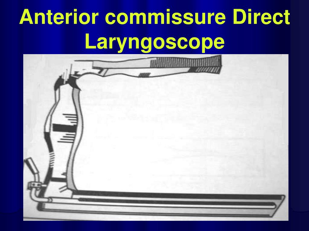
What type of laryngoscope is used for a microlaryngoscopy?
Microlaryngoscopy is performed with a rigid anterior commissure-type laryngoscope such as a Benjamin or Parsons placed in the vallecula (Fig. 2) and used to expose the endolarynx and subglottic airway.
Why is a posterior commissure laryngoscope important?
A posterior commissure laryngoscope can be quite helpful in providing appropriate visualization and exposure, because the laryngoscope has a notch to secure the ETT in anterior position. As in the rest of the larynx, angled telescopes can also be quite beneficial in helping to determine the extent of the RRP lesions.
What is the gold standard for laryngoscopy?
Operative direct microlaryngoscopy remains the gold standard for the assessment of the laryngeal epithelium and can be an invaluable tool in the assessment of certain voice disorders. Operative laryngoscopy allows for examination of the larynx through palpation of the laryngeal epithelium and the cricoarytenoid joint as well as offers the opportunity to perform diagnostic biopsy or phonomicrosurgery. Difficult laryngeal exposure may limit the utility of direct laryngoscopy in certain patients, especially in the population with head and neck cancer, although angled telescopes and curved or flexible instrumentation may help overcome these challenges.43
How severe is subglottic stenosis?
The severity of subglottic stenosis is graded using the Cotton-Myer grading system. A stenosis is grade 1 when 0% to 50% of the lumen is obstructed, grade 2 with 50% to 75% obstruction, grade 3 with 75% to 99% obstruction, and grade 4 when the airway lumen is completely obstructed. 13 Based upon the etiology, grade, and thickness of obstruction, various surgical techniques have been developed for the management of subglottic stenosis including laryngotracheal reconstruction with costal cartilage grafting, cricoid split, and cricotracheal resection. In neonates with severe stenosis, a tracheostomy may be required until definitive surgical management can be performed.
How to remove internal laryngocele cyst?
Internal laryngocele/saccular cysts should be removed by direct suspension microlaryngoscopy and CO2 laser.
How is rigid bronchoscopy performed?
Rigid bronchoscopy is performed using a combination of Hopkins rod telescopes and rigid bronchoscopes. The subglottis is evaluated initially. If subglottic stenosis is present, it is classified by the Cotton-Myer scale 1 and sized using appropriate endotracheal tubes ( Table 69-1 ). Additionally, the length of stenosis and the proximity to the vocal folds is assessed and documented. If a tracheotomy is in place, attention is paid to the evaluation of the suprastomal area, considering the possibility of suprastomal collapse, granuloma, intratracheal skin tract, and high tracheotomy. The trachea is evaluated to the level of the carina, looking for additional pathology, including tracheal stenosis, complete tracheal rings, tracheoesophageal fistula (TEF), TEF pouches, vascular compression, and tracheomalacia.
How many instruments are used in microlaryngeal surgery?
In general, microlaryngeal surgery is performed with two instruments at a time, one in each of the surgeon’s hands.
What is the vertical segment of a microlaryngeal probe?
The vertical segment on this probe is 4 millimeters in length and is shown here next to a penny for a sense of scale.
What are the distal ends of laryngoscopes used for?
The distal ends of several different laryngoscopes used in microlaryngeal surgery in adult patients. Note the various sizes and shapes, which are necessary to accommodate the variety of voice boxes (larynges) that exist across the anatomical spectrum.
What does the red arrow on the laryngoscope mean?
Red arrows indicate the breathing (endotracheal) tube, yellow arrows indicate the laryngoscope, and the green arrow shows the illuminated but unmagnified view of the vocal cords. An magnified view through the laryngoscope (see below) is necessary to provide maximal operative precision.
Why is the laryngoscope fixed?
It is typically then temporarily fixed into position in order to free the surgeon’s hands. The laryngoscope contains a light source, which illuminates the inner part of the laryngoscope, since room light is insufficient to view beyond the very outer portions of even the mouth.
What is a binocular microscope?
A binocular operating microscope provides high-powered magnification when working through the laryngoscope. Notice that it is crucial for the surgeon’s arms and wrists to be supported, in order to maximally stabilize the long instruments when performing laryngeal surgery.
What microscope do you use for vocal cord surgery?
At present, microlaryngoscopy (and, in particular, surgery on the vocal cords) is generally performed with either the use of: (1) a binocular operating microscope, or (2) a magnified, rigid telescope.
How to keep your temperature down after laryngeal surgery?
To help keep the temperature down, try to drink as much as possible . Hoarseness and weak voice are usual after laryngeal surgery. It is O.K. to talk quietly, unless the surgeon advises voice rest. No whispering, coughing, or throat clearing. Use a hard swallow or drink some water.
Can you smoke after a laryngoscopy?
Do not smoke or chew tobacco. Use this time to quit smoking permanently. After a laryngoscopy, it is not unusual to experience altered taste, numbness of the tongue, sore throat, jaw pain, or mild difficulty with swallowing. These symptoms usually go away in a couple of days.
