What is the lateral abdominal wall made up of?
The outermost muscular layer of the lateral abdominal wall is made up from the external oblique muscles. These two muscles arise from the outer parts of the lower 7 to 8 ribs on either side and extends anteroinferiorly to the anterior aspect of the iliac crests, pubic tubercle and to the linea alba in the midline.
What is the abdominal wall?
Abdominal wall. In medical vernacular, the term 'abdominal wall' most commonly refers to the layers composing the anterior abdominal wall which, in addition to the layers mentioned above, includes the three layers of muscle: the transversus abdominis (transverse abdominal muscle), the internal (obliquus internus) and the external oblique...
What are the layers of the anterolateral abdominal wall?
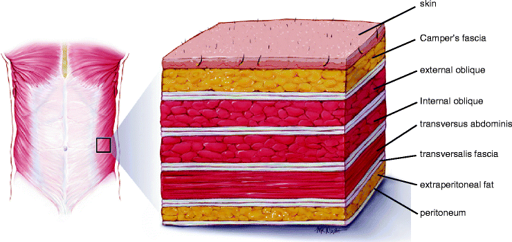
In human anatomy, the layers of the anterolateral abdominal wall are (from superficial to deep): Skin Subcutaneous tissue Fascia Camper's fascia - fatty superficial layer. Muscle External oblique abdominal muscle Transversalis fascia Extraperitoneal fat Peritoneum
How many muscles are there in the abdominal wall?
The following is a short description of these five muscles: The external oblique muscles are a pair of broad, thin, superficial muscles that lie on the lateral sides of the anterior abdominal wall. The location and structure of the external abdominal obliques gives them many different possible actions.
What does the abdominal wall consist of?
There are nine layers to the abdominal wall: skin, subcutaneous tissue, superficial fascia, external oblique muscle, internal oblique muscle, transversus abdominis muscle, transversalis fascia, preperitoneal adipose and areolar tissue, and peritoneum. Nerves, blood vessels, and lymphatics are present throughout.
Is the abdominal wall a muscle?
Your abdominal muscles are a set of strong bands of muscles lining the walls of your abdomen (trunk of your body). They're located toward the front of your body, between your ribs and your pelvis.
What are the 3 layers of the abdominal wall?
In medical vernacular, the term 'abdominal wall' most commonly refers to the layers composing the anterior abdominal wall which, in addition to the layers mentioned above, includes the three layers of muscle: the transversus abdominis (transverse abdominal muscle), the internal (obliquus internus) and the external ...
What is the abdominal wall lined with?
PeritoneumYour peritoneum is a membrane that lines the inside of your abdomen and pelvis (parietal layer). It also covers many of your organs inside (visceral layer). The space in between these layers is called your peritoneal cavity.
How thick is your abdominal wall?
Rectus abdominis muscle thicknesses were 9.58 mm (right) and 9.73 mm (left) at the xiphoid level and 10.26 mm (right) and 10.26 mm (left) at the umbilicus level. Subcutaneous fat thicknesses were 24.31 mm (right) and 23.39 mm (left). Rectus abdominismuscle thickness decreased with age and pregnancy.
How many layers are cut during C section?
Once the baby is delivered the uterus is closed with a double layer of stitching. Four of the five remaining layers are stitched with a single layer of stitching, but one layer is not restitched as it heals better – with no buckling and reduced chance of scar tissue developing, without restitiching.
What is the deepest muscle of the abdominal wall?
transversus abdoministransversus abdominis – the deepest muscle layer. Its main roles are to stabilise the trunk and maintain internal abdominal pressure. rectus abdominis – slung between the ribs and the pubic bone at the front of the pelvis.
How can I strengthen my abdominal wall?
Abdominal crunches are a classic core-strength exercise:Lie on your back and place your feet on a wall so that your knees and hips are bent at 90-degree angles. Tighten your abdominal muscles.Raise your head and shoulders off the floor. ... Return to the start position and repeat.
What are the four layers of the abdominal wall?
Classically the anterolateral abdominal wall has been described as separate layers from superficial to deep as follows:Skin.Subcutaneous tissues (further divided into the more superficial Camper's fascia and the deeper Scarpa's fascia)External oblique muscle.Internal oblique muscle.Transversus abdominis muscle.More items...•
What are the 7 layers of the abdominal wall?
Though its major part is muscular, the abdominal wall consists of at least seven layers: the skin, subcutaneous fat, deep fascia; abdominal muscles, transversalis fascia, extraperitoneal fat, and the parietal peritoneum....Lateral abdominal muscles:External oblique.Internal oblique.Transversus abdominis.
What is the abdominal muscle?
abdominal muscle, any of the muscles of the anterolateral walls of the abdominal cavity, composed of three flat muscular sheets, from without inward: external oblique, internal oblique, and transverse abdominis, supplemented in front on each side of the midline by rectus abdominis.
How can I strengthen my abdominal wall?
Abdominal crunches are a classic core-strength exercise:Lie on your back and place your feet on a wall so that your knees and hips are bent at 90-degree angles. Tighten your abdominal muscles.Raise your head and shoulders off the floor. ... Return to the start position and repeat.
What is abdominal wall pain?
Chronic abdominal wall pain (CAWP) refers to the pain originating from the abdominal wall which is often misdiagnosed as arising from a source inside the abdominal cavity, often resulting in inappropriate diagnostic investigations, unsatisfactory treatment, and considerable costs.
What is the function of the abdominal wall?
The major functions of the abdominal wall include: Providing a durable and flexible covering to prevent the abdominal viscera from leaving the abdominal cavity. Protecting internal abdominal organs from trauma/injury. Maintaining the anatomical position of the abdominal organs.
What is the abdominal wall?
In anatomy, the abdominal wall represents the boundaries of the abdominal cavity. The abdominal wall is split into the anterolateral and posterior walls.
What are the three layers of the abdominal wall?
In medical vernacular, the term 'abdominal wall' most commonly refers to the layers composing the anterior abdominal wall which, in addition to the layers mentioned above, includes the three layers of muscle: the transversus abdominis (transverse abdominal muscle), the internal (obliquus internus) and the external oblique (obliquus externus).
What are the layers of the abdominal wall?
It is composed of several layers, including skin, superficial fascia, subcutaneous fat, anterolateral and midline muscle groups, transversalis fascia, extraperitoneal fat and peritoneum. The anatomy is well demonstrated on CT and MRI. Ultrasound is useful in evaluating focal masses in the anterior abdominal wall but does not demonstrate ...
What is the rectus abdominis?
The rectus abdominis (commonly called the “sit-up” muscles) are paired paramedian strap muscles that extend from the ventral lower thorax to the pubis, separated from each other by the linea alba. These muscles are narrow and thick inferiorly, becoming broader and thinner superiorly.
Where do the posterior fibres of the iliac crest and pubic tubercle insert?
Insertion: iliac crest and pubic tubercle: The posterior fibres, from the lower ribs, pass vertically to insert into the anterior half of the liac crest. The middle and upper fibres pass anteriorly and forwards to end in the muscle’s aponeurosis.
Which layer of the thigh is thick and contains areolar tissue and variable amounts of fat?
The superficial layer is thick and contains areolar tissue and variable amounts of fat. It passes over the inguinal ligament to fuse with the underlying fascia of the thigh.
Which is thinner, the internal oblique or the external oblique?
The internal oblique lies internal to the external oblique and is thinner and less bulky than the external oblique
Which tendinous sheet medially contributes to the rectus sheath and linea alba?
The aponeurosis forms a strong tendinous sheet which medially contributes to the rectus sheath and linea alba.
What is the purpose of the abdominal wall?
The wall that encases the cavity of the abdomen has a number of things it is responsible for, including: 1 Keeping the abdominal viscera from being injured 2 Creating the wall that will keep the abdominal viscera within the cavity of the abdomen 3 Helping to keep the abdominal viscera in the proper position against gravity 4 Increasing intra-abdominal pressure that is used in actions such as vomiting or coughing 5 Working in a forceful manner to help push the abdominal viscera in an upward position
Which layer of the abdominal wall protects us from microbes and the elements?
There are superficial nerves and vessels that go between these two layers. 5. Skin. Skin is the outermost layer of the abdominal wall. It protects us from microbes and the elements, helps regulate body temperature, and permits the sensations of touch, heat, and cold.
What nerves run between the muscles of the abdominal wall?
In the layers of abdominal wall, there are a large number of nerves that go between the skin and muscles of the abdomen. They include: Thoracoabdominal nerves – There are five pair of these nerves and they run between the muscles of the abdominal wall to the muscles of the anterolateral wall of the abdomen. The cutaneous, lateral, and anterior ...
What are the muscles in the abdomen?
Muscles. There are two groups of five muscles that are located in the wall of the abdomen. The groups consist of vertical muscles and flat muscles. The Flat Muscles. These muscles laterally flex and rotate the trunk.
What is the layer above the umbilicus?
If it is above the umbilicus, it is made up of a single sheet of tissue; if it is below the umbilicus, it has two layers – the superficial layer that is fatty and the deep layer which has a lot of membranes. There are superficial nerves and vessels that go between these two layers. 5. Skin.
Which peritoneum is used for nerves?
While the visceral peritoneum shares the blood that is used by the nerves and lymphatic vessels of the organs of the abdomen, the parietal peritoneum utilizes the nerve supply and circulation from the layers of abdomen wall. 2.
Which peritoneum covers the organs?
The visceral peritoneum actually covers the organs and the parietal peritoneum surrounds the wall of the abdomen and includes the organs. The peritoneal cavity can be found in between the two layers with a fine layer of fluid that keeps the peritoneal surfaces lubricated.
What are the layers of the abdominal wall?
Layers of the abdominal wall as seen on high frequency ultrasound. Fat (F); external oblique muscle (EO); internal oblique muscle (IO); transversus abdominis muscle (TA); rectus abdominis (RA); peritoneum (arrow); aponeurosis of external oblique (white arrowhead) and anterior part of the aponeurosis of internal oblique contributing to the rectus sheath (black arrowhead).
What is the most important component of the anterior abdominal wall?
At its most inferior aspect the aponeurosis of external oblique folds back on itself to form the inguinal ligament and inguinal canal. This is an important component of the anterior abdominal wall since it contains two potential openings for the development of hernias. The first opening is the deep inguinal ring traditionally described as lying immediately lateral to the inferior epigastric artery, though in several studies (1,2,3) this has been shown to be variable. The second opening is the superficial inguinal ring located just superolateral to the pubic tubercle.
What is the posterior margin of the rectus abdominis?
The anterior margin of the sheath is formed by the external oblique aponeurosis and the anterior part of the internal oblique aponeurosis. The posterior margin is formed from the posterior part of the internal oblique aponeurosis and the transversus abdominis aponeurosis. The posterior layer ends at the arcuate line or linea semicircularis equidistant from the pubic symphysis and umbilicus. Additionally, the sheath ends superiorly at the superior insertions of the contributing muscles. The linea semilunaris indicates the most lateral extent of the rectus abdominis.
What are the three flat muscles in the abdominal wall?
MR anatomy of the abdominal wall demonstrating the three flat muscles (short arrow); the linea semilunaris (open arrow); rectus abdominis (black arrowhead); the linea alba (open arrowhead); the epigastric vessels (long arrow); the quadratus lumborum muscle (black arrow) and the erector spinae (white arrowhead).
What is the transversus abdominis plane?
Transversus abdominis muscle (TA); the transversus abdominis plane (arrowhead); internal oblique (IO); and external oblique (EO).The transversus abdominis plane is important in the anaethetisation of the abdominal wall post operatively since this is the plane through which the the intercostal and subcostal nerves pass.
Which muscle splits to cover the rectus abdominis anteriorly and posteriorly?
Axial CT image demonstrating the internal oblique muscle (arrow) and its aponeurosis (arrowhead) which splits to cover rectus abdominis anteriorly and posteriorly.
Which muscle is the outermost muscle of the lateral abdominal wall?
External oblique muscle. The outermost muscular layer of the lateral abdominal wall is made up from the external oblique muscle s. These two muscles arise from the outer parts of the lower 7 to 8 ribs on either side and extends anteroinferiorly to the anterior aspect of the iliac crests, pubic tubercle and to the linea alba in the midline.
What is the abdominal wall?
The abdominal wall is subdivided into the anterior wall, the right and left lateral walls, and the posterior wall. These walls are musculoaponeurotic, meaning they are composed of muscles and fascial layers, except for the posterior wall which is also made up by the lumbar vertebral column. This musculoaponeurotic wall functions to enclose ...
What are the two parts of the abdominal wall?
The abdominal wall is subdivided into the anterior wall, the right and left lateral walls, and the posterior wall.
Why is the transversus abdominis important?
And similar to both oblique muscles, the transversus abdominis helps to compress the abdominal contents in order to increase intra-abdominal pressure , which is helpful during forced expiration, defecation and labour.
What is the function of the musculoaponeurotic wall?
This musculoaponeurotic wall functions to enclose and protect the abdominal viscera, stabilize and contribute to movements of the trunk, and also increase the intra-abdominal pressure which is needed during urination , defecation, vomiting, and assisting in childbirth.
What is the external oblique?
The external oblique is innervated by the thoracoabdominal nerves, which are derived from the anterior rami of the T7 to T11 spinal nerves, ...
Where is the rectus abdominis located?
The rectus abdominis is a set of vertically oriented paired muscles that lies right at the midline of the anterior abdominal wall and originates at the pubic symphysis and pubic crests and inserts at the xiphoid process and fifth through seventh costal cartilages.
Which abdominal muscles are flat?
The external oblique muscle, the internal oblique muscle, and the transversus abdominis muscle are considered the flat abdominal muscles, and the fibers of each have varying orientations. All three of these abdominal muscles continue anteriorly and medially as aponeuroses. Aponeuroses are simply flat sheets of fibrous tissue ...
What are some common types of abdominal wall hernias?
The umbilical cord was once attached to the umbilicus (a.k.a. the belly button). This creates an area of weakness, and hernias in this area are called umbilical hernias. There are also several types of groin hernias. Inguinal hernias occur in the groin where the inguinal canal is located, near the pubic bone.
How do I know if I have a hernia?
Hernias are frequently visible as a bulge in the abdominal wall. The hernia may or may not cause discomfort or pain at the site. Sometimes hernias can enlarge over time. Other hernias, which were not painful in the past, can suddenly become painful.
Can I prevent a hernia?
Maintaining a healthy body weight, avoiding heavy lifting and not straining to urinate or pass stool helps prevent a hernia from developing. You can also exercise regularly to strengthen your abdominal wall muscles. If you already have a hernia it will not go away, but it may improve with weight loss down to a healthy weight.
When should I see a doctor?
Small, non-painful hernias which are not enlarging can be mentioned to your primary care physician at your next wellness exam. Hernias which are mildly painful or enlarging should be evaluated by a surgeon to see if your hernia should be repaired. Hernias which are severely painful may be a sign that the abdominal contents in the hernia have twisted and are dying due to the obstructed the blood supply.
How is a hernia diagnosed?
Many hernias can be diagnosed based on talking to the patient and performing a physical exam. Sometimes CT (computed tomography) scans or ultrasound are used to visualize the hernia for diagnosis or surgical planning.
How is a hernia repaired?
The goal of hernia surgery is to remove any trapped abdominal contents from the hernia, to place those structures back into the abdomen and then to close the abdominal wall hernia opening. Each hernia needs to be individually evaluated to determine the best approach. Some small, non-painful hernias may be watched for the development of symptoms.
What can I expect after hernia surgery?
Postoperative pain at the hernia and incision sites should be tolerable with prescription pain medication. Also, bruising and swelling are commonly experienced after surgery. Most people with sedentary jobs can return to work in 7-10 days and patients will be instructed to avoid heavy lifting or strenuous activity for at least 6-8 weeks.
Where does the abdominal wall originate?
Anatomy of the Abdominal Wall. The external oblique muscle originates from the 5th to 12th ribs and has a medio-caudal direction. The internal oblique muscle originates from the iliac crest and follows a medio-proximal direction. The direction of the fibres of both muscles rarely deviates more than 30° from the horizontal.
Which muscle is enclosed by the rectus abdominis?
Its fibres are directed horizontally. The aponeuroses of these three muscles form the sturdy rectus sheaths, which enclose the fourth abdominal wall muscle, the rectus abdominis, which inserts on the 5th, 6th and 7th ribs superiorly and on the pubic bone inferiorly.
What is the posterior sheath?
The posterior sheath is formed by the posterior leaf of the internal and the transversus abdominis aponeuroses and bears the superior and inferior epigastric arteries and their anastomotic network.
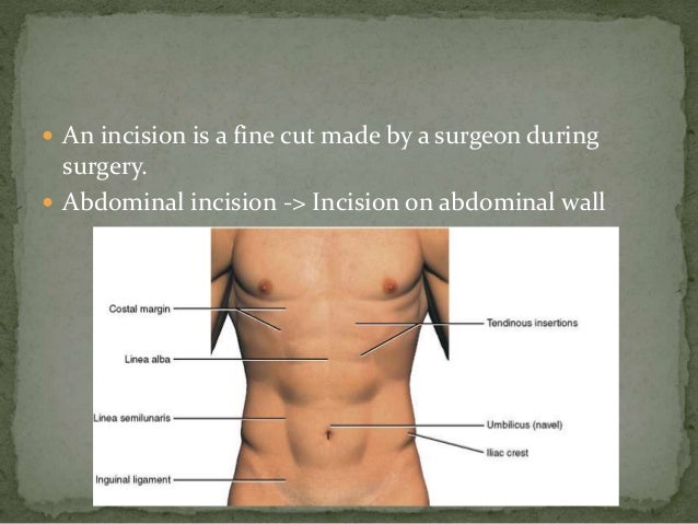
What is the choice of abdominal incision?
The choice of abdominal incision is mainly dependent on the area that needs to be exposed, the elective or emergency nature of the operation and the surgeon’s personal preference. However, type of abdominal incision may have a profound influence on the occurrence of postoperative wound complications.
Where is the anterior sheath located?
The recti are interrupted by three paired tendinous intersections anchoring them to the anterior sheath, broadly found close to the xiphisternum, at the level of the umbilicus and then halfway between the two.
Is there a posterior sheath above the costal margin?
2) There is no posterior sheath above the level of the costal margin, as the recti remain covered anteriorly by the external oblique aponeurosis and insert directly onto the underlying costal cartilages.
How many muscles are there in the abdomen?
There are five main muscles in your abdomen. Two are vertical (up and down) muscles located toward the middle of your body. Three are flat muscles stacked on top of each other, situated toward the sides of the trunk.
Where are the abdominal muscles located?
They’re located toward the front of your body, between your ribs and your pelvis.
What are the main muscles of the abdominal region?
There are five main muscles: pyramidalis, rectus abdominus, external obliques, internal obliques, and transversus abdominis. Ab strains and hernias are common, but several strategies can keep your abs safe and healthy.
What are the external obliques?
External obliques: The external obliques are a pair of muscles, one on each side of the rectus abdominis. They are the largest of the flat muscles and at the bottom of the stack. They run from the sides of your body toward the middle. The external obliques allow the trunk to twist side to side.
Which abdominal muscles are deepest?
Transversus abdominis: The transversus abdominis is at the bottom of the stack. This pair of muscles is the deepest of the flat muscles. They stabilize the trunk and help maintain internal abdominal pressure.
Where are the internal obliques located?
Internal obliques: The internal obliques are a pair of muscles on top of the external obliques, just inside your hip bones. Like the external obliques, they are on the sides of the rectus abdominis, running from the sides of your trunk toward the middle. They work with the external oblique muscles to allow the trunk to twist and turn.
What muscle is located in the lower abdomen?
Pyramidalis: This vertical muscle is small and shaped like a triangle. It’s located very low, in your pelvis. It helps maintain internal pressure in your abdomen.