Full Answer
How to pronounce Corti?
Pronunciation of Robert Corti with 1 audio pronunciations 0 rating rating ratings Record the pronunciation of this word in your own voice and play it to listen to how you have pronounced it.
What does Corti mean?
What does capelli corti mean in Italian? capelli corti. English Translation. short hair. More meanings for capelli corti. short-haired adjective. capelli corti, dai capelli corti. bobbed hair.
What does Corti mean in Italian?
Wikipedia (0.00 / 0 votes)Rate this definition: Cortino (Abruzzese: Curtènë) is a small town and comune in the central Italian region of Abruzzo in the Province of Teramo. It is located in the Gran Sasso e Monti della Laga National Park. The major part of the population lives in small hamlets dotting the comune.
What is the function of the organ of Corti?
- (1) Sound waves, transmitted by the perilymph, make the basilar membrane vibrate up and down. ...
- (2) Stereocilia of the OHCs, embedded to the tectorial membrane, bend when the basilar membrane rises, causing the OHCs to depolarise (by the influx of K+ ions).
- (3) Excited (depolarised) OHCs react by contracting (= electromotility). ...
What is the function of Corti?
The primary function of the organ of Corti is the transduction of auditory signals. Sound waves enter the ear via the auditory canal and cause vibration of the tympanic membrane.
What does Corti mean?
noun phrase. : a complex epithelial structure in the cochlea that contains thousands of hair cells, rests on the internal surface of the basilar membrane, and in mammals is the chief part of the ear by which sound waves are perceived and converted into nerve impulses to be transmitted to the brain.
What is the organ of Corti and what is it function?
The organ of Corti is a specialized sensory epithelium that allows for the transduction of sound vibrations into neural signals. The organ of Corti itself is located on the basilar membrane.
Why is it called organ of Corti?
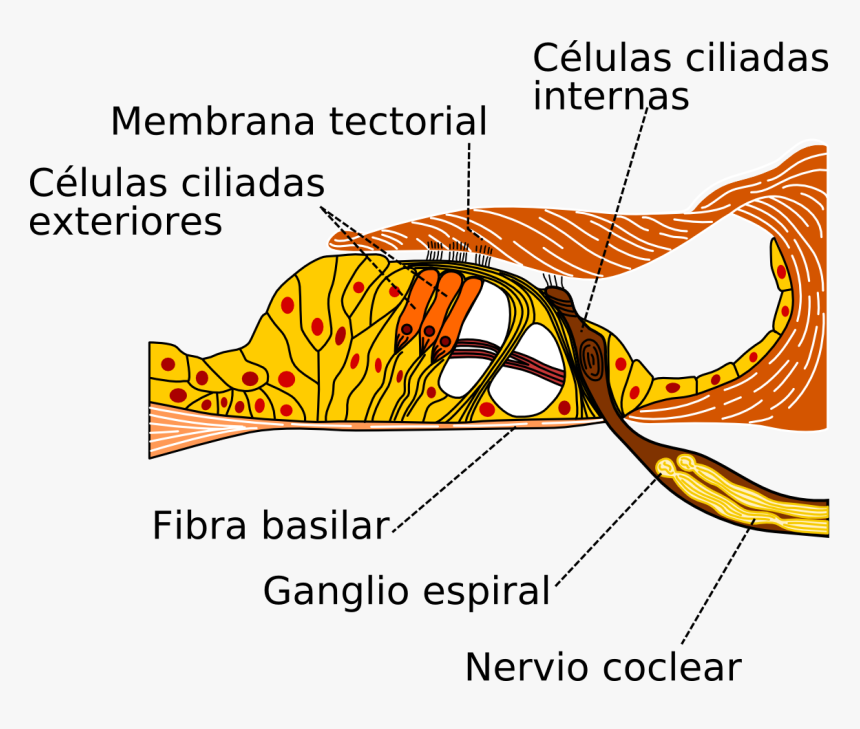
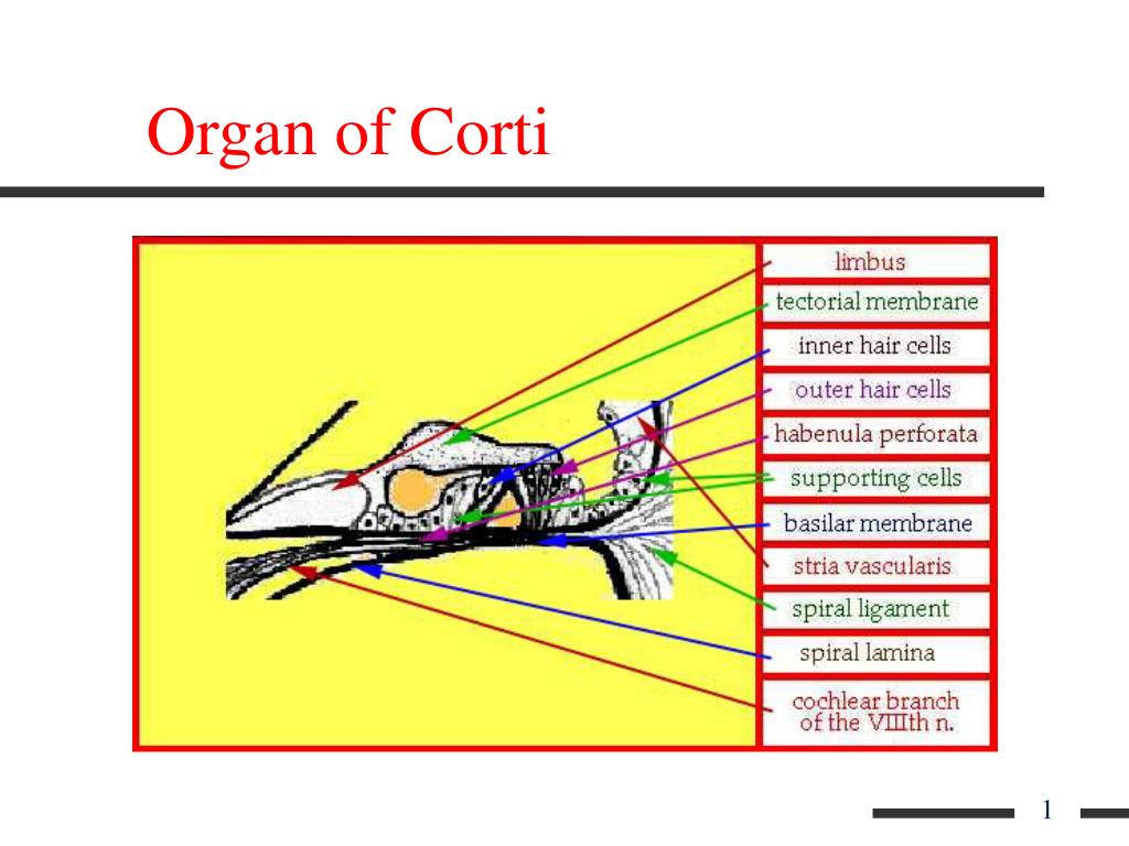
The organ of Corti is named after Italian anatomist Alfonso Corti, who first described it in 1851. Viewed in cross section, the most striking feature of the organ of Corti is the arch, or tunnel, of Corti, formed by two rows of pillar cells, or rods. The pillar cells furnish the major support of this structure.
What is the organ of Corti in psychology?
the organ of Corti is the receptor organ for hearing. It rests on the surface of the basilar membrane in the cochlea and contains hair cells, which transduce vibrations caused by sound waves into electrical impulses that can be interpreted by the brain.
What is the organ of Corti quizlet?
Organ of Corti: The true organ of hearing, a spiral structure within the cochlea containing hair cells that are stimulated by sound vibrations.
What part of the brain interprets sound?
The auditory brain receives the signals sent by the cochlea, interprets them and starts either reflexes or conscious perception, depending upon motivation and alertness. It is also responsible for the creation of memories, which is vital for future percepts!
What is your inner ear called?
inner ear, also called labyrinth of the ear, part of the ear that contains organs of the senses of hearing and equilibrium. The bony labyrinth, a cavity in the temporal bone, is divided into three sections: the vestibule, the semicircular canals, and the cochlea.
What is the organ of hearing?
The inner ear consists of a spiral shaped structure known as the cochlea (means snail-shell). Within the cochlea sits the organ of hearing where we have thousands of tiny cells, known as hair cells. The hair cells are stimulated and send messages to the auditory nerve.
What is the tunnel of Corti?
anatomy of the inner ear Corti is the arch, or tunnel, of Corti, formed by two rows of pillar cells, or rods. The pillar cells furnish the major support of this structure. They separate a single row of larger, pear-shaped inner hair cells from three or more rows of smaller, cylindrical outer hair cells.
How do we hear?
The Inner Ear As the fluid moves, 25,000 nerve endings are set into motion. These nerve endings transform the vibrations into electrical impulses that then travel along the eighth cranial nerve (auditory nerve) to the brain. The brain then interprets these signals, and this is how we hear.
What Chamber is the organ of Corti in?
The cochleaThe cochlea includes three chambers, and the organ of Corti is located in the scala media. A single row of inner hair cells is located on the medial side of the epithelium, whereas three rows of outer hair cells are located more laterally.
Which organ is larger, the basilar membrane or the corti?
The organ of Corti is larger and the basilar membrane on which it sits is longer as it gets further away from the base of the cochlea. This difference in size is consistent with the fact that different frequencies of sound result in greater vibrations of the organ of Corti depending on where along the length of the cochlea you are measuring. ...
Which organ is spiraling towards the top of the cochlea?
The organ of Corti is spiraling towards the top of the cochlea to the right. Stereocilia are on the top and radial fibers of the basilar membrane are seen on the bottom. The outer hair cells sit in a cup formed by a supporting cell. The supporting cells send out a narrow filament that angles towards the base of the cochlea.
What is the eighth nerve?
The nerve is made up of the neuronal projections that connect the hair cells with the brain and is called the eighth nerve because it is one of 12 nerves that come off the brain in the skull. The spiral shaped cochlea originates from one of the balance organs and contains the sensory epithelium for hearing. Heading.
What is the hearing organ called?
The Cochlea. The hearing organ in mammals is a spiraling structure called the "cochlea" from the Greek word for snail. It spirals out from the saccule (one of the balance organs). There are two and a half turns in the human cochlea and if you were to unwind the cochlea it would stretch to nearly an inch in length.
Where is the central axis of the spiraling cochlea?
The central axis of the spiraling cochlea is to the left of the drawing. Eighth nerve fibers pass through a bony shelf on their way to the hair cells (orange). The organ of Corti is made up of hair cells and supporting cells (purple and blue, respectively) that sit on a flexible basilar membrane which is anchored to the bony shelf on ...
Which cells are surrounded by supporting cells?
Neurons in the brain are surrounded by supporting cells. The cells in the muscles of the heart are close to one another. We now know that the spaces around the outer hair cell allow the cells to change their length during hearing. Figure 6 is a view of a portion of the third row of outer hair cells.
Why are the stereocilia bent?
The overlying tectorial membrane is not as flexible so the stereocilia are bent as the organ of Corti moves up and down against it. The electrical potential inside the hair cells changes as the stereocilia are bent. Heading.
What is the organ of the corti?
FMA. 75715. Anatomical terminology. The organ of Corti, or spiral organ, is the receptor organ for hearing and is located in the mammalian cochlea. This highly varied strip of epithelial cells allows for transduction of auditory signals into nerve impulses' action potential. Transduction occurs through vibrations of structures in ...
What is the function of the organ of Corti?
The function of the organ of Corti is to convert ( transduce) sounds into electrical signals that can be transmitted to ...
How does sound affect the corti?
Sound waves enter through the auditory canal and vibrate the tympanic membrane, also known as the eardrum, which vibrates three small bones called the ossicles . As a result, the attached oval window moves and causes movement of the round window, which leads to displacement of the cochlear fluid. However, the stimulation can happen also via direct vibration of the cochlea from the skull. The latter is referred to as Bone Conduction (or BC) hearing, as complementary to the first one described, which is instead called Air Conduction (or AC) hearing. Both AC and BC stimulate the basilar membrane in the same way (Békésy, G.v., Experiments in Hearing. 1960).
What happens if you have mutations in the genes expressed in or near the organ of Corti before the differentiation
Mutations in the genes expressed in or near the organ of Corti before the differentiation of hair cells will result in a disruption in the differentiation, and potential malfunction of , the organ of Corti.
How long is the cochlea?
If the cochlea were uncoiled, it would roll out to be about 33 mm long in women and 34 mm in men, with about 2.28 mm of standard deviation for the population . The cochlea is also tonotopically organized, meaning that different frequencies of sound waves interact with different locations on the structure.
Which part of the ear is the most stiff and narrow?
The base of the cochlea, closest to the outer ear, is the most stiff and narrow and is where the high-frequency sounds are transduced. The apex, or top, of the cochlea is wider and much more flexible and loose and functions as the transduction site for low-frequency sounds.
Which organ modulates the auditory signal?
The organ of Corti is also capable of modulating the auditory signal. The outer hair cells (OHCs) can amplify the signal through a process called electromotility where they increase movement of the basilar and tectorial membranes and therefore increase deflection of stereocilia in the IHCs.
What is the organ of corti?
The organ of Corti (Fig. 24.2C), the sensory epithelium resting upon the basilar membrane (for a review see Slepecky, 1996 ), senses mechanical vibration of incoming sound and converts it to action potentials. It is made up of two types of sensory cells ( Fig. 24.2C–G ): outer hair cells (OHCs) and inner hair cells (IHCs) and several types of supporting cells ( Fig. 24.2D ). The OHCs are arranged in three parallel rows ( Fig. 24.2D, F ), whereas the IHCs are in a single row ( Fig. 24.2D, F, G). At their apical end, the hair cells are provided with stereocilia in a typical ‘W’ pattern. The organ of Corti is overlain by the gel-like tectorial membrane, which is indirectly connected to the osseous spiral lamina through the spiral limbus. Only the stereocilia of the OHCs appear to be in contact with the tectorial membrane. Shearing movements between the basilar membrane with the sensory epithelium and the tectorial membrane cause receptor potentials to be produced in the hair cells, by means of deflections of their stereocilia (reviewed in Nobili et al., 1998 ). Sensory transduction in the cochlea has been studied for many years, and over the last 30 years it has been recognized that the IHCs ( Fig. 24.2G) act as the primary receptor cell (Markin and Hudspeth, 1995; Russell, 1983 ), whereas the OHCs act as motor cells that can convert membrane potential into a mechanical force (reviewed in Nobili et al., 1998 ).
Where does the organ of the corti develop?
The mammalian organ of Corti develops within the dorsal wall of the elongating cochlear duct, a structure composed of polarized, pseudostratified epithelial cells that first emerges from the ventromedial pole of the otocyst at ~ E12 in mice and attains its characteristic coiled configuration, albeit not full length, by E15 ( Morsli, Choo, Ryan, Johnson, & Wu, 1998 ). Two regions can be distinguished within the dorsal wall of the cochlear duct from which the organ of Corti develops, the greater and the lesser epithelial ridges (GER and LER, respectively, see Fig. 6 E14.5), regions which differ initially in their thickness. The IHCs with their associated supporting cells and those lying more medial (i.e., the cells of the spiral sulcus and spiral limbus) originate from the GER, while the outer hairs, Dieters’ cells, Hensen's cells, and the cells of Claudius and Boettcher originate from the LER.
How are OHCs and IHCs arranged?
The OHCs are arranged in three parallel rows ( Fig. 24.2D, F ), whereas the IHCs are in a single row ( Fig. 24.2D, F, G ). At their apical end, the hair cells are provided with stereocilia in a typical ‘W’ pattern.
What organ contains sensory hair cells?
The organ of Corti of the mammalian inner ear contains sensory hair cells and supporting cells in the auditory sensory epithelia. These cells are arranged to form a checkerboard-like cellular pattern. However, cellular and molecular mechanisms that produce this characteristic arrangement of cells had remained unknown for a long time. In the mouse organ of Corti, hair cells express nectin-1 and supporting cells express nectin-3; in addition, both cells express nectin-2 (Fig. 5 ). The trans -interaction between nectin-1 and -3 mediates the heterotypic adhesion between these two types of cells and establishes the checkerboard-like cellular pattern; this pattern is lost due to aberrant attachment between sensory hair cells in both nectin-1-deficient mice and nectin-3-deficient mice ( Togashi et al., 2011 ). Moreover, in the nectin-3-deficient mice, abnormal positioning of the kinocilium and misorientation and dysmorphology of the hair bundles are observed in aberrantly attached sensory hair cells ( Fukuda et al., 2014 ). Thus, the trans -interaction between nectin-1 and -3 is critical not only for the checkerboard-like cellular pattern formation but also for positioning of the kinocilium and the morphology and orientation of stereociliary bundle in hair cells.
Where is the cochlea located?
The cochlea includes three chambers, and the organ of Corti is located in the scala media. A single row of inner hair cells is located on the medial side of the epithelium, whereas three rows of outer hair cells are located more laterally.
What organs have lost their compact structure?
The mammalian organ of Corti has lost the compact structure that is so obviously a feature of all the other auditory and all vestibular receptor organs (Figs. 17 and 18). There are large extracellular spaces around the outer hair cells as well as between the tunnel rods in the organ of Corti, and without some structural provision for support, the organ would almost certainly collapse. The supporting cells have become specialized to provide the necessary stiffness by the presence of the “tonofilaments” which are arranged in bundles within the cytoplasm. These filaments were observed by early histologists; recent ultrastructural studies have confirmed their presence. One of the supporting elements, the tunnel rod, is a new phylogenetic development; there is no comparable structure in the pigeon or in most reptiles. Larsell, McCrady, and Larsell (1944) studied the embryonic development of the tunnel rods in the opossum pouch young. The 48-day-old pouch animal shows a transition from nondifferentiated epithelium in the apex to well-developed tunnel rods in the lower turns. The first evidence for differentiation is a slit between two tall columnar cells. This is the beginning of the tunnel opening which apparently becomes gradually widened at later stages. Tonofilaments are found in well-differentiated tunnel rods in the region of the upper first and lower second coils but not above and not below this region at this stage of development.
Overview
The organ of Corti, or spiral organ, is the receptor organ for hearing and is located in the mammalian cochlea. This highly varied strip of epithelial cells allows for transduction of auditory signals into nerve impulses' action potential. Transduction occurs through vibrations of structures in the inner ear causing displacement of cochlear fluid and movement of hair cells at the organ of Corti to produce electrochemical signals.
Structure
The organ of Corti is located in the scala media of the cochlea of the inner ear between the vestibular duct and the tympanic duct and is composed of mechanosensory cells, known as hair cells. Strategically positioned on the basilar membrane of the organ of Corti are three rows of outer hair cells (OHCs) and one row of inner hair cells (IHCs). Surrounding these hair cells are supporti…
Function
The function of the organ of Corti is to convert (transduce) sounds into electrical signals that can be transmitted to the brainstem through the auditory nerve. It is the auricle and middle ear that act as mechanical transformers and amplifiers so that the sound waves end up with amplitudes 22 times greater than when they entered the ear.
Development
The organ of Corti, in between the scala tympani and the scala media, develops after the formation and growth of the cochlear duct. The inner and outer hair cells then differentiate into their appropriate positions and are followed by the organization of the supporting cells. The topology of the supporting cells lends itself to the actual mechanical properties that are needed for the highly specialized sound-induced movements within the organ of Corti.
Clinical significance
The organ of Corti can be damaged by excessive sound levels, leading to noise-induced impairment.
The most common kind of hearing impairment, sensorineural hearing loss, includes as one major cause the reduction of function in the organ of Corti. Specifically, the active amplification function of the outer hair cells is very sensitive to damage from exposure to trauma from overly-loud soun…
Additional images
• Transverse section of the cochlear duct of a fetal cat.
• Diagrammatic longitudinal section of the cochlea
• Floor of ductus cochlearis
• Limbus laminæ spiralis and membrana basilaris
External links
• Dissecting the molecular basis of organ of Corti development PMC 3097286
• Organ of Corti 3D animation
• http://lobe.ibme.utoronto.ca/presentations/OHC_Electromotility/sld005.htm Diagram at University of Toronto