
However, there’s a difference between the two:
- An ultrasound is a tool used to take a picture.
- A sonogram is the picture that the ultrasound generates.
- Sonography is the use of an ultrasound tool for diagnostic purposes.
Is an ultrasound and a sonogram the same thing?
There is a slight difference in the terms but, simply, one is the process and the other is the product. An ultrasound is a procedure that uses sound waves to create images of organs and soft tissues. The sonogram is the actual image that is created by the ultrasound. So the difference is a matter of semantics.
Are sonograms and ultrasounds the same thing?
The term “ultrasound” refers to the procedure of using sound waves to create an image of an area inside the body, while a sonogram is the actual image that is produced by the ultrasound. Just as a camera captures a photo, an ultrasound exam produces a sonogram image. Basically, the former results in the latter.
Is sonography and ultrasound the same thing?
Sonography is the use of an ultrasound tool for diagnostic purposes. In short, an ultrasound is the process, while a sonogram is the end result. Sonography is a noninvasive, painless procedure.
What does a sonogram technician do?
- Important Facts About Sonogram Technicians
- Duties and Responsibilities. A sonogram technician works very closely with patients, performing medical imaging procedures on various parts of the body.
- Skills Required. ...
- Salary Info and Job Outlook. ...
Is an ultrasound and a sonogram the same thing?
Although the terms ultrasound and sonogram are technically different, they are used interchangeably and reference the same exam.
What is a sonogram used for?
Diagnostic ultrasound, also called sonography or diagnostic medical sonography, is an imaging method that uses sound waves to produce images of structures within your body. The images can provide valuable information for diagnosing and directing treatment for a variety of diseases and conditions.
What a sonogram can detect?
Ultrasound can detect cysts, tumors, obstructions or infections within or around your kidneys. Breast ultrasound: A breast ultrasound is a noninvasive test to identify breast lumps and cysts. Your provider may recommend an ultrasound after an abnormal mammogram.
Which is better ultrasound or sonography?
An ultrasound is the use of sound waves to create an image, and a sonogram is the image created by an ultrasound. Because both require professionals who learn how to perform ultrasounds, they are essentially the same career.
What abnormalities can be detected on an ultrasound?
What Kinds of Abnormalities can an Ultrasound Detect?The Nuchal translucency scan, typically at 12 to 14 weeks, is used to detect Down's syndrome, Edwards' syndrome, and Patau's syndrome.The general abnormality scan at 20 to 22 weeks detects problems such as spina bifida, ancencephaly, and heart abnormalities.More items...•
Can you see a tumor on an ultrasound?
An ultrasound (also known as ultrasonography, sonography, or sonogram) helps doctors look for tumors in certain areas of the body that don't show up well on x-rays. Doctors often use this procedure to guide a needle during a biopsy. Ultrasounds are usually quick and most don't require special preparation.
How accurate is a sonogram?
In 105 (43%), the accuracy of ultrasonic diagnosis was evaluated surgically. Sonography was proven correct in 60 (57%) patients who had undergone operation.
Can you see a cyst on an ultrasound?
Ultrasound imaging can help determine the composition of lump, distinguishing between a cyst and a tumour.
What are ultrasounds used for besides pregnancy?
In addition to pregnancy, ultrasound can be used to detect a wide range of digestive problems, including:Cysts.Gallstones.Abnormal enlargement of the spleen.Abnormal growths in the liver or pancreas.Liver cancer.Fatty liver disease.
Is a sonogram painful?
A sonohysterogram is an imaging procedure that allows your healthcare provider to see inside your uterus. It's simple, safe and relatively painless. And it can help shed light on what's causing issues like pelvic pain, irregular bleeding and infertility.
Can you eat or drink before a sonogram?
You may not eat or drink anything for 8 to 10 hours before the test. If you eat, the gallbladder and ducts will empty to help digest food and will not be easily seen during the test. If your test is scheduled in the morning, we suggest that you eat nothing after midnight the night before the test is scheduled.
When do you get your first sonogram?
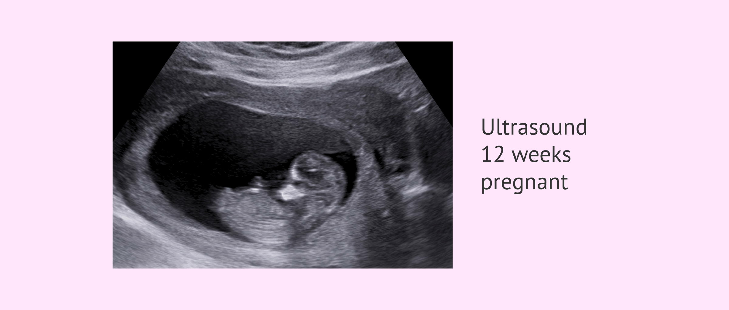
Your first ultrasound is called the “dating” or “viability” ultrasound. It's typically done between 7 and 8 weeks to verify your due date, to look for a fetal heartbeat, and to measure the length of the baby from “crown to rump.” At this ultrasound, you'll also learn whether you're having one baby, twins, or more!
Are Ultrasound and Sonogram the Same Thing?
Although “ultrasound” and “sonogram” are both used to refer to the same exam, they have different meanings. We’ll explore the differences and simil...
What Is a Sonogram?
A sonogram is an image produced by an ultrasound. In other words, it’s not the procedure itself but the product.
How Does a Sonogram Work?
A sonogram is produced by sound waves. During an ultrasound, the technologist will send sound waves to the part of the body under examination using...
What Is a Sonogram Used For?
As we’ll discuss more below, sonograms help doctors evaluate organs for infections, damage or disease. Pregnant women may get ultrasounds to genera...
What Is an Ultrasound?
A medical ultrasound is a type of imaging test. Unlike many other imaging tests, ultrasounds do not use radiation to produce images of the internal...
How Does an Ultrasound Work?
In general, an ultrasound machine measures sound waves that have bounced off the body’s internal tissues. The ultrasound’s computer processes these...
What Is an Ultrasound Used For?
An ultrasound has many uses in the medical world. Doctors may order an ultrasound to check for swollen, damaged or infected organs if a patient is...
What’s the Difference Between a Sonogram and an Ultrasound?
The easiest way to remember the difference between a sonogram and an ultrasound is to keep in mind that the ultrasound is the procedure, and the so...
How Are They Similar?
The sonogram and the ultrasound machine are both components of a highly useful medical exam. As a whole, an ultrasound and the sonogram it produces...
What is sonogram ultrasound?
See below for an in-depth explanation of the sonogram definition. When an Ultrasound is used, high-frequency sound waves are emitted from a transducer probe. This is then used to create images. The waves from the frequency bypass the outer skin and interact with a human’s inner structure before pinging back to the probe.
What is pelvic ultrasound?
A pelvic ultrasound is a noninvasive diagnostic exam that produces images that are used to assess organs and structures within the female pelvis. A pelvic ultrasound allows quick visualization of the female pelvic organs and structures. Pelvic examinations consist of ultrasound of the uterus, cervix, vagina, fallopian tubes, and ovaries.
How long does it take for ultrasound to work?
In most cases, the procedure doesn’t take longer than 30 minutes.
What is a sonogram?
Sonogram definition: a visual picture. Simply put, the image that has been produced from the scan. The live-feed provides the Ultrasound technician with a picture where they and the patient can preview the results. This can only be achieved after the CPU has processed the information from the scan.
Why do you need an ultrasound after a treatment?
Often, if you have a treatment done, an ultrasound would need to be taken after the treatment to ensure that everything is going along perfectly, and is working as expected.
What are the features of a kidney ultrasound?
When undergoing a kidney ultrasound, the results will be used to check the following: By checking all these necessary features, the scan can identify any tumors, cysts, abscesses, fluid collection, obstructions, or infections within or in close proximity to the kidneys.
Is there a difference between ultrasound and sonography?
It’s an alternative name for Ultrasound that is sometimes referred to as Sonography. Therefore highlighting there’s no difference between sonography vs ultrasound. This is not to be mistaken with a sonogram, which has an entirely different meaning. See below for an in-depth explanation of the sonogram definition.
How does a gel transducer work?
The gel helps remove air and connect the transducer to the skin. As a result, sound waves can travel directly to and from the transducer. Use of the transducer wand: The technologist will press the transducer against the gel-covered region of the patient’s skin.
How does a sonogram work?
During an ultrasound, the technologist will send sound waves to the part of the body under examination using a wand-shaped device called a transducer. The sound waves will bounce back once they contact the tissues being studied, travel through the transducer and enter the ultrasound computer.
Why do you need a gel before an ultrasound?
The gel is an important part of the ultrasound procedure because sound waves do not travel well through the air. The gel helps remove air and connect the transducer to the skin.
What is the function of a transducer?
The transducer sends high-frequency sound waves into the body. As the sound waves bounce off of organs, tissues and fluids, the transducer picks up subtle changes in the pitch and direction of the sound waves. This information is measured instantly and displayed on the computer.
What is an ultrasound machine?
In general, an ultrasound machine measures sound waves that have bounced off the body’s internal tissues. The ultrasound’s computer processes these sound waves and turns them into real-time images. If you need to get an ultrasound, you can expect the following steps:
What happens when sound waves hit a tissue?
When sound waves hit a tissue, they reflect back into the ultrasound machine and transform into a white or gray image, de pending on the intensity of the sound wave. Extremely dense tissues such as bones and kidney stones produce echoes easily and appear bright white on a sonogram.
What is the role of a radiologist in ultrasound?
A radiologist, who is a doctor trained to analyze imaging results, will interpret the sonogram. They will then share their findings with the doctor who requested the exam. An ultrasound procedure is quick and painless and requires little preparation.
Why do we use ultrasound for sonography?
Sonography is performed using an ultrasound tool. Sonogram helps in evaluating organs for infections, damage or disease and in case of pregnancy, to generate images of the foetus. Ultrasound helps the doctors to get sonograms and thus insights into the inner working of a human’s body for diagnostic purposes.
Why are sonograms important?
They’ve played a major role in providing basis for evaluation of internal organs of the body and determine and eventually cure the infections or diseases caused in the internal organs or soft tissues in the body.
How does a sonogram work?
A Sonogram is a visual image produced by a process of ultrasound and it uses the sound waves to generate the picture. The pictures are produced owing to the fact that the sound waves reflect and bounce back once they hit a surface. To go into detail, the tighter and harder the surface is, the more the sound waves bounce back.
What is the definition of sonogram?
Ultrasound. Definition. A Sonogram refers to a visual image produced, that is the end result of another process. Ultrasound is the procedure of using soundwaves to create images of the internal body. Result vs Process. A Sonogram is the end result of Ultrasound.
What is ultrasound in medical terms?
Ultrasound is a process that makes use of sound waves to generate sonograms. In other words, it is an imaging test in the field of medicine. They do not make use of harmful radiations, rather put into use high-frequency sound waves, which are neither harmful nor painful.
Why do sound waves turn black?
For instance, sound waves easily pass through fluids and thus they will present a completely black picture when the come in contact with urine, water or other liquids. Moreover, upon hitting a tissue, they are bound to portray a greyish or whitish picture, directly depending on the intensity of the sound waves.
What is ultrasound test?
An ultrasound, as is evident, is a process; it is a type of imaging test, whose end result is a sonogram. In simple words, Sonogram, also called sound writing, is the output produced by Ultrasound, which is also called diagnostic sonography. This technology is greatly useful in determining the infections or diseases in the internal parts ...
