Dorsal Visual Pathway. This information is integrated into a coherent spatial representation of the world. The dorsal pathway is also hypothesised to be an 'action' pathway, in that it computes spatial relations between the organism and the environment, which allows for the organism's effective interaction with the environment.
What is the dorsal pathway in the brain?
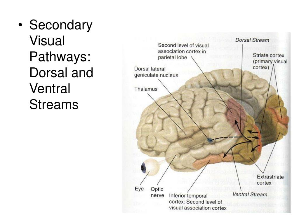
Dorsal visual pathway: this pathway extends from the primary visual cortex (V1) in the occipital lobe to the parietal lobe. The dorsal pathway is subdivided by the intraparietal sulcus (IPS) into several main sectors including the superior parietal lobule, inferior parietal lobule, and the supramarginal gyrus.
Are dorsal object representations dissociable from the dorsal pathway?
Taken together, the findings reviewed thus far indicate that dorsal object representations are dissociablefromthosegeneratedintheventralpathway,andplayanindependentandfunctional role in visual perception. In light of the above, we propose that visual perception should not be.
What is the dorsal column-medial lemniscus pathway?
In the spinal cord, this pathway travels in the dorsal column, and in the brainstem, it is transmitted through the medial lemniscus hence the name dorsal column-medial lemniscus pathway. [1]
What does the dorsal column do in the spinal cord?
Nerve Connections of the Dorsal Column The dorsal column, also known as the medial lemniscal pathway, is an ascending pathway of the spinal cord (meaning it is responsible only for sending information from receptors and elsewhere in the peripheral nervous system up toward the brain) and is located on the posterior portion of the spinal cord.

What pathway is dorsal or ventral?
The ventral stream (or “vision-for-perception” pathway) is believed to mainly subserve recognition and discrimination of visual shapes and objects, whereas the dorsal stream (or “vision-for-action” pathway) has been primarily associated with visually guided reaching and grasping based on the moment-to-moment analysis ...
What is the dorsal pathway known as?
According to one widely-accepted hypothesis, the dorsal stream (so named because of the path it takes along the dorsal side of the brain) carries information related to movement and spatial relationships between objects in the visual field. It is sometimes called the "where" pathway. See also: ventral stream.
Where is the dorsal visual pathway?
Dorsal visual pathway: this pathway extends from the primary visual cortex (V1) in the occipital lobe to the parietal lobe. The dorsal pathway is subdivided by the intraparietal sulcus (IPS) into several main sectors including the superior parietal lobule, inferior parietal lobule, and the supramarginal gyrus.
What does damage to the dorsal pathway cause?
Dorsal damage can cause: Trouble with spatial perception and perception of complex movement. Trouble with spatial orientation and navigation. Impaired spatial guidance of motor activities (saccadic and pursuit eye movements; reaching, grasping and pointing; walking over steps; navigating crowds and obstacles)
What is the pathway of the brain?
What is a neural pathway? In brief, a neural pathway is a series of connected neurons that send signals from one part of the brain to another. Neurons come in three main types: motor neurons that control muscles; sensory neurons that are stimulated by our senses; and inter-neurons that connect neurons together.
What is the function of the dorsal visual pathway?
The dorsal visual pathway is a functional stream originating in primary visual cortex and terminating in the superior parietal lobule that is responsible for the localization of objects in space and for action-oriented behaviors that depend on the perception of space.
What are the two visual pathways in the brain?
Beyond area V1 (shown at occipital pole) and V2 of the cortex, the visual pathway is segregated into two separate pathways—dorsal (red arrows) and ventral (green arrows).
What are visual pathway disorders?
Damage to the visual pathway somewhere between optic nerve and visual cortex, including optic chiasm, optic tract, and optic radiations. Site of damage often localizable by ophthalmoscopy, pupil reactions, and pattern of visual field defects.
What is ventral in biology?
Medical Definition of ventral 1 : of or relating to the belly : abdominal. 2a : being or located near, on, or toward the lower surface of an animal (as a quadruped) opposite the back or dorsal surface. b : being or located near, on, or toward the front or anterior part of the human body.
What is the ventral stream responsible for?
From their model, the ventral stream processes visual information for the purpose of visual perception (“vision for perception”), while the dorsal stream processes visual information for the purpose of executing movements (“vision for action”).
Where does the dorsal column send neuronal information?
Just like all other sensory, motor, and cognitive pathways in the body, however, the dorsal column is continuously sending an incredible amount of neuronal information from the receptors in the skin, muscles, and more, straight to the brain, through the brainstem, and other structures such as the thalamus .
What is the dorsal column?
The dorsal column, also known as the medial lemniscal pathway, is an ascending pathway of the spinal cord (meaning it is responsible only for sending information from receptors and elsewhere in the peripheral nervous system up toward the brain) and is located on the posterior portion of the spinal cord. It is responsible for transmitting sensory information related to
Which organ sends impulses from the spinal cord to the thalamus?
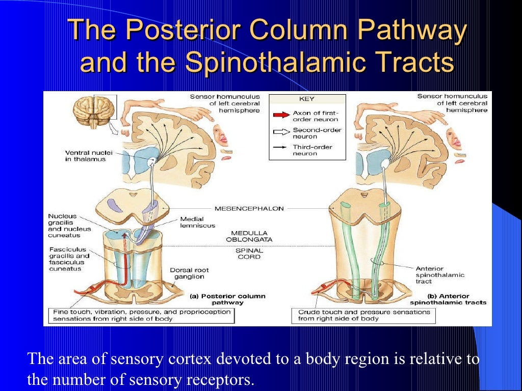
Pathway of Action Impulses Through the Dorsal Column. Speaking of the thalamus, the fasciculus gracilis and fasciculus cuneatus also send impulses from the spinal cord up to the thalamus for the sensations of touch, pressure, and proprioception (also known as kinesthesia).
What is the function of the dorsal column?
It relays information related to many different types of touch, motor functionality, and more. The nerve signals that travel through the dorsal column also travel a bit differently than most others, in that they decussate at a different point in their transmission pathway, and the stages of transmission and points of synapsing are different ...
Which pathway regulates proprioception?
Proprioception (specifically as it relates to skeletal muscles) that is regulated by the medial lemniscal pathway is transmitted by two distinct types of fibers (that, of course, divide even further into more types of fibers!): Extrafusal fibers: fibers that surround the majority of muscle spindles.
Where does the medial lemniscal pathway synapse?
Going back to the fact that the medial lemniscal pathway doesn’t synapse where it enters the spinal cord: It instead extends vertically and synapses in the medulla oblongata, specifically, onto one of the two structures known as the nucleus cuneatus or the nucleus gracilis. Laterally to medially, these are organized like so: N.C. – N.G. – N.G. – N.C. This part of the synapse is known as the first-order neuron.
Why is the dorsal column stimulated?
In fact, because of the nerve connections that are regulated by the dor sal column, along with its wide variety of functions, the stimulation of the dorsal column is used as a form of treatment for chronic pain like complex regional pain syndrome (CRPS). Just like all other sensory, motor, and cognitive pathways in the body, however, ...
Who is the author of Separate Visual Pathways for Perception and Action?
Goodale, M. A., & Milner, A. D. (1992). Separate visual pathways for perception and action. Trends in Neurosciences, 15 (1), 20–25. PubMed Google Scholar
Which lobe of the visual cortex is responsible for the localization of objects in space?
The dorsal visual pathway is a functional stream originating in primary visual cortex and terminating in the superior parietal lobule that is responsible for the localization of objects in space and for action-oriented behaviors that depend on the perception of space.
What is the function of the dorsal column?
The primary function of the dorsal column pathway is to convey sensory information regarding fine touch, two-point discrimination, conscious proprioception , and vibration sensations from our skin and joints, excluding the head.
Which pathway is the ascending tract of the spinal cord?
The dorsal column pathway is one of the ascending tracts i.e. the neural pathways by which sensory information from the peripheral nerves is transmitted to the cerebral cortex. In the spinal cord, this pathway travels in the dorsal column, and in the brainstem, it is transmitted through the medial lemniscus hence the name dorsal column-medial lemniscus pathway.
What are the two types of neurons that are axonal?
The pseudounipolar neurons contain peripheral (distal) and central (proximal) axonal processes. The peripheral (distal) axons receive various signal inputs from the skin via the receptors associated with the dorsal column medial lemniscus pathway. These receptors classify as two types: tactile mechanoreceptors and conscious proprioception.
Why is 2PD considered an integrative test?
It is also regarded as an integrative test because it requires a high degree of sensory processing. 2PD sense may be impaired by both damage to the medial lemniscus pathway and peripheral nerve damage. The therapist may use calipers or simply a reshaped paperclip to do the testing.
Which neuron is the second order neuron?
Both nucleus cuneatus and nucleus gracilis in medulla represent the second-order neuron of the DCML pathway. The axons of second order neuron from nucleus gracilis and nucleus cuneatus are known as internal arcuate fiber which cross at the level of medulla forming medial lemniscus and then travels through the brainstem with a preserved somatotopic arrangement. The medial lemniscus terminates and synapses in the thalamus particularly, in the ventral posterolateral (VPL) nucleus of the thalamus with the preservation of the somatotopy.
Where do proximal axons enter the spinal cord?
The proximal axons of the dorsal root ganglia enter the spinal cord through the medial dorsal root entry zone. Once in the spinal cord, the central axonal process gives off small collateral branches that will terminate in the spinal gray matter to facilitate spinal reflexes. The majority of the central axonal process, however, will leave the dorsal horn gray matter without synapsing and enter the dorsal funiculus to help constitute either the fasciculus gracilis or the fasciculus cuneatus in ipsilateral side.
What is the third order neuron?
Third order neuron. Ventral posterolateral (VPL) neurons are third-order neurons of the pathway, and its axons will project laterally out of the thalamus and course somatotopically through the posterior limb of the internal capsule and then terminating in the primary somatosensory cortex of the postcentral gyrus.
What is the dorsal column-medial lemniscus pathway?
The dorsal column-medial lemniscus pathway (DCML) is a sensory pathway of the central nervous system. It conveys sensation of fine touch, vibration, pressure, two-point discrimination and proprioception (position) from the skin and joints.
Which pathway receives information from receptors in the skin and joints?
Dorsal column. Sensory pathway that receives information from receptors in the skin and joints. Nerve tracts in the white matter of the dorsal columns of the spinal cord (first-order neurons) carries this information to the medulla. Medial lemniscus. Continuation of the dorsal column, this pathway starts within the brainstem, ...
Which nerves synapse with second-order neurons?
The dorsal column fibers of the fasciculus gracilis ascend, without crossing, until they reach the gracilis nucleus. Here, they synapse with second-order neurons. The axons of the second-order neurons ( internal arcuate fibers) run anteromedially and cross (sensory decussation) the midline of the midbrain to the contralateral side. After decussation, the internal arcuate fibers form the medial lemniscus which ascends towards the ventral posterolateral nucleus of the thalamus and then projects onwards to the primary somatosensory cortex.
Which part of the medulla is the axon?
Together these fasciculi ascend the spinal cord to reach the lower (closed) part of the medulla oblongata. In the medulla, axons in the gracile fasciculus synapse with the gracile nucleus and axons in the cuneate fasciculus synapse with neurons in the cuneate nucleus.
What are the most important conscious cutaneous receptors for the DCML system?
The most important conscious cutaneous receptors for the DCML system are: Ruffini endings (bulbous corpuscles); detect tension deep in the skin and connective tissue or fascia. Lamellar corpuscles (Pacinian corpuscles); detect rapid vibrations (200-300Hz).
How to tell if you have a lemniscus pathway lesion?
The clinical signs of dorsal column-medial lemniscus pathway lesion include: Astereognosis: the patient cannot recognize an object by its shape and weight using touch alone. Agraphesthesia: the patient cannot recognize, by touch, a number or letter drawn in the palm of the patient’s hand by the examiner.
Which column runs from the spinal cord to the medulla?
The dorsal (posterior) column, which runs from the spinal cord to the medulla, and the medial lemniscus which runs as a continuation of the dorsal column, from the medulla to the cortex. In the cortex the DCML pathway projects onto the primary somatosensory cortex of the postcentral gyrus. Here sensation location is 'mapped' using ...
Which axis is the dorsal pathway?
the dorsal pathway, and are differentially shaped along the perceptual (caudal–medial)–motor (rostral–lateral) axis, with the more-caudal–medial regions being responsive to the visual prop- erties of the input and the more-rostral–lateral regions tuned more in the service of the visuomotor output. In light of the emerging evidence, visual perception should be studied not simply as a function of one (ventral) ‘what’ pathway, but instead as the joint outcome of the processing and coordination of different ‘what’ regions in both cortical visual pathways.
How are the dorsal and ventral pathways connected?
The dorsal and the ventral pathway are anatomically connected to each other by several major groups of axons. The parietomedial temporal pathway travels the caudal part of the inferior parietal lobule (cIPL, dorsal pathway) to the hippocampal formation and to parahippocampal areas which are part of the ventral pathway [114]. In addition to these directconnections,thecIPLisconnectedtotheseregionsthroughasetofindirectconnectionsthattravelthroughlimbic regions [115,116]. Importantly, the hippocampal formation is known to be involved in complex spatial processing and navigation, and the dorsal pathway input might also be engaged in such computations [42]. The lateral surfaces of the dorsalandventralpathway areconnectedbytwoadditionaltracts:theposterior arcuatefasciculus(pAF)andthevertical occipital fasciculus (VOF) [51,117]. In addition to the anatomical connections between the two pathways, imaging studies have documented strong functional connections between the two pathways. For example, based on the anatomical infrastructure of the VOF [52], regions in intermediate visual areas in the dorsal (V3A/B) and ventral (hV4/VO-1) pathways are connected and exchange visual information. Moreover, strong functional connectivity between the two pathways was found in higher- level regions such as the posterior parietal cortex and the lateral occipital complex. Interestingly, this functional connectivity was modulated by the validity of the perceptual input (i.e., possible vs[15_TD$IF]. impossible objects) [30]. Another study,whichutilizedeffectivefunctionalconnectivityanalysis,showedthatdorsalpathwayactivationwascorrelatedwith activationintheanteriorventralpathway[54]inaperceptualtaskthatincludedactionobservation.Takentogether,these findings further suggest that the two pathways are interconnected both functionally and anatomically, and these connections might be important in theprocess ofobject recognition. Future investigationemployingadvanced analytical methods (e.g., dynamic causal modeling [118]) may further clarify how object representations are shaped in the two pathways as a function of the connections between the two pathways.
What is the lateralization of tool sensitivity to the left hemisphere?
The lateralization of tool-sensitivity to the left hemisphere [119] in right-handed individuals raises important questions about hemispheric differences in dorsal object representations [120,121]. Hemispheric differences are known to modulate visual representations in the ventral pathway. For example, greater face sensitivity is usually observed in the right hemisphere, while the left ventral pathway exhibits greater sensitivity to written words [122,123], although this lateralization is graded rather than absolute. It is not clear to what extent similar lateralization applies to perceptual representations in the posterior part of the dorsal pathway. Neuropsychological findings provide some support for hemispheric specialization of the dorsal pathway. In a recent study, perceptual impairments following a parietal lesion were observed among patients with right, but not left, hemisphere lesions [62]. These observations are compatible with early neuropsychological investigations (see text for details [63]) and also with previous studies demonstrating that visual functions associated with the parietal cortex, such as visual attention [124] and mental rotation abilities [125], are usually more dominant in the right than left hemisphere. Hence, visual representations in the dorsal pathway may be shaped not only as a function of anatomy within each hemisphere (see main text for details) but also between hemispheres (left, motor; and right, spatial).
What is the reverse pattern of perceptual impairment?
The reverse, but complementary, pattern is that perceptual impairment – specifically of 3D objects and global form perception – results from dorsal pathway lesions, as shown in humans and non-human primates [27,43,60–63]. For example, in humans, patients with posterior parietal lesions exhibited marked perceptual deficits, particularly for 3D objects defined from binocular and monocular depth cues [63]. Correspondingly, non-human primate lesion studies thataimedtoexplorethecausalroleofdorsalregionsin3Dperceptionshowedthatdeactivation of the dorsal area CIP (caudal intraparietal) led to perceptual impairments related to 3D disparity perception as well as to decreased inferotemporal ventral activation [27].
Which lobe of the visual cortex is the dorsal pathway?
Dorsal visual pathway: this pathway extends from the primary visual cortex (V1) in the occipital lobe to the parietal lobe. The dorsal pathway is subdivided by the intraparietal sulcus (IPS) into several main sectors including the superior parietal lobule, inferior parietal lobule, and the supramarginal gyrus.
Which visual system is subserved by the ventral occipitotemporal pathway?
The cortical visual system is almost universally thought to be segregated into two anatomically and functionally distinct pathways: a ventral occipitotemporal pathway that subserves object perception, and a dorsal occipitoparietal path- waythatsubservesobjectlocalizationandvisuallyguidedaction.Accumulating evidence from both human and non-human primate studies, however, chal- lenges this binary distinction and suggests that regions in the dorsal pathway contain object representations that are independent of those in ventral cortex andthatplayafunctionalroleinobjectperception.Wereviewheretheevidence implicating dorsal object representations, and we propose an account of the anatomical organization, functional contributions, and origins of these repre- sentations in the service of perception. Two Cortical Visual Pathways
Which part of the parietal cortex generates object representations?
In both human and non-human pri- mates, the posterior portion of the dor- sal pathway generates object-based representations that are unrelated to action planning or execution. Patients with extensive lesions to the ventral pathway still generate object representations in the dorsal pathway, and evince perceptual sensitivity to object structural information. Neuropsychological investigations with patients, and lesion studies with non- human primates, have demonstrated that a lesion to the posterior part of the parietal cortex leads to perceptual deficits, particularly in 3D perception and in the perception of structure from motion.
Where is the dorsal cavity located?
The dorsal cavity lies close to the spine in the posterior portion of the body. The dorsal cavity contains the spinal column, central nervous system (i.e., brain and spinal cord), and meninges (i.e., tissue that surrounds the brain and spinal cord).
What are the most important facts to know about dorsal and ventral?
These terms can also be referred to as posterior and anterior surfaces. Ventral and dorsal can be used to describe the position of organs in relation to one another. For example, one could say, “The small intestine is ventral to the kidneys”, which means the small intestine is in front of the kidneys. These anatomical terms can also describe different body cavities. The dorsal cavity contains the spinal cord, central nervous system, and spinal column, whereas the ventral cavity consists of the thoracic, abdominal, and pelvic cavities.
What are the dorsal and ventral cavities?
What are the dorsal and ventral body cavities? The dorsal and ventral body cavities, two of the largest body compartments in humans, are anatomical spaces that contain various organs and other structures. The dorsal cavity lies close to the spine in the posterior portion of the body. The dorsal cavity contains the spinal column, ...
What is the difference between ventral and dorsal?
In general, ventral refers to the front of the body, and dorsal refers to the back. These terms are also known as anterior and posterior, respectively.
What is the paired anatomical term for the ventral and dorsal?
The anatomical position of a human body is defined as a body standing upright with the head facing forward, arms down at the sides with the palms turned forward, and feet parallel facing forward.
What is the dorsal side of the penis?
For instance, the dorsal part of the penis is the area that is closest to the abdomen when erect. Similarly, for the feet, the dorsal side is the top of the foot , or the area facing upwards when standing upright.
What is the ventral cavity?
On the anterior side of the body, the ventral cavity is made up of the thoracic cavity, abdominal cavity, and pelvic cavity. The thoracic cavity contains the heart, lungs, breast tissue, thymus gland, and blood vessels. Inside the abdominal cavity are the stomach, liver, gallbladder, pancreas, small intestine, colon, appendix, and kidneys.
Introduction
- The dorsal column pathway is one of the ascending tracts i.e. the neural pathways by which sensory information from the peripheral nerves is transmitted to the cerebral cortex. In the spinal cord, this pathway travels in the dorsal column, and in the brainstem, it is transmitted through the medial lemniscus hence the name dorsal column-medial lemni...
Course
- First order neuron
The cell body of the dorsal root ganglia, which is composed of pseudounipolar neurons, characterizes the first-order neuron of the pathway. The pseudounipolar neurons contain peripheral (distal) and central (proximal) axonal processes. The peripheral (distal) axons receiv… - Second order neuron
Both nucleus cuneatus and nucleus gracilis in medulla represent the second-order neuron of the DCML pathway. The axons of second order neuron from nucleus gracilis and nucleus cuneatus are known as internal arcuate fiber which cross at the level of medulla forming medial lemniscu…
Function
- The primary function of the dorsal column pathway is to convey sensory information regarding fine touch, two-point discrimination, conscious proprioception, and vibration sensations from our skin and joints, excluding the head. The dorsal column nuclei also transmit visceral nociceptive information to the contralateral ventral posterior lateral (VPL) thalamic nucleus, which in turn rel…
Clinical Relevance
- Posterior cord syndrome, also known as posterior spinal artery syndrome- an infraction to posterior spinal artery.
- Tabes Dorsalis: It is a slow degenerative disease that represents a late manifestation of untreated syphilis. It can cause deleterious neurological deficits due to its involvement in the posterior...
- Subacute combined degeneration of the spinal cord (SCD): SCD is characterized by axonal myelin abnormalities in the mentioned pathways as a result of B12 deficiency, leading to a compromise in nerv...
- Brown-Sequard syndrome: It is a hemisection involving either the left or right side of the spinal cord, is a syndrome that usually happens at the cervical level and affects the posterior colum…
Assessment
- Romberg testis part of a neurological exam used clinically to test for the integrity of the posterior column pathway. In the assessment, the integrity of the sensation it carries are tested i.e. fine touch, 2-point discrimination, conscious proprioception, and vibration sensation. 1. Fine touch:Fine touch can be tested with harp pointed object like touch prick (pin prick) at different d…
Treatment