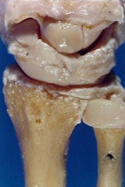
It’s located closest to the pinky finger. It helps to stabilize the wrist and allows the joint to bear more weight. Although the second bone of the forearm, the ulna, articulates with the radius, it’s separated from the wrist joint by a disc of fibrocartilage called the articular disk.
What is the function of the articular disc?
The presence of an articular disk also permits a more even distribution of forces between the articulating surfaces of bones, increases the stability of the joint, and aids in directing the flow of synovial fluid to areas of the articular cartilage that experience the most friction. What is the function of articular disc?
What is the articulation of the wrist joint?
The wrist joint generally refers to the radiocarpal joint, which is the articulation between the distal end of the radius and the articulating surface of the scaphoid, lunate, and triquetral bones. Other articulations in the wrist area include the distal radius and ulnar and the carpal bones. What joints have articular discs?
What prevents the wrist bone from articulating with the carpal bone?
It is prevented from articulating with the carpal bones by a fibrocartilaginous ligament, called the articular disk, which lies over the superior surface of the ulna. Together, the carpal bones form a convex surface, which articulates with the concave surface of the radius and articular disk. Fig 1 – Articular surfaces of the wrist joint.
What is the function of the articular disc in TMJ?
The articular disc in the TMJ has an important functional role. It fills the space between the condyle and the temporal bone, and acts as a stress absorber and distributors during the jaw activity.

What is the most common fracture of the carpal bone?
Scaphoid Fracture. The scaphoid bone of the hand is the most commonly fractured carpal bone – typically by falling on an oustretched hand (FOOSH). In a fracture of the scaphoid, the characteristic clinical feature is pain and tenderness in the anatomical snuffbox.
Why is the scaphoid at risk of necrosis?
The scaphoid is at particular risk of avascular necrosis after fracture because of its so-called ‘retrograde blood supply’ which enters at its distal end. This means that a fracture to the middle (or ‘waist’) of the scaphoid may interrupt the blood supply to the proximal part of the scaphoid bone rendering it avascular.
What is the synovial joint of the wrist?
The wrist is an ellipsoidal (condyloid) type synovial joint, allowing for movement along two axes. This means that flexion, extension, adduction and abduction can all occur at the wrist joint.
Which two organs produce adduction?
Adduction – Produced by the extensor carpi ulnaris and flexor carpi ulnaris
How many ligaments are there in the wrist?
Ligaments. There are four ligaments of note in the wrist joint, one for each side of the joint. Palmar radiocarpal – Found on the palmar (anterior) side of the hand. It passes from the radius to both rows of carpal bones.
What is the name of the joint between the forearm and the hand?
The Wrist Joint. The wrist joint (also known as the radiocarpal joint) is a synovial joint in the upper limb, marking the area of transition between the forearm and the hand. In this article, we shall look at the structures of the wrist joint, the movements of the joint, and the relevant clinical syndromes.
Where does the wrist get its blood from?
The wrist joint receives blood from branches of the dorsal and palmar carpal arches, which are derived from the ulnar and radial arteries (for more information, see Blood Supply to the Upper Limb)
What are the ligaments of the wrist?
The ligaments of the wrist joint are quite variably described in the literature, which can lead to a degree of confusion in regards to their anatomy. A notable feature of the ligaments of the wrist is that none of them are truly extracapsular; most of them are rather defined as thickenings of the joint capsule, providing it with additional support. The palmar ligaments are notably more numerous than those of the dorsal wrist joint, with almost the entire palmar portion of the joint capsule being composed of individual ligaments. The palmar ligaments tend to converge distally, presenting as an apex-distal ‘V’ when viewed collectively.
Which ligaments are more numerous than the dorsal wrist joint?
The palmar ligaments are notably more numerous than those of the dorsal wrist joint, with almost the entire palmar portion of the joint capsule being composed of individual ligaments. The palmar ligaments tend to converge distally, presenting as an apex-distal ‘V’ when viewed collectively.
What is the radiocarpal joint?
The radiocarpal joint is a synovial joint formed between the radius, its articular disc and three proximal carpal bones; the scaphoid, lunate and triquetral bones. Technically, the radiocarpal joint is considered to be the only articular component of the wrist joint; many references, however, may also include adjacent joints, ...
What is the outer layer of a radiocarpal joint?
The outer portion of the capsule is composed of fibrous connective tissue which provides structural support to the joint, while the inner layer is composed of a synovial membrane responsible for the secretion of synovial fluid, keeping the joint lubricated.#N#Proximally, the capsule is usually independent of that of distal radioulnar joint, and it attaches to the distal aspects of radius and ulna. Distally, the joint capsule attaches on the margins of the proximal articular surfaces of the involved carpal bones.
What are the primary movements of the radiocarpal joint?
The primary movements of the radiocarpal joint are flexion, extension, abduction and adduction. This article will discuss the anatomy and function of the radiocarpal joint. Key facts about the radiocarpal joint. Type.
Where does the palmar ulnocarpal ligament originate?
The palmar ulnocarpal ligament arises from the anterior margin of the triangular fibrocartilage complex, the palmar radioulnar ligament and ulnar styloid process. It divides into three parts and courses distally obliquely towards the capitate, lunate and triquetrum bones, forming the unlocapitate, ulnolunate and ulnotriquetral divisions, respectively.
Where is the radiolunate ligament located?
Long radiolunate ligament: runs in parallel along the ulnar border of the radioscaphocapitate ligament, from the distal radius to the lunate bone. This ligament was formerly known as the radiolunotriquetral ligament, however the amount of fibres continuing to the triquetrum is negligible.
What is a TFCC tear?
A TFCC tear is a tear of the triangular fibrocartilage complex, which consists of :
What is the ligament on the back of the wrist called?
There are a number of ligaments in the wrist, however, the ligaments that are of most importance are the scapholunate ligament (on the back of the wrist) and what is known as the TFCC or triangular fibrocartilage complex.
What are the symptoms of a TFCC tear?
TFCC tear symptoms. Symptoms of a TFCC tear include wrist pain on the little pinky finger side. There will be tenderness over the back of the wrist. Pain worsens when bending the wrist sideways so the little finger moves towards the forearm (called ulnar deviation). There is likely to be swelling in the wrist, reduced grip strength ...
What is the procedure to examine the damage to the wrist?
If however, the injury is more severe then an arthroscopic evaluation of the wrist would be required which is an operation to examine exactly what the damage to the wrist is. Keyhole surgery is done and a very small camera is inserted into the back of the wrist to image the ligaments and examine the injury.
How is keyhole surgery done?
Keyhole surgery is done and a very small camera is inserted into the back of the wrist to image the ligaments and examine the injury. These ligaments can then be tightened up and repaired with the minimum of invasive surgery.
How long does it take to get a torn ulna out of your wrist?
It involves trimming the torn piece of cartilage. In cases where the ulna is too long, the end of the bone may be shaved away. The wrist is then immobilized for 2-4 weeks.
What sports cause degenerative tears?
Sports in which this injury is common include racket and bat sports like tennis and baseball and gymnastics due to weight-bearing on the hands. It also occurs in Water skiing. Degenerative tears occur due to repetitive loading over a long period and are usually found in the older population.
What is a TFCC tear?
A TFCC tear is any injury or damage to the TFCC. There are two types of TFCC tear: Type 1. These tears result from physical injury, such as when a person overextends or over-rotates their wrist, or when they fall on their hand with it extended. Type 2.
Why is TFCC tear important?
Accurate classification of a TFCC tear is important for guiding treatment decisions.
How to tell if TFCC tears are a symptom?
Other symptoms can include: stiffness or weakness in the wrist. pain when touching or moving the wrist. a limited range of motion in the hand or wrist. wrist swelling. a clicking or popping sound when moving the wrist.
What is the triangular fibrocartilage complex?
Summary. The triangular fibrocartilage complex is a structure in the wrist. Sustaining an injury or tear to this area can cause pain along the outside of the wrist and limit its range of motion. The triangular fibrocartilage complex (TFCC) is a network of ligaments, tendons, and cartilage that sits between the ulna and radius bones on ...
How long does it take for a TFCC tear to heal?
Recovery time for a TFCC tear depends on the type, severity, and treatment of the injury. A case study. Trusted Source. from 2016 suggests that TFCC tears that do not require surgery can take up to 12 weeks to fully heal. Following surgery, a TFCC tear may take around 3 months. Trusted Source.
How to diagnose TFCC tear?
To diagnose a TFCC tear, a doctor will usually begin by asking the person about their symptoms and medical history. They may then perform a physical examination of the wrist area. During the physical exam, the doctor may: Carefully apply pressure to the outer edge of the wrist to isolate the source of the pain.
What is the role of TFCC?
The TFCC connects the bones in the hand and forearm, forming the wrist. The TFCC connects the bones in the hand to the bones in the forearm to form the wrist. It plays an important role in: moving the wrist. rotating the forearm. supporting the forearm when the palm is gripping an object.
