What is the fovea centralis made of?
Fovea centralis. The fovea centralis is a small, central pit composed of closely packed cones in the eye. It is located in the center of the macula lutea of the retina.
What is the fovea centralis of the eye?
The fovea centralis is located at the center of the retina's macula and contains cones that enable central and daytime vision. Learn more about the definition and function of the fovea centralis of the eye and the effects of macular degeneration on eyesight. Updated: 01/11/2022 What Is the Fovea Centralis?
What is the function of the fovea?
The fovea or fovea centralis is a small depression at the center of the retina that's responsible for central vision. It's the point at which visual acuity is at its highest. Visual acuity is the ability to identify the details of objects when you look at them. The fovea contains many cones (the cells that receive visual information).
What happens if the fovea centralis is damaged?
The fovea centralis consists of only cones and is therefore crucial to central vision. Degeneration of the macula and fovea centralis leads to age-related macular degeneration, or AMD, which can cause blindness.

What is the primary function of the fovea centralis?
Anatomical Parts The fovea is responsible for sharp central vision (also called foveal vision), which is necessary in humans for reading, driving, and any activity where visual detail is of primary importance.
What is the structure and function of the fovea?
A fovea is a pitted invagination in the inner retina that overlies an area of densely packed photoreceptors specialized for high acuity vision. A fovea contains particularly high numbers of photoreceptors and neurons, and provides the highest visual resolution (Walls, 1942).
What is the fovea centralis quizlet?
What is the fovea centralis & why is it important? A small pit in the retinal layer that contains cones only is located lateral to the optic disk in each eye. Anything that must be viewed (discriminative vision) is focused on the fovea bcuz its the area of greatest visual acuity.
What is the meaning of fovea centralis?
fovea centralis in British English (sɛnˈtrɑːlɪs ) noun. a small depression in the centre of the retina that contains only cone cells and is therefore the area of sharpest vision.
Is the Fovea Centralis a blind spot?
The blind spot (Fovea centralis) This seemingly poor design of the retina, which produces the blind spot in our visual field, is referred to by experts as the inverted eye. The blind spot is located about 15 degrees on the nasal side of the fovea.
Why is the Fovea Centralis the area of the sharpest vision?
The resolution or sharpness in vision is because of the high concentration of cone cells in the fovea. The fovea has the densest concentration of photoreceptor cells that are known as cones. Rods are completely absent from the fovea.
Where is the fovea centralis and why is it important?
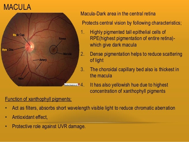
The fovea centralis is located in the center of the macula lutea, a small, flat spot located exactly in the center of the posterior portion of the retina. As the fovea is responsible for high-acuity vision it is densely saturated with cone photoreceptors.
What is the fovea centralis and why is it important quizlet?
The fovea centralis is a small, central pit composed of closely packed cones in the eye. It is located in the center of the macula lutea of the retina. The fovea is responsible for sharp central vision such as reading and driving.
What differentiates the fovea centralis of the retina from the rest of the retina?
The foveal center or 'foveola' contains the highest density of cone photoreceptors in the retina. Cone photoreceptors function in bright light and support high acuity and color vision.
Why are there only cones in the fovea?
Rods are responsible for vision at low light levels (scotopic vision). They do not mediate color vision, and have a low spatial acuity. Cones are active at higher light levels (photopic vision), are capable of color vision and are responsible for high spatial acuity. The central fovea is populated exclusively by cones.
Does the fovea contain rods and cones?
In the fovea, there are NO rods... only cones. The cones are also packed closer together here in the fovea than in the rest of the retina. Also, blood vessels and nerve fibers go around the fovea so light has a direct path to the photoreceptors.
What is the fovea made up of?
conesThe fovea centralis is made up entirely of cones. Cones are a type of photoreceptor that allows for sharp vision and visual acuity.
What is the fovea made up of?
conesThe fovea centralis is made up entirely of cones. Cones are a type of photoreceptor that allows for sharp vision and visual acuity.
What is found in the fovea?
The fovea is a depression in the inner retinal surface, about 1.5 mm wide, the photoreceptor layer of which is entirely cones and which is specialized for maximum visual acuity. Within the fovea is a region of 0.5mm diameter called the foveal avascular zone (an area without any blood vessels).
What is the function of the retina?
The retina is a layer of photoreceptors cells and glial cells within the eye that captures incoming photons and transmits them along neuronal pathways as both electrical and chemical signals for the brain to perceive a visual picture.
What does fovea look like?
The human fovea is densely packed with cones. It looks like a little pit on the retina because the cells that are above the retinal surface, such as retinal ganglion cells, horizontal cells, and amacrine cells, are swept away so that the cones are at the surface.
Anatomy
The central fovea appears as a small flat spot at the retina's center. It's about 1.5 mm in diameter and contains about 199,000 cones/mm squared. 2
Fovea Problems & Diseases
Left untreated, a variety of eye diseases may impair the fovea, resulting in vision loss.
Why Routine Eye Exams are Important
Routine treatment is important because it helps your eye doctor detect any issues before they become serious.
What is the fovea centralis?
Fovea centralis. The fovea is a tiny part of the eye’s anatomy that makes a huge difference in our eyesight. Resting inside the macula, the fovea (also called “fovea centralis”) provides our absolute sharpest vision.
Why is the fovea important?
Because the fovea is such an essential part of a person’s vision, it’s important to prevent and/or monitor the conditions that may jeopardize its function. Conditions that may affect the fovea include:
Why is the fovea anatomy so tricky?
Fovea anatomy can be tricky because the retina and macula are also light-sensitive parts of the eye that create sharp vision. So, where does the fovea come into play, and how is it different from the macula and retina?
What is the term for fluid buildup in the macula?
Macular edema — Fluid buildup in the macula. Retinal detachment — When the retina lifts or tears away from the back of the eye. Retinoblastoma — Cancer of the eye that begins in the retina. Retinal vein occlusion — When a vein or artery in the retina becomes blocked.
What are cone cells responsible for?
Cone cells are responsible for producing color and fine details , while rods provide peripheral vision, movement and shades of grey. Rods are mostly located outside the macula, and the cones are located inside. The fovea eye pit does not have any rods or other neurons, only millions of tightly packed cones.
Which part of the eye produces sharper vision?
Think of it as “sharp, sharper, sharpest”: The retina is the tissue that lines the back of the eyeball and produces sharp vision whenever light hits it correctly. The macula is the center portion of the retina that produces even sharper vision with its rods and cones. The fovea is the pit inside the macula with only cones, so vision can be at its sharpest.
Which part of the eye works together to provide the best central and peripheral vision and visual acuity?
The retina, macula and fovea work together to provide the best central and peripheral vision and visual acuity possible.
What is the fovea?
The fovea is responsible for sharp central vision (also called foveal vision), which is necessary in humans for activities for which visual detail is of primary importance , such as reading and driving. The fovea is surrounded by the parafovea belt and the perifovea outer region.
Where is the fovea found?
Other animals. The fovea is also a pit in the surface of the retinas of many types of fish, reptiles, and birds. Among mammals, it is found only in simian primates. The retinal fovea takes slightly different forms in different types of animals.
How do pigments enhance the acuity of the fovea?
The pigments also enhance the acuity of the fovea by reducing the sensitivity of the fovea to short wavelengths and counteracting the effect of chromatic aberration. This is also accompanied by a lower density of blue cones at the center of the fovea. The maximum density of blue cones occurs in a ring about the fovea.
What is the size of the fovea?
Structure. The fovea is a depression in the inner retinal surface, about 1.5 mm wide, the photoreceptor layer of which is entirely cones and which is specialized for maximum visual acuity. Within the fovea is a region of 0.5mm diameter called the foveal avascular zone (an area without any blood vessels).
Why is the foveal focus dark?
The combined effects of the macular pigment and the distribution of short wavelength cones results in the fovea having a lower sensitivity to blue light (blue light scotoma). Though this is not visible under normal circumstances due to "filling in" of information by the brain, under certain patterns of blue light illumination, a dark spot is visible at the point of focus. Also, if mixture of red and blue light is viewed (by viewing white light through a dichroic filter), the point of foveal focus will have a central red spot surrounded by a few red fringes. This is called the Maxwell's spot after James Clerk Maxwell who discovered it.
How far is the Perifovea from the Fovea?
The parafovea extends to a radius of 1.25 mm from the central fovea, and the perifovea is found at a 2.75 mm radius from the fovea centralis. The term fovea comes from the from Latin foves 'pit'.
How many cone cells are there in the fovea?
On average, each square millimeter (mm) of the fovea contains approximately 147,000 cone cells, or 383 cones per millimeter. The average focal length of the eye, i.e. the distance between the lens and fovea, is 17.1 mm. From these values, one can calculate the average angle of view of a single sensor (cone cell), which is approximately 31.46 arc seconds .
What structures bend the light so that it focuses the light ideally on the fovea?
Cornea and Len s: Both these structures bend the light so that it focuses the light ideally on the fovea. Glasses are used to help get the light perfectly focused on th... Read More
Which part of the retina is uniquely evolved to maximize central visual acuity for tasks like reading and recognizing people?
Central vision: This part of the central retina is uniquely evolved to maximize central visual acuity for tasks like reading and recognizing people's faces. It does n... Read More
What is the center of the macula?
Fovea: The macula is small central portion of the retina. The center of the macula is the fovea centralis, an area where all of the photoreceptors are cones;... Read More
Which macula allows us to see best?
The macula FC is the: on the macula that allows us to see best because because of the topography of that area is devoid of large blood vessels and high density of cones and... Read More
Which spot in the retina allows you to perceive fine detail for reading or face recognition?
Central vision: This is the spot at the exact optical center of the retina which allows you to perceive fine detail for reading or face recognition for instance. Rea... Read More
Is the fovea separate from the retina?
Not really: The fovea is separate from the retina . Each carry about 50% input from he eye to the brain.

Overview
Function
In the primate fovea (including humans) the ratios of ganglion cells to photoreceptors is about 2.5; almost every ganglion cell receives data from a single cone, and each cone feeds onto between one and 3 ganglion cells. Therefore, the acuity of foveal vision is limited only by the density of the cone mosaic, and the fovea is the area of the eye with the highest sensitivity to fine details. Cones in the central fovea express opsins that are sensitive to green and red light. These cones are the '…
Structure
The fovea is a depression in the inner retinal surface, about 1.5 mm wide, the photoreceptor layer of which is entirely cones and which is specialized for maximum visual acuity. Within the fovea is a region of 0.5mm diameter called the foveal avascular zone (an area without any blood vessels). This allows the light to be sensed without any dispersion or loss. This anatomy is responsible for the depression in the center of the fovea. The foveal pit is surrounded by the foveal rim that cont…
Other animals
The fovea is also a pit in the surface of the retinas of many types of fish, reptiles, and birds. Among mammals, it is found only in simian primates. The retinal fovea takes slightly different forms in different types of animals. For example, in primates, cone photoreceptors line the base of the foveal pit, the cells that elsewhere in the retina form more superficial layers having been displaced away from the foveal region during late fetal and early postnatal life. Other foveae may s…
Additional images
• Illustration showing main structures of the eye including the fovea
• Structures of the eye labeled
• This image shows another labeled view of the structures of the eye
• Schematic diagram of the macula lutea of the retina, showing perifovea, parafovea, fovea, and clinical macula
See also
• Eye movement
• Gaze-contingency paradigm
• Macular degeneration
• Foveated imaging