What is the principle of simple stain?
Simple Staining: Principle, Procedure and Results. These stains will readily give up a hydroxide ion or accept a hydrogen ion, which leaves the stain positively charged. Since the surface of most bacterial cells and cytoplasm is negatively charged, these positively charged stains adhere readily to the cell surface.
What is the principle of Gram staining?
Principle of Gram staining. The structure of the organism’s cell wall determines whether the organism is gram psitive or negative. When stained with a primary stain and fixed by a mordant, some bacteria are able to retain the primary stain by resisting declorization while others get decolorized by a decolorizer.
What happens when basic stain is applied to bacteria?
When basic stain is applied, there is an attraction between the negatively charged cell surface and positively charged chromophore, which leads to staining of the cell (Figure 3.2). In negative staining, the background is coloured and bacteria remains colourless.
What is positive staining in microbiology?
In positive staining, the surface of the bacterial cell takes on the colour of the stain. When basic stain is applied, there is an attraction between the negatively charged cell surface and positively charged chromophore, which leads to staining of the cell (Figure 3.2).
What is the principle of the H&E staining?
The principle behind H & E stain is the chemical attraction between tissue and dye. Hematoxylin, a basic dye imparts blue-purple contrast on basophilic structures, primarily those containing nucleic acid moeties such as chromtatin, ribosomes and cytoplasmic regions rich in RNA.
What is the principle of staining in histopathology?
Staining is used to highlight important features of the tissue as well as to enhance the tissue contrast. Hematoxylin is a basic dye that is commonly used in this process and stains the nuclei giving it a bluish color while eosin (another stain dye used in histology) stains the cell's nucleus giving it a pinkish stain.
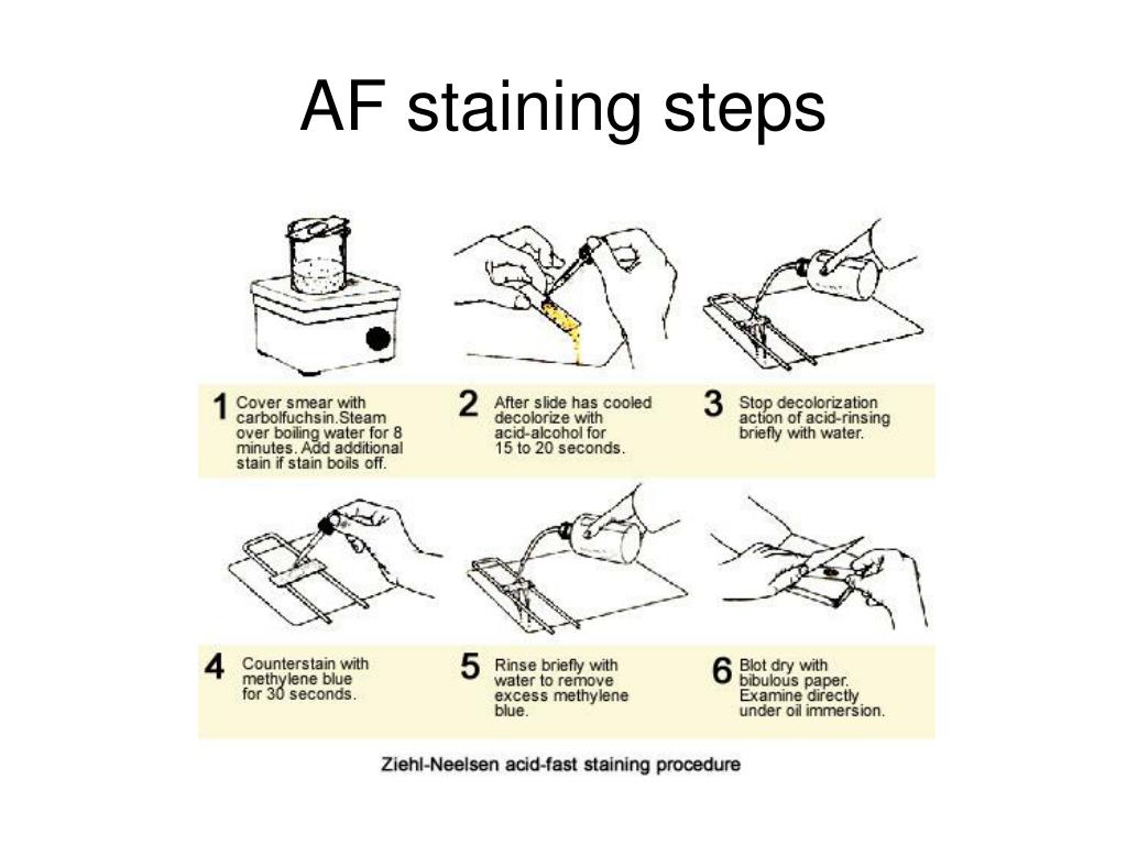
What is the procedure of ZN staining?
Ziehl Neelsen Acid-fast stainStep 2: Smear Preparation (Review) ... Cover the smear with carbolfuchsin dye. ... Dry heat for 2 minutes.Cool and rinse with water. ... Wash the top and bottom of slide with water and clean the slide bottom well.Counterstain with Methylene Blue for 30 seconds to 1 minute.
What is the purpose of acid-fast staining?
The acid-fast stain is a laboratory test that determines if a sample of tissue, blood, or other body substance is infected with the bacteria that causes tuberculosis (TB) and other illnesses.
What are the 3 types of differential staining?
Article Summary: The Gram, Ziehl Neelsen acid fast, and endospore stains are differential tests used to help identify bacteria. Here's how they work.
What are the 3 types of special stains?
Special StainsMassons Trichrome. The trichrome stain helps to highlight the supporting collagenous stroma in sections from a variety of organs. ... Verhoff's Elastic Stain. ... Reticulin Stain. ... Giemsa Stain.
Why H2SO4 is used in ZN staining?
The waxy material is hydrophobic to aqueous solution but not to phenolic solution of basic fuchsin. Hence strong carbol fuchsin is able to stain the cell. Upon staining, they tend to resist decolourization by 20% H2SO4 (sulphuric acid).
What is the mordant used in ZN staining?
In the Ziehl–Neelsen stain, heat acts as a physical mordant while phenol (carbol of carbol fuschin) acts as the chemical mordant. Since the Kinyoun stain is a cold method (no heat applied), the concentration of carbol fuschin used is increased.
What is the difference between Gram stain and Ziehl-Neelsen stain?
The main difference between the two acid-fast staining methods is the use of heat during the primary staining process. The Ziehl-Neelsen method uses heat to infuse the primary stain into the acid-fast cells.
What are the 3 main steps of an acid fast stain?
Procedure of Acid Fast Stain Cover the filter paper with the primary stain, carbolfuchsin. Leave the slide on the water bath for 3 to 5 minutes. Continue to apply stain if the filter paper begins to dry. Remove the filter paper and rinse the slide with water until the solution runs clear.
What is another name for acid-fast staining?
Ziehl–Neelsen staining is a type of acid-fast stain, first introduced by Paul Ehrlich. Ziehl–Neelsen staining is a bacteriological stain used to identify acid-fast organisms, mainly Mycobacteria.
Why is carbol fuchsin used in acid-fast staining?
Carbol fuchsin is used as the primary stain dye to detect acid-fast bacteria because it is more soluble in the cells' wall lipids than in the acid alcohol.
What is the importance of staining?
The purpose of staining is to increase the contrast between the organisms and the background so that they are more readily seen in the light microscope.
What is the purpose of staining cells?
Why Stain Cells? The most basic reason that cells are stained is to enhance visualization of the cell or certain cellular components under a microscope. Cells may also be stained to highlight metabolic processes or to differentiate between live and dead cells in a sample.
What is staining technique?
Staining, in microbiology, can be defined as a technique which is used to enhance and contrast a biological specimen at the microscopic level. Stains and dyes are used to highlight the specimen at the microscopic level to study it at higher magnification for histopathological studies and diagnostic purposes.
What are the three major groups of stains used for histopathology?
Acid and Basic dyes I.e. a technique called the Mallory staining technique uses three acidic dyes: aniline blue, acid fuschin and orange G, which selectively stain collagen, cytoplasm and red blood cells respectively.
Why does a Gram negative organism get decolorized?
Since Gram negative organism have thin peptidoglycan layer(1-2 layers)and have additional lipopolysaccharide layerwhich gets dissolved due to the addition of alcohol, so gram negative organism fails to retain the complex and gets decolorized as the complex is washed away.
What color does a Gram positive cell lose after decolorization?
After decolorization, the Gram-positive cell remains purple and the Gram-negative cell loses its purple color. Counterstain, which is usually positively-charged safranin or basic fuchsin, is applied last to give decolorized Gram-negative bacteria a pink or red color. Requirements and preparation of reagents.
What determines whether an organism is gram psitive or negative?
The structure of the organism’s cell wall determines whether the organism is gram psitive or negative. When stained with a primary stain and fixed by a mordant, some bacteria are able to retain the primary stain by resisting declorization while others get decolorized by a decolorizer.
What is the interaction between iodine and CV+?
Iodine (I), used as mordant interacts with CV+ and forms large complexes of crystal violet and iodine (CV–I) within the inner and outer layers of the cell.
What is the color of gram positive?
Observe under microscope. Interpretation of Gram staining. The staining results of gram stain are as follows : Gram Positive: Dark purple.
What is the role of iodine in the cell membrane?
Iodine (I), used as mordant interacts with CV+ and forms large complexes of crystal violet and iodine (CV–I) within the inner and outer layers of the cell. When a decolorizer such as alcohol or acetoneis added, it interacts with the lipids of the cell membrane.
How long to leave crystal violet stain on a smear?
Cover the smear with crystal violet stain and leave for 1 minute.
How long does it take for a smear to stain?
Staining. Cover the smear with methylene blue and allow the dye to remain in the smear for approximately one minute (Staining time is not critical here; somewhere between 30 seconds to 2 minutes should give you an acceptable stain, the longer you leave the dye in it, the darker will be the stain).
How to prepare a smear?
Preparation of a smear 1 Using a sterilized inoculating loop, transfer loopful of liquid suspension containing bacteria to a slide (clean grease free microscopic slide) or transfer an isolated colony from a culture plate to a slide with a water drop. 2 Disperse the bacteria on the loop in the drop of water on the slide and spread the drop over an area the size of a dime. It should be a thin, even smear. 3 Allow the smear to dry thoroughly. 4 Heat-fix the smear cautiously by passing the underside of the slide through the burner flame two or three times. It fixes the cell in the slide. Do not overheat the slide as it will distort the bacterial cells.
What is a simple stain?
The simple stain can be used as a quick and easy way to determine cell shape, size and arrangements of bacteria. True to its name, the simple stain is a very simple staining procedure involving a single solution of stain.
How to fix a smear on a slide?
Allow the smear to dry thoroughly . Heat-fix the smear cautiously by passing the underside of the slide through the burner flame two or three times. It fixes the cell in the slide. Do not overheat the slide as it will distort the bacterial cells.
How to clean a slide that has been stained?
Wipe the back of the slide and blot the stained surface with bibulous paper or with a paper towel.
Why do bacteria stain?
If methyene blue is used, some granules in the interior of the cells of some bacteria may appear more deeply stained than the rest of the cell, which is due to presence of different chemical substances.
How to study a smear?
Choose an area of the smear in which the cells are well spread in a monolayer. Center the area to be studied, apply immersion oil directly to the smear, and focus the smear under oil with the 100X objective.
What happens when a bacterial stain is positive?
When basic stain is applied, there is an attraction between the negatively charged cell surface and positively charged chromophore, which leads to staining of the cell (Figure 3.2).
Why is the background of a negative stain colorless?
It is because the acidic dyes are repelled by the negatively charged bacterial surface. The background gets stained and the cell remains colourless. This technique is useful for revealing the cell shape, size and demonstrating capsule (Figure 3.3).