What does the dorsal root ganglia consist mainly of?
The dorsal root ganglion, more recently referred to as the spinal ganglion, is a collection of neuronal cell bodies of sensory neurons . It is the most common type of sensory ganglion in the human body . Each cell body in the ganglion belongs to what is considered to be a pseudounipolar neuron. A pseudounipolar neuron consists of a ...
Does the ganglia contain sensory neurons?
Ganglia. A ganglion is a group of neuron cell bodies in the PNS. These ganglia contain the cell bodies of neurons with axons that are sensory endings in the periphery, such as in the skin, and that extend into the CNS through the dorsal nerve root.
Where are the ganglia, spinal, and cranial nerves?
Those ganglia can be found both in head and neck (and they are part of the cranial nerves) and in the trunk, close to the thoracic and abdominal/pelvic organs. Their preganglionic neurons are located in the cranial nuclei of the brainstem , and in the lateral horn of the sacral spinal
What is the singular form of ganglia?
ganglia The singular form of loci is locus To change a term ending in um to a plural form, you change um to a Medical terms derived from a person's name are called eponyms True or False: A combining vowel is not used if the ending or suffix begins with a vowel.
What is the spinal ganglion composed of?
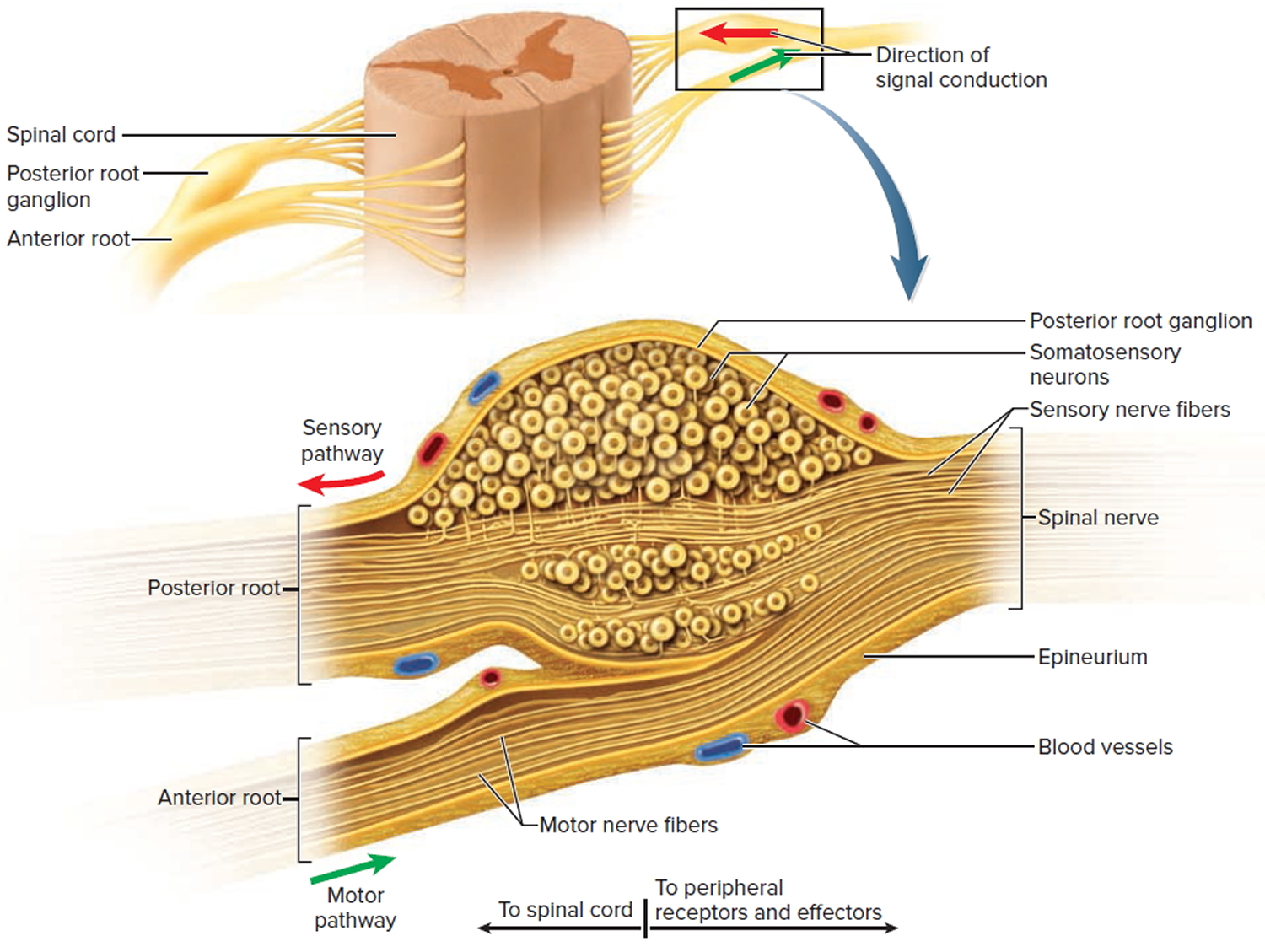
The spinal ganglion contains the cell bodies of sensory neurons situated in the posterior root of each spinal nerve (except for spinal nerve C1). These neurons are known as first-order neurons of the somatosensory system and carry sensations related to touch, vibration, proprioception, pain and temperature.
What type of tissue is spinal ganglion?
A dorsal root ganglion (or spinal ganglion; also known as a posterior root ganglion) is a cluster of neurons (a ganglion) in a dorsal root of a spinal nerve. The cell bodies of sensory neurons known as first-order neurons are located in the dorsal root ganglia.
What is the type of the nerve cells in spinal ganglia?
description. A spinal ganglion, for instance, is a cluster of nerve bodies positioned along the spinal cord at the dorsal and ventral roots of a spinal nerve. The dorsal root ganglia contain the cell bodies of afferent nerve fibres (those carrying impulses toward the central nervous system);…
How is spinal ganglion formed?
Development. The spinal ganglia are derived from the neural crest under the influence of nerve growth factor. The neural crest arises at the transition between the neural plate and the ectoderm at the time of formation of the neural groove.
What is ganglion and its structure?
A ganglion is a collection of neuronal bodies found in the voluntary and autonomic branches of the peripheral nervous system (PNS). Ganglia can be thought of as synaptic relay stations between neurons. The information enters the ganglia, excites the neuron in the ganglia and then exits.
What is found in the ganglia of spinal nerves quizlet?
The spinal ganglia or dorsal root ganglia contain the cell bodies of sensory neurons entering the cord at that region. a group of fibers (axons) outside the CNS. The spinal nerves contain the fibers of the sensory and motor neurons.
Which neuron is present in ganglia?
The cell bodies of somatic sensory and visceral sensory neurons are found in the dorsal root ganglia (spinal ganglia) of spinal nerves, and on the ganglia of selected cranial nerves. These structures are hence known as sensory ganglia.
What are the types of ganglia?
There are two types of ganglia in the PNS: sensory ganglia: - cell bodies of sensory neurons. autonomic ganglia: cell bodies of efferent neurons from the autonomic nervous system.
What is the difference between a nerve and a ganglion?
Both nerves and ganglia are structures found in the nervous system. However, a ganglion refers to a collection of nerve cells outside of the CNS whereas a nerve is the axon of a neuron. An afferent neuron, by the way, carries impulses whereas an efferent neuron is involved in motor functions.
What are three neuronal cell types that can be contained within a ganglion?
The three most common types of ganglion cells in primates are called midget cells, parasol cells, and small bistratified cells, and together they comprise approximately 70% of all ganglion cells.
What are the two main types of cells in the dorsal root ganglion?
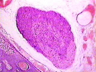
Two types of cells can be seen in a slide of a dorsal root ganglion. These are the neuron cells and the satellite cells. inside each nucleus. The neuronal cell bodies are seen to be highly granular.
Which has its cell body in a ganglion?
postganglionic neuron. Ganglions are collections of cell bodies of nerve fibers located in the peripheral nervous system (PNS). Postganglionic neurons start from the ganglion, that's why they have their cell bodies in the ganglion.
What are the two main types of cells in the dorsal root ganglion?
Two types of cells can be seen in a slide of a dorsal root ganglion. These are the neuron cells and the satellite cells. inside each nucleus. The neuronal cell bodies are seen to be highly granular.
What is a ganglion histology?
A ganglion (pl. ganglia) is a mass of nerve cell bodies found outside of the central nervous system (CNS) along with some glial cells and connective tissue. Ganglia have both afferent and efferent nerve fibers. As they exist outside of the CNS, they are sometimes referred to as peripheral ganglia.
What are the different types of ganglia?
There are two types of ganglia in our bodies—sensory and motor. Sensory ganglia are ovoid in shape and contain oval cell bodies with nuclei that form in a circular pattern. In the spine, motor ganglia form a long chain from the base of the skull down to the tail end of the spine.
What is ganglion cell?
Ganglion cells are the projection neurons of the vertebrate retina, conveying information from other retinal neurons to the rest of the brain. Their perikarya are the largest of any retinal neurons and are located along the inner margin of the retina, in the ganglion cell layer.
How many pairs of ganglia are there?
23 pairs of ganglia can be found: 3 in the cervical region (which fuse to create the superior, middle and inferior cervical ganglions), 12 in the thoracic region, 4 in the lumbar region, four in the sacral region, and a single, and the unpaired ganglion impar mentioned above.
Which ganglia contains sensory neurons?
Dorsal root ganglia contain clusters of sensory neuron cell bodies which transmit messages relating to pain, touch, and temperature from the PNS, towards the CNS. Satellite glial cells separate and inhibit interaction between cell bodies in the ganglion. Most of the body’s sensory neurons are contained here.
What nerve sends nerve fibers to the brain?
Facial nerve (CN VII) The neurons from the lacrimal and superior salivatory nuclei of the brainstem send fibers in the pterygopalatine ganglion and submandibular ganglion. The postganglionic fibers go on to innervate the lacrimal gland and glands in the nasal mucosa.
What is the sympathetic chain ganglia?
Sympathetic chain ganglia, also known as paravertebral ganglia, are the autonomic ganglia of the SNS. They consist of a paired chain of ganglia found ventral and lateral to the spinal cord. The ganglia extend from the upper neck to the coccyx, where the two chains fuse to form the unpaired ganglion impar.
What is a ganglion in 2021?
Reading time: 13 minutes. A ganglion is a collection of neuronal bodies found in the somatic and autonomic branches of the peripheral nervous system (PNS) . Ganglia can be thought of as synaptic relay stations between neurons.
What are preganglionic and postganglionic neurons?
Preganglionic vs postganglionic neurons 1 Have short preganglionic fibers, and long postganglionic fibers 2 Contain lightly myelinated preganglionic fibers, and unmyelinated postganglionic fibers 3 Originate within the lateral horn of the spinal cord, in the thoracic and upper lumbar regions (T1 to L2,3)
Which ganglion transmits sensory information?
Inside the inferior (or the nodose) ganglion there are cell bodies of neurons that transmit general sensory information from the mucosa of the larynx , pharynx , and soft palate .
How are spinal ganglia formed?
The spinal ganglia are derived from the neural crest, under the influence of several factors, the best known is nerve growth factor. The neural crest arises at the transition between the neural plate and ectoderm at the time of formation of the neural groove. The neural plate border is determined by a gradient of bone morphogenic protein. The neural crest cell type is determined by two gene sets: neural plate border specifier genes and neural crest specifier genes. The first set determines cells that border the neural plate, consequently the neural crest area. The second set is needed for neural crest cell survival. Neural plate border cells change in neural crest cells by Wnt genes, fibroblast growth factors, and retinoic acid. Migration and differentiation are organized by neural crest effector genes. The first steps in migration are induced by neural crest genes like snail, slug, and twist by downregulating cadherin expression. Tight junction components like occludin are downregulated and seemingly connexin is upregulated ( Fig. 4 ). Neural crest cells produce not only neurons but also glia, and a variety of cells contributing to head–neck, heart, peripheral nervous system, and skin structures). The migration of neural crest cells depends on local membrane-bound proteins, but they first have to pass the basal lamina using metalloproteases. Receptor–ligand coupling is important in migration. The specialized coupling for trunk ganglia are: Eph receptors and ephrins ligands, Slit (protein) and Robo (receptor), and semaphorins and neuropilins. The migration pathways are: (1) ventromedial, determined by Slit and Robo, and semaphorins and neuropilins, while (2) the dorsal path-way is regulated by Eph-ephrins. The ventromedial pathway is used for DRG and sympathetic ganglia formation. The dorsal pathway is involved in production of melanocytes ( Fig. 4 ). Note that cranial ganglia are differently constituted, not only from neural crest cells as the DRGs are but also from placode cells.
What are the dorsal root ganglia?
The dorsal root ganglia (or spinal ganglia) are described as nodule-like structures found on the posterior roots of each spinal nerve, which contain the soma (or cell bodies) of the afferent sensory nerves carrying sensory signals back to the central nervous system ( Figure 33.1) ( Standring, 2008 ). These structures develop from neural crest cells migrating into the rostral mesoderm, and each is described as oval and reddish in nature with size being dependent on its root for the corresponding level ( Geffen & Goldstein, 1996 ). Histologically, the ganglia are described as containing the cell bodies of the pseudounipolar sensory neurons, which are spherical in nature and lack dendrites ( Robinson, 1969; Standring, 2008 ). Because the cell bodies do not have dendrites and are not directly involved with the conduction of the sensory signal, the ganglion also has glial cells (satellite cells) within it to electrically insulate the cell bodies. Nerve fibers and connective tissue are also noted on histology, but the predominant image is the circular cell bodies of the sensory nerves with the glial cells noted interspersed between the cell bodies.
What is the cause of spinal cord disease?
Neurosyphilis may cause disease of the spinal cord, roots, and ganglia. The onset of symptoms varies depending on the lesion, from acute in spinal cord infarction to subacute in gummatous spinal cord compression; symptoms may be chronic in hypertrophic spinal pachymeningitis or tabes dorsalis, also known as progressive locomotor ataxia. The site most often involved in tabes dorsalis is lumbosacral and the mechanism is progressive neuronal degeneration secondary to chronic meningitis with vasculopathy and ischemia. The latency from initial infection is 15–30 years. Penicillin has dramatically reduced the incidence of tabes dorsalis ( Merritt et al., 1946; Aho et al., 1969; Towpik and Nowakowska, 1970; Heathfield, 1976 ). Tabes dorsalis peaks in the second decade of life for the juvenile form, and in the fifth to seventh decades for the adult form. The clinical manifestations are those of a posterior myelopathy with involvement of sensory and autonomic tracts and sparing of motor tracts. It is common to find associated abnormalities like cranial nerve and pupillary dysfunction. The onset is usually with paresthesias in girdle or band-like distribution around the trunk and/or the lower extremities that are episodic and of variable intensity. Pain is the most frequent complaint and typically of sudden onset, lancinating, stabbing, or lightning, and recurrent in clusters lasting minutes to hours. Visceral crisis refers to attacks of sudden pain lasting days to weeks, referred to the stomach (nausea and vomiting), bowels (diarrhea or constipation), larynx (hoarseness, stridor), bladder, or uterus ( Stokes et al., 1944 ). On neurological examination, there may be patchy areas of increased sensitivity to pinprick, temperature, or touch and of delayed perception (referred to as Hitzig's zones) or areas of analgesia. Vibratory and position sense loss is frequent over the sacrum and lower extremities. Sensory ataxia with positive Romberg's sign is also frequent ( Merritt et al., 1946 ). Reflexes are decreased or absent. The muscle tone is decreased, resulting in hyperextensible joints. The gait is typically broad-based, stamping, and worse in the dark. Repetitive trauma may result in Charcot's joints ( Merritt et al., 1946) and malum perforans ( Andrejevic and Jovic, 1970 ). Charcot’s arthropathy of the spine can cause cauda equina syndrome from posterior nerve root compression ( Ramani and Sengupta, 1973 ). Dysautonomia is frequent with orthostatic hypotension, impotence, bladder dysfunction (urinary retention, overflow incontinence), and bowel incontinence.
What is the diameter of a jejunal ganglioneuroma?
The jejunal ganglioneuroma measured 6 cm in maximal diameter and was intramural, i.e. located between the two layers of the muscularis externa; it was causing significant reduction in the luminal diameter. 27 In the horse with ganglioneuromatosis, there were numerous sessile to pedunculated, firm, nodular, white or red, 3–15 mm diameter masses protruding from the serosal surface of the antimesenteric wall of the small colon. 28 Those masses were located within the muscularis externa and were multifocally adherent to the colonic mesentery.
Where is the ganglion located?
Ganglion located along the dorsal root of a spinal nerve , containing the cell bodies of afferents innervating the viscera (sympathetic afferents), as well as the skin and skeletal muscle (somatic afferents).
Where are sensory neurons located?
Sensory neurons are located in the spinal ganglia within the dorsal roots along the spinal cord (see Figure 1-7) and in the ganglia of CN V. The receptors for temperature, pressure, touch, and noxious stimuli (nociception) are located on or near body surfaces. Axons are located in peripheral nerves and enter the spinal cord via the dorsal roots (see Figure 1-7 ). After entering the spinal cord, axons synapse on interneurons that stimulate limb flexion and inhibit limb extension in the ipsilateral limb and facilitate extension and inhibit flexion in the contralateral limb. This is the sensory component of withdrawal and crossed extensor reflexes ( Figure 1-10 ). Fibers are also projected to the brain for conscious perception of sensory information ( Figure 1-11 ).
What is the function of ganglia?
Ganglia play an essential role in connecting the parts of the peripheral and central nervous systems.
Where are the basal ganglia located?
The basal ganglia are located in the brain stem, thalamus, and cerebral cortex areas of the brain. Being in the brain, they are part of the central nervous system, not the peripheral nervous system, as other ganglia are. This group of structures is important in regulating voluntary movements. In addition to playing a role in motor control, this ...
What do sensory neurons do?
They also deliver information about body position and sensory feedback relating to organs. For example, if your stomach hurts, the sensory neurons of the peripheral nervous system are sending a message through the sensory ganglia to your central nervous system that something is not right.
What is the plural of ganglion?
Ganglia is the plural of the word ganglion. Ganglia are clusters of nerve cell bodies found throughout the body. They are part of the peripheral nervous system and carry nerve signals to and from the central nervous system. They are divided into two broad categories, the sensory ganglia and the motor ganglia ...
What are the dorsal roots of spinal nerves?
Dorsal roots of spinal nerves. Roots of some cranial nerves like the trigeminal nerve. One portion of these sensory ganglia connects to the peripheral nervous system. The other is connected to the central nervous system via the brain or spinal cord. Motor ganglia are part of the autonomic nervous system (ANS).
Which ganglia are ovoid?
Sensory ganglia are ovoid in shape and contain oval cell bodies with nuclei that form in a circular pattern. In the spine, motor ganglia form a long chain from the base of the skull down to the tail end of the spine. Motor ganglia contain irregularly shaped cell bodies.
Which organs receive information from the central nervous system?
Motor ganglia receive information from the central nervous system to regulate and control involuntary movements and functions. Involuntary functions include those of organs such as the heart and lungs. Motor ganglia also send information to the central nervous system from these organs.
Where does the spinal ganglia come from?from sciencedirect.com
The spinal ganglia are derived from the neural crest under the influence of nerve growth factor. The neural crest arises at the transition between the neural plate and the ectoderm at the time of formation of the neural groove.
What are the segments of the spinal ganglia called?from sciencedirect.com
Although initially segmental strands of cells are present along the entire trunk region due to their migration in between somites, later they concentrate into a number of separate cell groups, parallel to the neural tube and from then on are called DRGs. In early stages, the embryonic nerve cells (neuroblasts) of these future spinal ganglia give rise to outgrowth of processes toward the neighboring periphery (ie, dermatomes, myotomes, and sclerotomes) and of centrally directed processes toward the spinal division of the neural tube.
What Causes Ganglion Cysts?from laspine.com
Normally, special cells around joints that connect bones (diarthrodial joints) produce a lubricating substance in connective tissues (synovial membranes) that allows a joint to move without pain from friction. Age-related wear or deterioration from repetitious movements sometimes causes the surface of a joint to wear away.
What Are the Symptoms of Spinal Ganglion Cysts?from laspine.com
When a ganglion cyst develops in the spine, it may develop in a location that’s close to a nerve root or disc. This compression can result in noticeable discomfort. It’s also possible for patients who have slippage of one disc on top of the one below it (degenerative spondylolisthesis) to develop cysts. Patients may experience the following symptoms with spinal ganglion cysts:
What causes numbness and numbness in the spine?from sinicropispine.com
Spinal ganglion cysts that compress nerves or discs can cause a range of symptoms, including localized pain, radiating pain, numbness, muscle weakness, inhibited gait, stiffness and in rare cases, bladder issues.
How to treat ganglion cysts?from sinicropispine.com
A surgeon may also be able to drain the cyst with a needle and administer a steroidal agent to shrink the cyst.
Why are the spinal ganglia more vulnerable than the CNS?from sciencedirect.com
Neurons of spinal ganglia may be more vulnerable than those of the CNS to various chemicals and toxins because of the fenestrated endothelium in the ganglia.
What is the other name of ganglia?
Dorsal root ganglia (also known as the spinal ganglia) contain the cell bodies of sensory (afferent) neurons. Cranial nerve ganglia contain the cell bodies of cranial nerve neurons.
What are the types of ganglia?
There are two types of autonomic ganglia: the sympathetic and the parasympathetic based on their functions. … In summary, autonomic ganglia can be divided into three groups:
What is the difference between ganglion and nerve?
Ganglion is collection of cell bodies of neurons outside the CNS , while nerves are the axons of neurons that may be afferents carrying sensations or efferents carrying motor commands .
What is ganglion cells?
Ganglion cells are the final output neurons of the vertebrate retina . Ganglion cells collect information about the visual world from bipolar cells and amacrine cells (retinal interneurons). This information is in the form of chemical messages sensed by receptors on the ganglion cell membrane.
How many ganglia do humans have?
They contain the cell bodies of neurons that innervate the structures and surface of the body wall and extremities. In humans there are usually 24 paravertebral ganglia in each chain.
What is a ganglion in the neck?
The superior cervical ganglion (SCG) is part of the autonomic nervous system (ANS), more specifically it is part of the sympathetic nervous system, a division of the ANS most commonly associated with the fight or flight response. The ANS is composed of pathways that lead to and from ganglia, groups of nerve cells.
What is ganglion in spinal cord?
A spinal ganglion, for instance, is a cluster of nerve bodies positioned along the spinal cord at the dorsal and ventral roots of a spinal nerve . The dorsal root ganglia contain the cell bodies of afferent nerve fibres (those carrying impulses toward the central nervous system);…
Where does the spinal ganglia come from?from sciencedirect.com
The spinal ganglia are derived from the neural crest under the influence of nerve growth factor. The neural crest arises at the transition between the neural plate and the ectoderm at the time of formation of the neural groove.
What are the segments of the spinal ganglia called?from sciencedirect.com
Although initially segmental strands of cells are present along the entire trunk region due to their migration in between somites, later they concentrate into a number of separate cell groups, parallel to the neural tube and from then on are called DRGs. In early stages, the embryonic nerve cells (neuroblasts) of these future spinal ganglia give rise to outgrowth of processes toward the neighboring periphery (ie, dermatomes, myotomes, and sclerotomes) and of centrally directed processes toward the spinal division of the neural tube.
What Causes Ganglion Cysts?from laspine.com
Normally, special cells around joints that connect bones (diarthrodial joints) produce a lubricating substance in connective tissues (synovial membranes) that allows a joint to move without pain from friction. Age-related wear or deterioration from repetitious movements sometimes causes the surface of a joint to wear away.
What Are the Symptoms of Spinal Ganglion Cysts?from laspine.com
When a ganglion cyst develops in the spine, it may develop in a location that’s close to a nerve root or disc. This compression can result in noticeable discomfort. It’s also possible for patients who have slippage of one disc on top of the one below it (degenerative spondylolisthesis) to develop cysts. Patients may experience the following symptoms with spinal ganglion cysts:
What causes numbness and numbness in the spine?from sinicropispine.com
Spinal ganglion cysts that compress nerves or discs can cause a range of symptoms, including localized pain, radiating pain, numbness, muscle weakness, inhibited gait, stiffness and in rare cases, bladder issues.
How to treat ganglion cysts?from sinicropispine.com
A surgeon may also be able to drain the cyst with a needle and administer a steroidal agent to shrink the cyst.
Why are the spinal ganglia more vulnerable than the CNS?from sciencedirect.com
Neurons of spinal ganglia may be more vulnerable than those of the CNS to various chemicals and toxins because of the fenestrated endothelium in the ganglia.
How many nuclei are in the basal ganglia?
The basal ganglia consist of five pair s of nuclei: caudate nucleus, putamen, globus pallidus, subthalamic nucleus, and substantia nigra. These nuclei are grouped into broader clusters; Striatum, which further consists of the: Dorsal striatum, made by the caudate nucleus and putamen.
What is the function of the basal ganglia?
The function of the basal ganglia is to fine-tune the voluntary movements. They do so by receiving the impulses for the upcoming movement from the cerebral cortex, which they process and adjust. They convey their instructions to the thalamus, which then relays this information back to the cortex.
What is the striatum made of?
Dorsal striatum, made by the caudate nucleus and putamen. Ventral striatum, composed of nucleus accumbens and olfactory tubercle (this part of the striatum is considered part of the limbic system) Globus pallidus, that consists of an internal segment (GPi) and an external segment (GPe) Subthalamic nucleus.
What is the name of the structure that separates the striatum?
Together with the globus pallidus, the striatum forms a structure called corpus striatum. The striatum is the main input unit of the basal ganglia.
Where is the striatum located?
The striatum is a complex nucleus located deep in subcortical structures of the forebrain, inside the insular lobe .#N#In the introduction, we mentioned that the striatum is composed of the dorsal and ventral parts. The ventral striatum is considered part of the limbic system, thus we will not describe it furthermore.
Is the subthalamic nucleus anatomical?
The subthalamic nucleus is not an anatomical part of the basal ganglia. However, given their functional connection, the subthalamus is listed as a functional part of the basal ganglia.
Is the subthalamus part of the basal ganglia?
However, given their functional connection, the subthalamus is listed as a functional part of the basal ganglia. The subthalamic nucleus lies at the junction of the diencephalon and midbrain, ventral to the thalamus and ventro-lateral to the red nucleus.
Which canal innervates the meninges, vertebrae and spinal ligaments?
reenters the vertebral canal and innervates the meninges, vertebrae and spinal ligaments
How many layers are in a wrap?
wraps fascicles, composed of up to 20 layers of overlapping, squamous epithelium-like cells