
What are the planes of flexion in the thoracic spine?
The angles of the joints in the thoracic spine allow for motion in all planes: rotation, flexion/extension, and lateral flexion. However, the ribs block excessive lateral flexion from occurring. Sagittal plane flexion and extension is also available in the region of the thoracic spine.
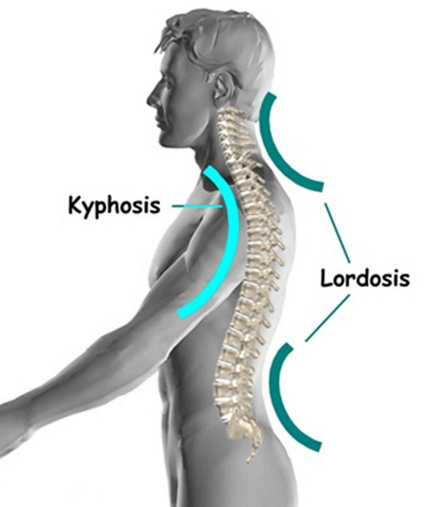
What is thoracic extension of the spine?
When the term “extension” is used about the thoracic spine, the meaning is a reduction in relative flexion. While there may not be true extension of the thoracic spine, this reduction in flexion is important for movement of the scapula and arm.
How much can the thoracic spine flex?
From neutral, the thoracic spine can flex to approximately 35° of additional flexion. Thus, in full flexion, the thoracic spine demonstrates up to 75° of flexion. Conversely, the thoracic spine only moves 20° to 25° of extension. Therefore, if the normal position is 40°, the fully extended is still 15-20° of flexion.
How far can the thoracic spine flex from neutral?
From neutral, the thoracic spine can flex to approximately 35° of additional flexion. Thus, in full flexion, the thoracic spine demonstrates up to 75° of flexion. Conversely, the thoracic spine only moves 20° to 25° of extension.

What is flexion of the thoracic spine?
From neutral, the thoracic spine can flex to approximately 35 ° of additional flexion. Thus, in full flexion, the thoracic spine demonstrates up to 75 ° of flexion. Conversely, the thoracic spine only moves 20 ° to 25 ° of extension.
What muscles do thoracic flexion?
Muscles of the Spinal ColumnTHORACIC MUSCLESFUNCTIONLongissimus ThoracisExtension, lateral flexion of vertebral column, rib rotationIliocostalis ThoracisExtension, lateral flexion of vertebral column, rib rotationSpinalis ThoracisExtends vertebral columnSemispinalis ThoracisExtends & rotates vertebral column1 more row•May 15, 2019
What is thoracic movement?
The primary movement of the thoracic spine is rotation. The other movements — flexion, extension, and side bending — are considerably smaller in range in comparison to the movement of the lower cervical (neck) and lumber (low back) areas.
How is thoracic spine flexion measured?
0:191:1847 Thoracic and Lumbar Lateral Flexion Goniometer and Tape ...YouTubeStart of suggested clipEnd of suggested clipWith the thoracic and lumbar spines positioned at the end of passive range measure the distance fromMoreWith the thoracic and lumbar spines positioned at the end of passive range measure the distance from the floor to the tip of the long finger.
What causes thoracic spine stiffness?
The most common reason is poor posture and not moving enough. Both of these are often caused by prolonged sitting at your desk with your back rounded, especially if your arms are stretched forward for things like computer work.
Why is thoracic mobility important?
Thoracic mobility can be thought of the available movement or motion of this region of our body, and is very important for achieving good posture to ensure you don't get pain from poor posture, and it is also essential for most sporting performances.
What does thoracic mean?
Definition of thoracic : of, relating to, located within, or involving the thorax.
What part of the body is thoracic?
The thoracic spine is located in the upper and middle part of the back. Twelve vertebrae are located in the thoracic spine and are numbered T-1 to T-12. Each number corresponds with the nerves in that section of the spinal cord: T-1 through T-5 nerves affect muscles, upper chest, mid-back and abdominal muscles.
What is the function of the thoracic?
Supporting your chest and abdomen: Your thoracic spine helps stabilize your rib cage, and your rib cage, in turn, helps stabilize your thoracic spine. Together, your thoracic spine and ribcage protect your heart and lungs.
What is normal range of motion for thoracic spine?
20° to 45°The normal ROM of forward flexion (forward bending) in the thoracic spine is 20° to 45°.
What is normal thoracic ROM?
Range of Motion ROM Using a Goniometer (Geelhoed et al, 2006): Flexion: 20-45 degrees. Extension: 25-45 degrees. Lateral Flexion: 20-40 degrees.
How do you test for thoracic mobility?
1:163:47Screening for thoracic spine mobility in athletes - YouTubeYouTubeStart of suggested clipEnd of suggested clipOkay a quick test to determine. If he has compensatory motion through a thoracic spine is to doMoreOkay a quick test to determine. If he has compensatory motion through a thoracic spine is to do shoulder active shoulder elevation and to see to see what happens it's Justin elevates his shoulder.
What three muscles perform flexion at the vertebral column?
They include the longissimus, iliocostalis, and spinalis muscles. Their attachments subdivide these muscles, and they all have a common tendinous origin. They play a role in the movement of the thoracic cage and flexion of the upper vertebral column and head.
What muscles perform flexion of the vertebral column?
The quadratus lumborum aids in lateral flexion of the vertebral column. The quadratus lumborum originates on the ilium and inserts on the last rib as well as lumbar vertebrae. When these muscles contract, they depress (or pull down) the ribs, and they also aid in lateral flexion of the vertebral column.
What muscles do lumbar flexion?
The primary muscles involved with producing lumbar flexion are the Rectus Abdominis, internal oblique, and external obliques. The nerves that innervate the Rectus Abdominis are the Intercostals (7-11) and Subcostal (T12).
Which muscles are responsible for flexing the lumbar spine?
These muscles include the large paired muscles in the lower back, called erector spinae, which help hold up the spine, and gluteal muscles. The flexor muscles are attached to the front of the spine and enable flexing, bending forward, lifting, and arching the lower back.
What muscles do thoracic flexion?
Spinal Muscles: A Comprehensive Guide THORACIC MUSCLES FUNCTION Longissimus Thoracis Extension, lateral flexion of vertebral column, rib rotation Iliocostalis Thoracis Extension, lateral flexion of vertebral column, rib rotation Spinalis Thoracis Extends vertebral column Semispinalis Thoracis Extends & rotates vertebral column.
What is thoracic flexibility?
From neutral, the thoracic spine can flex to approximately 35 ° of additional flexion. Thus, in full flexion, the thoracic spine demonstrates up to 75 ° of flexion. Conversely, the thoracic spine only moves 20 ° to 25 ° of extension.
What limits thoracic flexion?
The positioning of the ribs and spinous processes greatly limits flexion and extension of the thoracic vertebrae. Thoracic vertebrae have superior articular facets that face in a posterolateral direction.
What is normal thoracic range of motion?
Normal ranges of motion for the cervical spine include 50 degrees of flexion, 60 degrees of extension, 45 degrees of lateral, or side bending, and 80 degrees of rotation. The ranges of motion for the thoracic spine include 30 degrees of rotation and 50 degrees of kyphosis.
What are the symptoms of thoracic spine nerve damage?
What Are the Symptoms of Thoracic Spine Nerve Damage? Significant leg weakness or loss of sensation. Loss of feeling in genitals or rectal region. No control of urine or stool. Fever and lower back pain. A fall or injury that caused the pain.
Why do we need thoracic rotation?
Thoracic mobility can be thought of the available movement or motion of this region of our body, and is very important for achieving good posture to ensure you don’t get pain from poor posture, and it is also essential for most sporting performances. Posture. Upper limb movements. Lower limb movements.
Why is my thoracic spine so tight?
The causes of thoracic spine syndrome can vary significantly. The most common reason is poor posture and not moving enough. Both of these are often caused by prolonged sitting at your desk with your back rounded, especially if your arms are stretched forward for things like computer work.
Which joint is unique to the thoracic spine?
Costovertebral joints, unique to the thoracic spine - consists of the head of the rib articulating with:
What are the characteristics of the thoracic vertebrae?
The primary characteristic of the thoracic vertebrae is the presence of costal facets. There are 6 facets per thoracic vertebrae: 2 on the transverse processes and 4 demifacets. The facets of the transverse processes articulate with the tubercle of the associated rib. The demifacets are bilaterally paired and located on the superior and inferior posterolateral aspects of the vertebrae. They are positioned so that the superior demifacet of inferior vertebrae articulates with the head of the same rib that articulates with the inferior demifacet of a superior rib. For example, the inferior demifacets of T4 and the superior demifacets of T5 articulate with the head of rib 5.
How many facets are there in the thoracic vertebrae?
The primary characteristic of the thoracic vertebrae is the presence of costal facets. There are 6 facets per thoracic vertebrae: 2 on the transverse processes and 4 demifacets. The facets of the transverse processes articulate with the tubercle of the associated rib.
What is the name of the spinous process found at T1?
Vertebral prominens name given to long prominent spinous process found at T1. T11 and T12 are atypical - contain a single pair, “whole,” costal facet that articulate with the 11 and 12 ribs, respectively. They also lack facets on the transverse processes.
What is the normal sagittal plane alignment of the thoracic spine?
The sagittal plane alignment of the Thoracic spine is on average 35% (normal range is 20° to 50°).
How many thoracic arteries are there?
There are 12 in the thoracic region.
How many types of joints are there in the thoracic spine?
There are 3 types of joints present in the thoracic spine:
What is a thoracic extension?
Thoracic extension involves concurrent posterior rotation (external torsion) and depression of the posterior ribs with elevation of the anterior ribs. Bending to the side is a combination of spinal segments side bending, ribs on the same come together while ribs on the opposite side separate.
What is physical therapy?
home exercises that stretch and strengthen the back, shoulder and stomach muscles, massage, postural education, joint mobilisations, heat and/or ice therapy etc.
What is joint hypermobility?
Joint hypermobility is usually addressed with therapeutic exercise to improve neuromuscular control. Joint hypomobility may be addressed with manual interventions including joint mobilization and manipulation.
How to help a stiff upper back?
Exercises - home exercise program. A good way to help clients to combat a painful/stiff upper back is to make an appropriate home exercise program (HEP) ie the right stretching and strengthening routine. Be sure to read this link to help get better adherence to a HEP.
Is thoracic spine mobility dependent on the clinician?
Most interventions to address thoracic spine mobility are dependent on the clinician providing the intervention.
What plane is the thoracic spine oriented?
The superior and inferior facets of the vertebrae are oriented ~60 degrees from the horizontal plane and ~20 degrees from the frontal plane. Since the thoracic spine connects cervical to lumbar, the junctional regions are important considerations as well. When transitioning from one region, there is no immediate change between cervical ...
What is the critical zone of the thoracic spine?
As Dr. Chris Fox wrote in a previous post on thoracic spine anatomy, " In the thoracic spine, T4-9 is known as the critical zone because the vertebral canal is narrowest here; it also has reduced blood supply (Egan et al, 2011). T6 is a tension point; here motion of the spinal core versus canal converge in different directions." Improving the neural mobility in the thoracic spine can help improve movement and decrease pain in the joints above and below.
How many vertebrae are in the thoracic spine?
The thoracic spine is comprised of 12 vertebrae. These vertebrae have similar characteristics to cervical and lumbar spine- a vertebral body, pedicles directed posterior from the body, lamina that connect to form a spinous process, vertebral facets, and costal demi-facets (Neumann, 2010). The superior and inferior facets ...
Which vertebrae are similar to the cervical spine?
The superior thoracic vertebrae bare qualities similar to the cervical spine and the inferior thoracic vertebrae resemble the lumbar spine. This may explain why stiffness is often noted in these regions. Additionally, the thoracic spine houses the sympathetic nervous system.
What is T6 in the spine?
T6 is a tension point; here motion of the spinal core versus canal converge in different directions.". Improving the neural mobility in the thoracic spine can help improve movement and decrease pain in the joints above and below.
Which nerve is located anterior to the costotransverse joints?
Additionally, the thoracic spine houses the sympathetic nervous system. The sympathetic nerve trunk lies anterior to the costotransverse joints. Clinically, this may help explain unusual symptoms that can be reproduced by neural tension tests such as the SLUMP TEST or Straight Leg Raise Test.
Is thoracic spine pain a musculoskeletal pain?
In physical therapy school, the thoracic spine is often glossed over during the musculoskeletal courses. This may be due to the low incidence and prevalence of thoracic spine pain. The incidence of thoracic spine pain is only ~15-19% across the population. This is relatively low compared to lumbar spine pain, which has an 80% prevalence in adults.
Why is a thoracic extension important?
Proper Thoracic Extension is important not only for proper posture, but also to prevent neck, shoulder, upper back, lower back and even hip pain. If you don’t have proper Thoracic Extension, your body is going to take the path of least resistance ...
What stretch to do for a thoracic extension?
Half Wall Hang – A great stretch for your lats that will also work on your Thoracic Extension is the Half Wall Hang. With this stretch, you’ve got to make sure to focus on extending your upper and mid back as you relax over. It is very easy with this stretch to simply extend your low back instead of your Thoracic spine, especially if your Thoracic Extension is limited, so really focus on keeping your abs engaged as you extend.
How to improve thoracic extension?
The first step to improving your Thoracic mobility and extension is to roll out all the muscles that get tight from sitting in flexion for most of the day. That means you really need to focus on rolling out and loosening up your chest and lats.
What is the best way to stretch your bicep?
These stretches will loosen tight muscles caused by sitting in flexion for most of the day and will even start to put your spine through extension. Standing Chest Stretch – The Standing Chest Stretch is a great way to stretch your chest, shoulders and bicep.
What happens if you don't have a thoracic extension?
If you don’t have proper Thoracic Extension, your body is going to take the path of least resistance and seek out extra extension from other areas – areas that may not have the ability to provide the extension needed and therefore become overworked and injured.
What exercises can help with back extension?
No one move alone will solve your problem. You must included Foam Rolling, Stretching, Activation and Strength Training in your workout routine.
How to roll out chest?
To roll out your chest, you can use a larger foam ball against the ground or a smaller ball against the wall in a doorway. The smaller ball in the doorway can dig in a little bit more if you can handle the extra firmness and pressure.
How to get the thoracic spine to flex?
Move slowly and keep the spine neutral. Perform 12-15 repetitions. Overhead squat with tubing —this exercise will attempt to pull the arms and shoulders into extension and the thoracic spine into flexion.
How to do thoracic extension?
Try to emphasize thoracic flexion over the stability ball and then extend into a neutral position. Move slowly and don’t hyperextend the spine. Perform 12-15 reps.
What is the Thoracic Spine?
The thoracic spine is the twelve vertebrae of the middle segment of the vertebral column. While all vertebrae have rib components, they are usually small and make up the transverse processes in regions other than the thorax (Drake, Vogl & Mitchell, 2015).
What happens if the lumbar spine does not rotate?
If the thoracic spine does not rotate, the lumbar spine is forced to rotate more, which could play a role in low back pain or injury. As Bogduk (2005) describes, rotation of more than 3 ° at any lumbar segment could damage the articular surface and tear collagen fibers that make up the disc between each vertebra.
How many thoracic vertebrae rotate?
In many cases, the last two to three thoracic vertebrae begin to demonstrate movement characteristics of the lumbar spine. On average, each thoracic vertebra can rotate approximately 3 °. Therefore, the entire thoracic spine should demonstrate between 30 -35 ° of total rotation to each side (Neumann, 2010).
How much does the lumbar spine rotate?
However, the lumbar spine also rotates, albeit less than the thoracic. The lumbar vertebrae demonstrate approximately 2 ° of rotation between each, resulting in 10 ° of rotation to each side. Thus, the total thoracolumbar rotation should be approximately 45 ° in each direction.
What does "extension" mean in a scapula?
When the term “extension” is used about the thoracic spine, the meaning is a reduction in relative flexion. While there may not be true extension of the thoracic spine, this reduction in flexion is important for movement of the scapula and arm.
What is the body of a thoracic vertebra?
The body of a thoracic vertebra is somewhat “heart-shaped,” and is larger than the cervical but smaller than the lumbar vertebrae in size. The body also has small, smooth, and somewhat concave costal facets for the attachment of the ribs. Ribs are generally inserted between two vertebrae, such that each vertebra contributes to articulating with half of the articular surface. Each vertebra therefore has a pair of superior articular facets that face posteriorly and a pair of inferior articulating facets that face anteriorly (except for T12). This means that the rib will articulate with the inferior costal facet of the upper vertebrae and the superior costal facet of the lower vertebrae. Transverse processes arise from the arch found behind he superior articular processes and pedicles, and are thick and strong with a clubbed end and a small concave surface for the articulation with the tubercle of a rib. These processes are directed obliquely backward towards the spinous process and lateralward.
Where are the thoracic vertebrae located?
The thoracic vertebrae are located in the middle section of the vertebral column, specifically inferior to the cervical vertebrae and superior to the lumbar vertebrae. These vertebrae span the large majority of the chest cavity area.
How many vertebrae are there in the thoracic spine?
Two muscles also interact with those twelve vertebrae, these being the spinalis and longissimus.
How long is the reading time for the thoracic vertebrae?
Last reviewed: May 31, 2021. Reading time: 15 minutes. The twelve thoracic vertebrae are strong bones that are located in the middle of the vertebral column, sandwhiched between the cervical ones above and the lumbar vertebrae below. Like typical vertebrae, they are separated by intervertebral discs. However, they are various anatomical features ...
Why do thoracic vertebrae increase in size?
Thoracic vertebrae increase in size as they descend towards the lumbar vertebrae; this is because the lower vertebrae must be able to support more of the body’s weight when a person is standing due to the effects of gravity. To summarize, the main anatomical components of a thoracic vertebra are: Body. Spinous process.
What is the ribs of a vertebra?
Ribs are generally inserted between two vertebrae, such that each vertebra contributes to articulating with half of the articular surface. Each vertebra therefore has a pair of superior articular facets that face posteriorly and a pair of inferior articulating facets that face anteriorly (except for T12).
What are the distinguishing features of the thoracic vertebrae?
Distinguishing features of the thoracic vertebrae include the presence of facets on the sides of the bodies for articulation with the heads of the ribs, and facets on the transverse processes of all, except the 11th and 12th vertebrae, for articulation with the tubercles of the ribs.

Introduction
Bones
Joints
Structure and Function
Nerves
Ligaments
Intervertebral Disc
- The intervertebral disc (IVD) is important in the normal functioning of the spine. 1. It is a cushion of fibrocartilageand the principal joint between two vertebrae in the spinal column. 2. There are 12 in the thoracic region. 3. IVDs allow the spine to be flexible without sacrificing a great deal of strength and provide a shock-absorbing effect wi...
Muscles
Pathophysiology