What does the oculomotor nerve do?
What is the oculomotor nerve?
- Olfactory nerve (CN I) enables sense of smell.
- Optic nerve (CN II) enables vision.
- Trigeminal nerve (CN V) enables sensation in your face.
- Vestibular and cochlear nerves (CN VII) enable balance and hearing.
What muscle is innervated by the trochlear nerve?
The trochlear nerve (/ˈtrɒklɪər/), also called the fourth cranial nerve or CN IV, is a motor nerve (a somatic efferent nerve) that innervates just one muscle: the superior oblique muscle of the eye, which operates through the pulley-like trochlea.
Which nerve innervates all your hamstring muscles?
- Origin: Lower, medial surface of the ischial tuberosity
- Insertion: Medial tibia (pes anserinus)
- Function: Knee flexion, hip extension and medial rotation of the tibia (with knee flexion)
- Innervation: Tibial nerve
- Vascular supply: Perforating branches of the deep femoral artery
What are cranial nerves provide both sensory and motor innervation?
Trigeminal. Both sensory and motor. Pons. Three Parts: V 1 ( ophthalmic nerve) is located in the superior orbital fissure V 2 ( maxillary nerve) is located in the foramen rotundum. V 3 ( mandibular nerve) is located in the foramen ovale . Receives sensation from the face and innervates the muscles of mastication .
See 7 key topics from this page & related content
See 7 key topics from this page & related content
Where does the oculomotor nerve begin and end?
What is the anatomy of cranial nerve III? CN III starts in the midbrain. It travels through many structures in your head until it reaches the back of your eyes.
How many muscles does the oculomotor nerve innervate?
These nerve axons will arise from the oculomotor nucleus and innervate skeletal muscles associated with the eye. There are seven extrinsic eye muscles (muscles that lay outside of the eye itself) that move the superior eyelid and the eyeball.
What does the oculomotor nerve control?
Cranial nerve 3, also called the oculomotor nerve, has the biggest job of the nerves that control eye movement. It controls 4 of the 6 eye muscles in each eye: Medial rectus muscle (moves the eye inward toward the nose) Inferior rectus muscle (moves the eye down)
Is the oculomotor nerve sensory or motor?
motorThe trochlear, abducens, accessory, and hypoglossal nerves are only motor nerves; the trigeminal nerve is both sensory and motor; the oculomotor nerve is both motor and parasympathetic; the facial glossopharyngeal, and vagus nerves have sensory, motor, and parasympathetic components (Standring, 2008).
What extraocular muscles are innervated by the oculomotor nerve?
The oculomotor nerve innervates the superior, inferior, and medial recti, as well as the inferior oblique and levator palpebrae superioris muscles.
Which muscles are innervated by which nerves?
Muscles Innervated by Cranial NervesCranial NerveMuscleGlossopharyngeal nerve (CN IX)Stylopharyngeus muscleVagus nerve (CN X)Muscles of the palate and pharynx except tensor palati muscle (CN V3) Stylopharyngeus muscle (CN IX) All muscles of the larynxAccessory nerve (CN XI)Sternocleidomastoid muscle Trapezius muscle6 more rows•Apr 9, 2020
What muscles does cranial nerve 5 innervate?
This branch supplies motor innervation to the facial muscles involved in mastication which include the masseter, temporalis muscle, and the lateral and medial pterygoids.
Which of the indicated muscles is not innervated by oculomotor nerve?
The oculomotor nucleus originates at the level of the superior colliculus. The muscles it controls are the striated muscle in levator palpebrae superioris and other extraocular muscles except for the superior oblique muscle and the lateral rectus muscle.
Where does the oculomotor nerve start?
The oculomotor nerve begins at the brainstem, which is a structure low in the back of your brain that connects the brain to the spinal column. In the brainstem, two clusters of neurons called nuclei give rise to the oculomotor nerve.
Which nerve is responsible for the movement of the eye?
Motor function means movement, and the oculomotor nerve is responsible for much of the movement associated with your eyes.
What nerve divides into superior and inferior branches?
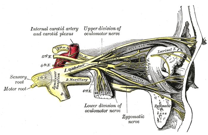
Once the oculomotor nerve is inside the orbit, it divides into its superior and inferior branches.
What nerves are involved in eye movement?
Anatomy. Function. Associated Conditions. Treatment. The oculomotor nerve enables most of your eye movements, some aspects of vision, and raising the eyelid. It's the third cranial nerve and works with cranial nerves four ( trochlear) and five ( trigeminal) to coordinate eye movement. The oculomotor nerve contains both motor ...
What causes oculomotor palsy?
It's caused by compression of the nerve at the junction of the posterior communicating artery and the internal carotid artery. Symptoms of congenital oculomotor palsy include: A pupil that's "fixed" (doesn't change size in response to light) on the same side as the compression.
Which nerve innervates the parasympathetic fibers?
The parasympathetic fibers from the oculomotor nerve innervate two muscles inside the iris:
Which nerve branches out to innervate the muscles?
As it travels through your head toward the eyes, the oculomotor nerve branches out to innervate (supply nerve function to) various muscles.
What is the oculomotor nerve?
The oculomotor nerve is the third of 12 pairs of cranial nerves in the brain. This nerve is responsible for eyeball and eyelid movement.
Which nerve controls the eye?
The oculomotor nerve involves two separate components, each of which has a distinct function. The somatic motor component supplies four extraocular muscles in the eye and the upper eyelid’s levator palpebrae superioris with motor (movement) fibers. It controls the muscles that allow for visual tracking and fixation by the eye.
Which motor component controls parasympathetic innervation?
The visceral motor component controls parasympathetic innervation (nerves related to involuntary actions) of the ciliary muscles and constrictor papillae, aiding in accommodation and pupillary light reflexes. Accommodation is the ability of the eye to keep an object in focus as the object’s distance from the eye changes. Pupillary light reflexes are automatic changes in dilation (size) of the pupil, which regulate the amount of light that enters the eye, making sure the light is enough to see but not too bright.
Can oculomotor nerves be paralyzed?
The oculomotor nerve can become paralyzed in a condition known as oculomotor nerve palsy. This condition can result from multiple sclerosis or other demyelinating diseases, direct trauma, space-occupying lesions (such as brain cancer), microvascular disease (such as diabetes), or spontaneous subarachnoid hemorrhage (bleeding into the space between two of the membranes that cover the brain). A berry aneurysm is a type of subarachnoid hemorrhage.
Where does the oculomotor nerve originate?
The oculomotor nerve originates from the third nerve nucleus at the level of the superior colliculus in the midbrain. The third nerve nucleus is located ventral to the cerebral aqueduct, on the pre-aqueductal grey matter. The fibers from the two third nerve nuclei located laterally on either side of the cerebral aqueduct then pass through ...
Which nerves innervate the eye?
The oculomotor nerve is derived from the basal plate of the embryonic midbrain. Cranial nerv es IV and VI also participate in control of eye movement.
Why is it easier to detect damage to the oculomotor nerve?
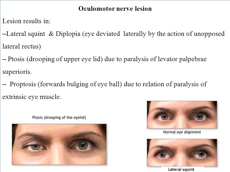
Since the oculomotor nerve controls most of the eye muscles, it may be easier to detect damage to it. Damage to this nerve, termed oculomotor nerve palsy, is known by its down and out symptoms, because of the position of the affected eye (lateral, downward deviation of gaze).
What causes paralysis of the oculomotor nerve?
Paralysis of the oculomotor nerve, i.e., oculomotor nerve palsy, can arise due to: 1 direct trauma, 2 demyelinating diseases (e.g., multiple sclerosis ), 3 increased intracranial pressure (leading to uncal herniation )#N#due to a space-occupying lesion (e.g., brain cancer) or a#N#spontaneous subarachnoid haemorrhage (e.g., berry aneurysm ), and 4 microvascular disease, e.g., diabetes.
What causes oculomotor nerve palsy?
Disease. Paralysis of the oculomotor nerve, i.e., oculomotor nerve palsy, can arise due to: microvascular disease, e.g., diabetes. In people with diabetes and older than 50 years of age, an oculomotor nerve palsy, in the classical sense, occurs with sparing (or preservation) of the pupillary reflex.
Which branch of the oculomotor nerve supplies the superior rectus and levator palpebrae?
Superior branch . The superior branch of the oculomotor nerve or the superior division, the smaller, passes medially over the optic nerve. It supplies the superior rectus and levator palpebrae superioris .
Which cranial nerve innervates the extrinsic eye muscles?
t. e. The oculomotor nerve is the third cranial nerve (CN III). It enters the orbit through the superior orbital fissure and innervates extrinsic eye muscles that enable most movements of the eye and that raise the eyelid.
Where do the oculomotor nerve fibers come from?
The fibers of the oculomotor nerve arise from the medial part of the cerebral peduncle. We can distinguish a medial or interpeduncular group of fibers, and a lateral group of fibers that emerge from the anterior surface of the peduncle. These fibers unite to form a nerve cord whose course we will examine.
Which system separates the oculomotor nerves?
With the basilar arterial system. The basilar trunk separates the two oculomotor nerves, which then pass between the posterior cerebral and superior cerebellar arteries. In the cavernous sinus. The oculomotor nerve lies on the most cephalic part of the wall of the cavernous sinus. In the superior orbital fissure.
How many myelinated nerve fibers are in the oculomotor nerve?
Anatomy and pathophysiology. The oculomotor nerve is composed of about 23 000 myelinated nerve fibers which extend from cell bodies in the midbrain to their target muscles in the orbit. A number of authors have described the portion of this nerve comprising autonomic fibers, some of which are pupillomotor axons.
What nerve is responsible for elevating the upper eyelid?
The oculomotor nerv e is the cranial nerve responsible for innervating the superior, inferior, and medial recti. It functions to rotate the eye medially, upwards, and downwards while also being responsible for elevating the upper eyelid. The nerve tract can be described with five segments: the nucleus, fascicles, subarachnoid cistern, cavernous sinus, and intra-orbital segments. Injury to the oculomotor nerve can happen anywhere along this tract. The four mechanisms of injury that lead to oculomotor nerve palsy are sudden displacement, gradual displacement, ischemia, and direct involvement.
What nerves control the eye?
The oculomotor, trochlear, and abducens nerves control actions of the intraocular (pupillary sphincter) and extraocular muscles. These nerves are observed for symmetry of eye movement, globe position, asymmetry or drooping of the eyelid (ptosis), and twitching or fluttering of the lids or globes. With the lights dimmed, anisocoria or differences in the sizes of the pupils should be ruled out. Pupillary light response is evaluated for quickness and symmetry. Normally, the pupils average about 3 mm in diameter in a room with adequate light. They can range, however, from about 6 mm in children to < 2 mm in elderly patients. Also, one patient's pupils may be different from each other by about 1 mm in size. This is called physiologic anisocoria. The pupils are normally round and regularly shaped, constricting quickly from direct illumination, and slightly less to illumination of the pupils on the opposite side. This is called the consensual response. They dilate again quickly when illumination is removed. Normal pupils constrict when the eyes converge to focus on a closer object, such as the tip of the nose. This is known as accommodation.
Which nerve runs forward, laterally and very slightly cephalad towards the lateral side of the posterior clin?
On leaving the peduncle, the oculomotor nerve runs forward, laterally and very slightly cephalad towards the lateral side of the posterior clinoid process (Fig. 12.1 ). Before the posterior clinoid process, it crosses the dura mater to enter the lateral wall of the cavernous sinus.
Is oculomotor palsy rare?
Oculomotor palsy is rare in childhood [29]. We had a 7-year-old child who presented with right-sided oculomotor paresis due to an arachnoid cyst located in the interpeduncular and ambient cistern more to the right side ( Fig. 4.6A–C ). Cysts in this location may behave similarly to a posterior communication artery aneurysm that compresses the oculomotor nerve with ophthalmoplegia, including ptosis and ipsilateral pupillary dilatation.
Which cranial nerves are responsible for innervation of the extraocular muscles?
Of the cranial nerves with motor functions, CN III, CN IV, and CN VI are the ocular motor nerves, which provide innervation to the extraocular muscles.
Which nerves are involved in the motor nucleus of the cranial nerves?
The corticobulbar tract (otherwise known as the corticonulcear tract) is responsible for influencing the motor nuclei of a number of cranial nerves, including the: oculomotor (III)
What nerves are affected by trochlear palsy?
Trochlear nerve palsy. Damage to the trochlear nerve interrupts motor input to the superior oblique muscle. If the oculomotor and abducens nerves are unaffected, the actions of the recti and inferior oblique muscles will be unopposed.
Why do I have oculomotor paralysis on the same side of the lesion?
Because of this, patients present with oculomotor paralysis on the same side of the lesion due to damage to the ipsilateral oculomotor nerve, and contralateral hemiplegia due to damage to the upper motor neuron fibers of the corticospinal tract within the crus cerebri. Oculomotor deficits may include:
Which nerve is damaged by oculomotor nerve palsy?
Oculomotor nerve palsy. Damage to the oculomotor nerv e interrupts motor input to the majority of extraocular muscles, including most of the recti and the inferior oblique, as well as the levator palpebrae superioris, the sphincter pupillae, and the ciliary muscles.
Where do corticobulbar fibers come from?
Corticobulbar motor fibers arise from the frontal eye fields (a region in the caudal portion of middle frontal gyrus), motor cortex (the precentral gyrus), and somatosensory cortex (the postcentral gyrus) where they travel from the cortex through the internal capsule and to the CN nuclei in the brainstem.
What are the components of the reflex?
This reflex has two main components, an afferent (sensory) component involving the optic nerve and an efferent (motor) component involving the oculomotor nerve . The sequence begins with light-induced activation of retinal fibers (the afferent component of the reflex).
Where does the oculomotor nerve originate?
The oculomotor nerve originates from 2 nuclei in the midbrain [1]: Oculomotor nucleus. Accessory parasympathetic nucleus (Edinger-Westphal nucleus) The oculomotor nerve exits the brainstem near midline at the base of the midbrain just caudal to the mammillary bodies. It passes through the cavernous sinus and proceeds through ...
What is the function of the oculomotor nerve?
Somatic (voluntary) functions of the oculomotor nerve include elevation of the upper eyelid via innervation of the levator palpebrae superioris muscle. Other essential functions include coordination of eye muscles for visual tracking and gaze fixation.
What nerve helps to coordinate eye position?
The oculomotor nerve helps to adjust and coordinate eye position during movement. Several movements assist with this process: saccades, smooth pursuit, fixation, accommodation, vestibulo-ocular reflex, and optokinetic reflex.
What nerves are involved in innervation of the eyelid?
Innervation to the upper eyelid (somatic) Innervation of the eye muscles that allow for visual tracking and gaze fixation (somatic) Like all other nerve fibers in the human body, the oculomotor nerve can become impaired in disease states which can lead to lifelong impairment in normal vision.
Why is oculomotor nerve palsy important?
Recognizing oculomotor nerve dysfunction (oculomotor nerve palsy) is crucial because it can be a sign of underlying or uncontrolled chronic disease states. It is an essential clinical symptom that can be life-saving in certain circumstances.
Where does the parasympathetic nerve originate?
The parasympathetic nerve supply of the oculomotor nerves develops from the caudal midbrain and rostral hindbrain neural crest cells. Some cells may also originate from the ectodermal placode caudal to the eye. [3][4] These cells migrate ventrolaterally and rostrally toward the optic vesicle.
Where is the oculomotor nucleus located?
Both the oculomotor nucleus and Edinger-Westphal nucleus are both located in the medial midbrain, which is supplied by the paramedian branches of the upper basilar artery and the proximal posterior cerebral artery. Nerves. The oculomotor nerve helps to adjust and coordinate eye position during movement.