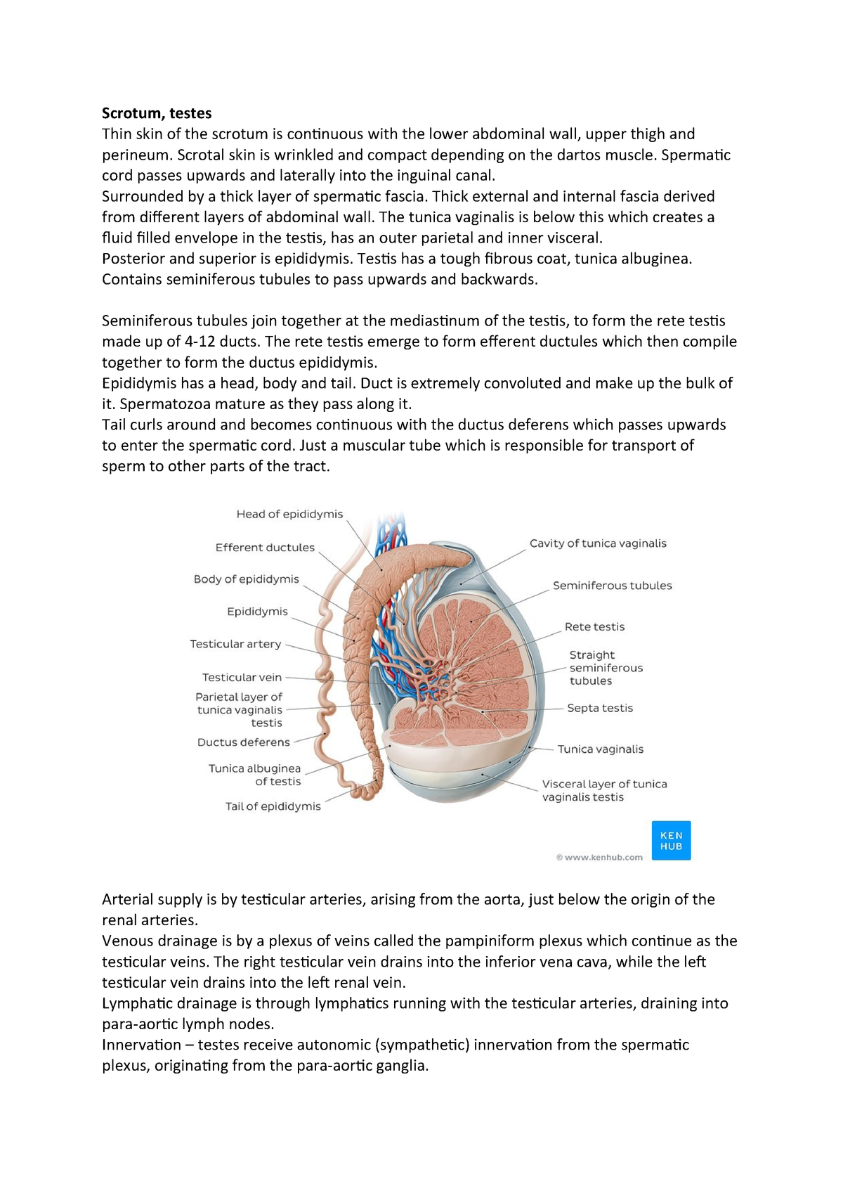
What are the tubules of the testes surrounded by?
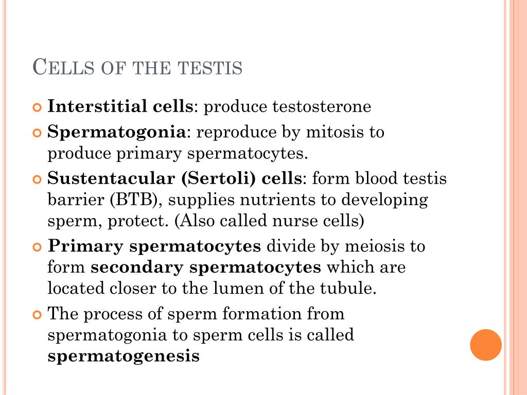
The tubules are surrounded by the connective tissue stromal cells which contain testosterone secreting Leydig (interstitial) cells. The tubules are lined with a layer of seminiferous epithelium, which contains supporting Sertoli (sustentacular) cells, and spermatogenic cells.
How many seminiferous tubules are in the testis?
Put your knowledge to the test with our reproductive system labeling diagrams and quizzes. Each of the 200-300 lobules of the testis are filled with one to four highly convoluted seminiferous tubules which each course towards the mediastinum testis.
What is the most superficial layer of the testis?
The most superficial layer of the testis is a capsule made of dense fibrous connective tissue called the tunica albuginea, which perpendicularly gives rise to the septa that divide the tissue of the testis into lobules.
What is the function of the seminiferous tubules?
The seminiferous tubules are where the production, maturation and transport of the sperm cells takes place. The seminiferous tubules are located in the male testicles, which are the two oval-shaped organs on located beneath the penis. In each testicle, there are approximately 800 seminiferous tubules.

What are the 3 layers of the testes?
There are three layers to the tunica, the tunica vasculosa, tunica albuginea and tunica vaginalis.Tunica vasculosa. The tunica vasculosa is the inner layer of the tunica and consists of blood vessels and connective tissue. ... Tunica albuginea. ... Tunica vaginalis.
What is the seminiferous tubules of the testes?
The seminiferous tubules are the basic units of the testicles where the SSCs proliferate and differentiate through cyclic events (mitosis, meiosis, postmeiotic spermatid development, and spermiogenesis) to generate spermatozoa in a process called spermatogenesis [19].
Which structures are found within the seminiferous tubules of the testes?
coiled tubules known as the seminiferous tubules; the sperm cells are produced within the walls of the tubules. Within the walls of the tubules, also, are many randomly scattered cells, called Sertoli cells, that function to support and nourish the immature sperm cells by giving them nutrients and blood products.…
Where are seminiferous tubules located quizlet?
Seminiferous tubules are located in the testis. Made up of columnar Sertoli cells surrounded by spermatogenic cells on the epithelial interior and stem cells exteriorly. The seminiferous tubules function to produce sperm, maintain sperm, and store the sperm.
Where is the seminiferous tubules located?
Abstract. Seminiferous tubules of testis open into a continuous chamber of flattened and irregular-shaped anastomosing channels called the rete testis. Positioned at one pole of the testis (hilus), the rete is adjacent to the connective tissue capsule covering of the testis, known as the tunica albuginea.
Where can you find seminiferous tubules?
the testisSeminiferous tubules are special tubular structures in the testis that produce sperm continuously throughout the male life cycle.
What surrounds seminiferous tubules?
The seminiferous tubules are surrounded by a basal lamina composed of extracellular matrix that serves to separate them from the interstitial compartment, provides structural integrity to the tubules, and regulates the function of cells in contact with it.
Where are seminiferous tubules of each lobe empty sperms?
Each lobe contains four tightly coiled seminiferous tubules (this is the location of actual sperm production). The seminiferous tubules empty sperm into the testicular network where they travel to the epididymis. The epididymis is located outside of the testis (see figures 1-8 and 1-9).
What is the tunica albuginea made of?
type I collagenThe tunica albuginea is composed primarily of tough type I collagen with a minority component of more flexible type III collagen and elastin. It is arranged in a bilayer, with inner circular layers and outer longitudinal layers (see Fig.
What cells are located in the epithelium of seminiferous tubules?
The seminiferous tubules are lined by a complex stratified epithelium containing two distinct populations of cells, spermatogenic cells, that develop into spermatozoa, and Sertoli cells which have a supportive and nutrient function.
What cells are located in the epithelium of seminiferous tubules quizlet?
The insides of the tubules are lined with seminiferous epithelium, which consists of two general types of cells: spermatogenic cells and Sertoli cells.
What type of cell did you locate outside of the seminiferous tubule?
The regions outside the seminiferous tubules are called interstitial spaces, which contain Leydig's cell.
What is the main function of seminiferous tubules?
They play a role in the production of sperms. They help in the maintenance of sperms. They also play a role in storage of the sperms. During the process of meiosis, the Sertoli cells that line the seminiferous tubules undergo the process of differentiation to be converted into sperm.
What is mean by seminiferous tubules?
Definition of seminiferous tubule : any of the coiled threadlike tubules that make up the bulk of the testis and are lined with a layer of epithelial cells from which the spermatozoa are produced.
What is a seminiferous?
Medical Definition of seminiferous : producing or bearing seed or semen the seminiferous epithelium of the testis.
What do seminiferous tubules secrete?
They secrete androgen-binding protein, a binding protein which increases the concentration of testosterone inside the seminiferous tubules. Embryologically, they also secrete the anti-Müllerian hormone (AMH) necessary for the female Müllerian ducts to regress.
Where are seminiferous tubules formed?
The seminiferous tubules are formed from the testis cords that develop from the primitive gonadal cords, formed from the gonadal ridge .
What type of cells are in the tubule?
The epithelium of the tubule consists of a type of sustentacular cells known as Sertoli cells, which are tall, columnar type cells that line the tubule. In between the Sertoli cells are spermatogenic cells, which differentiate through meiosis to sperm cells. Sertoli cells function to nourish the developing sperm cells.
What hormones do a sex slave secrete?
Embryologically, they also secrete the anti-Müllerian hormone (AMH) necessary for the female Müllerian ducts to regress.
Which side of the scrotum is the longitudinal section?
Longitudinal section through the left side of the scrotum and the left testis (Seminiferous tubules visible in center, but not labeled). Seminiferous tubule (transverse section). Photomicrograph of section through rat testis, showing seminiferous tubules.
What happens to DNA during spermatogenesis?
During spermatogenesis, the DNA of spermatogenic cells in the seminiferous tubules is subject to damage from such sources as reactive oxygen species. The genomic integrity of spermatogenic cells is protected by DNA repair processes. Deficiencies in the enzymes employed in these repair processes may lead to infertility.
Why are testis outside the body?
in order for the proteins to not denature(stopping meiosis), testis are outside body
Where does seminal fluid go?
seminal fluid only goes into urethra, but sperm actually goes into body cavity
What is the function of the epididymis?
allows epididymis to produce substances(glycoproteins and lipids) that make sperm mature and maintains structure of tubules
What is the basement membrane made of?
acellular basement membrane that surrounds the outside of the tubules. made of peritubular myoid cells
What is a defect in sperm transport?
defect in sperm transport(ductus deferens is blocked), male being over breed/over used-sperm not viable for long
How long is sperm viable?
end of epididymis. sperm is almost done maturing by the end of this, more of a site for storage"waiting" sperm is viable here for 60 days
Which organ communicates with hormones produced in the pituitary?
communicates with hormones produced in the pituitary; testis produces testosterone that goes into bloodstream to act on other muscles;temperaturecommunication to testis for warmth(neuro-endocrine redlex acts;impulse to brain to contract cremaster muscle)
What is the stratified seminiferous epithelium?
Stratified seminiferous epithelium lining these tubules contains Sertoli cells and several layers of germ cells. A basal lamina and a thin layer of connective tissue surrounds each tubule. 600x. The convoluted portions of the seminiferous tubules form the main mass of the testis and are the site of spermatogenesis.
Where do primary spermatocytes form?
Primary spermatocytes are diploid cells that form in the basal compartment but quickly move through the blood-testis barrier to the adluminal compartment. Many primary spermatocytes are seen because they remain in prophase of the first meiotic division for an extended period.
What is a spermatid?
Spermatids are haploid cells formed when secondaries complete the second meiotic division. Early spermatid nuclei are spherical and smaller than spermatogonia nuclei. Spermatids do not divide but differentiate by condensing and elongating their nuclei, developing a flagellum and losing excess cytoplasm. Cytoplasmic continuity between adjacent spermatids is lost during this process.
What is the second meiotic division?
Secondary spermatocytes complete the second meiotic division very quickly, so these cells are not frequently seen in sections. Spermatids (early) >. Spermatids are haploid cells formed when secondaries complete the second meiotic division. Early spermatid nuclei are spherical and smaller than spermatogonia nuclei.
How are sertoli cells identified?
Therefore, Sertoli cells must be identified primarily by their nuclei, which are oval, euchromatic, infolded and display a prominent nucleolus. Sertoli cells form the blood-testis barrier and do not divide.
What is the tunica propria?
The tunica propria is composed of the basal lamina of the seminiferus epithelium, three to five layers of myoid cells and collagen fibrils. The myoid cells secrete the fibrils and contract in peristaltic waves to propel spermatozoa through the seminiferous tubules. Sertoli cells >.
Overview
Structure
The epithelium of the tubule consists of a type of sustentacular cells known as Sertoli cells, which are tall, columnar type cells that line the tubule.
In between the Sertoli cells are spermatogenic cells, which differentiate through meiosis to sperm cells. Sertoli cells function to nourish the developing sperm cells. They secrete androgen-binding protein, a binding protein which increases the concentration of testosterone inside the seminifer…
Function
Spermatogenesis, the process for producing spermatozoa, takes place in the seminiferous tubules. During spermatogenesis, the DNA of spermatogenic cells in the seminiferous tubules is subject to damage from such sources as reactive oxygen species. The genomic integrity of spermatogenic cells is protected by DNA repair processes. Deficiencies in the enzymes employed in these repair processes may lead to infertility.
Additional images
• Seminiferous tubule (right) with sperm (black, tiny, ovoid). H&E stain.
• Longitudinal section through the left side of the scrotum and the left testis (Seminiferous tubules visible in center, but not labeled).
• Seminiferous tubule (transverse section).
See also
• Leydig cells
External links
• Histology image: 17802loa – Histology Learning System at Boston University
• Image
• Diagram