Explore
- High blood pressure
- Diabetes
- High cholesterol
- Family history of stroke or heart disease
- Recent transient ischemic attack (TIA) or stroke
- Abnormal sound in carotid arteries (bruit), detected by your doctor using a stethoscope
- Coronary artery disease
Why would I need a carotid ultrasound?
- Blood clot in the carotid arteries that can slow or block blood flow to the brain
- Carotid artery dissection, which is a split in the layers of the carotid artery wall. ...
- Carotid artery stenosis, which is a narrowing of the carotid arteries due to a buildup of plaque inside them. ...
- Congenital malformations, which are abnormalities that are present at birth
What to expect after carotid ultrasound?
Your doctor will recommend carotid ultrasound if you have the following medical conditions:
- Stenosis of the carotid arteries
- Transient ischemic attack (TIA)
- Stroke
- High blood pressure
- Diabetes
- Abnormal sound in carotid arteries, detected by your doctor using a stethoscope
- High cholesterol in the blood
- Coronary artery disease
- Heart disease
- Family history of stroke or heart disease
Who needs carotid ultrasound?
Who Should Get a Carotid Artery Ultrasound Strokes are the fifth leading cause of death in the United States, and they are the main reason why you would need to get a carotid artery screening. This test is advisable if you have a family history of stroke, high blood pressure, high cholesterol or are overweight or a smoker.
Why is carotid artery ultrasound advised?

Why would a doctor order a carotid Doppler?
A carotid ultrasound is performed to test for narrowed carotid arteries, which increase the risk of stroke. Carotid arteries are usually narrowed by a buildup of plaque — made up of fat, cholesterol, calcium and other substances that circulate in the bloodstream.
What does a carotid Doppler test show?
A carotid artery Doppler ultrasound is a diagnostic test used to check the circulation in the large arteries in the neck. This exam shows any blockage in the veins by a blood clot or “thrombus” formation.
When do you use the carotid Doppler?
You may need a carotid artery ultrasound when your healthcare provider wants to look for blood clots or plaque (fat and cholesterol deposits) on your carotid artery walls. These plaque deposits can limit ― and eventually block ― the flow of blood to your brain, face and neck.
What are the most common reasons for doing Doppler ultrasound?
A Doppler ultrasound may help diagnose many conditions, including:Blood clots.Poorly functioning valves in your leg veins, which can cause blood or other fluids to pool in your legs (venous insufficiency)Heart valve defects and congenital heart disease.A blocked artery (arterial occlusion)More items...
What are some symptoms of a blocked carotid artery?
Carotid Artery Blockage SymptomsBlurred vision or vision loss.Confusion.Memory loss.Numbness or weakness in part of your body or one side of your body.Problems with thinking, reasoning, memory and speech.
Who needs carotid ultrasound?
Your doctor may recommend a carotid ultrasound if you: Have had a stroke or mini-stroke recently. During a mini-stroke, you may have some or all of the symptoms of a stroke. However, the symptoms usually go away on their own within 24 hours.
How accurate is a carotid Doppler?
Results: ICA Doppler ultrasound sensitivity and specificity resulted respectively of 97% (confidence interval [CI] 95%) and 100% ([CI] 95%), while negative likelihood ratio was 0.03 (CI 95%).
How long does it take for a carotid Doppler?
The carotid Doppler usually takes 30 to 45 minutes to complete.
How long does it take to get results from carotid ultrasound?
It may take 3 to 4 days before you know the results of your test. What is a Carotid Artery Duplex? A Carotid Artery Duplex is an ultrasound test that uses high frequency sound waves (ultrasound) to show how well blood is flowing through the carotid arteries. The carotid arteries are located in the neck.
What happens if a Doppler test is positive?
Normal test results indicate thatyou have no narrowing or blockages in your arteries. It also means that the blood pressure in your arteries is normal. Abnormal blood flow patterns, including narrowing or closing of the arteries, can indicate: blockage in the arteries, which may be due to a buildup of cholesterol.
How long does it take to get results from a Doppler test?
Get your results in 2-5 days from an accredited laboratory with free shipping.
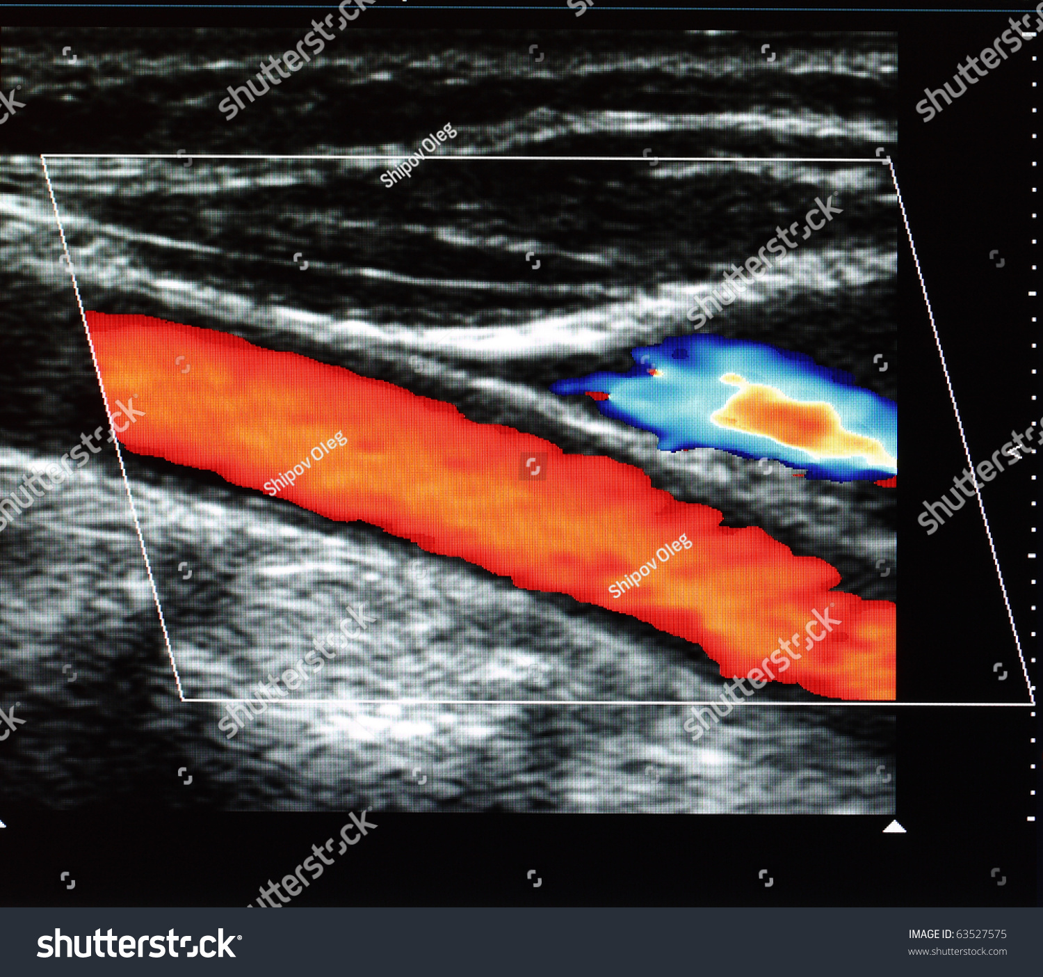
What does a blocked carotid artery look like on ultrasound?
0:344:18Identifying carotid artery disease with ultrasound - YouTubeYouTubeStart of suggested clipEnd of suggested clipAnd will be shown in the lighter shades of red on the colour flow map a tight stenosis or highMoreAnd will be shown in the lighter shades of red on the colour flow map a tight stenosis or high velocity jet just beyond the stenosis may also cause aliasing to occur in the colour flow.
How accurate is a carotid Doppler?
Results: ICA Doppler ultrasound sensitivity and specificity resulted respectively of 97% (confidence interval [CI] 95%) and 100% ([CI] 95%), while negative likelihood ratio was 0.03 (CI 95%).
What are normal carotid Doppler results?
Normal Results A normal result means there is no problem with the blood flow in the carotid arteries. The artery is free of any significant blockage, narrowing, or other problem.
What is the treatment for a blockage in the carotid artery?
Carotid endarterectomy, the most common treatment for severe carotid artery disease. After making an incision along the front of your neck, the surgeon opens the affected carotid artery and removes the plaques. The artery is repaired with either stitches or a graft.
What percentage of carotid artery blockage requires surgery?
If a carotid artery is narrowed from 50% to 69%, you may need more aggressive treatment, especially if you have symptoms. Surgery is usually advised for carotid narrowing of more than 70%. Surgical treatment decreases the risk for stroke after symptoms such as TIA or minor stroke.
Why Do I Need a Carotid Ultrasound?
A physician prescribes a carotid ultrasound for a variety of reasons, including if
What Do Carotid Arteries Do?
Your large carotid arteries supply blood to the brain. These arteries can narrow due to arteriosclerosis or other causes and impede blood flow, which can lead to transient ischemic attack ( a mini-stroke) or cerebral vascular accident (a stroke).
What happens after a carotid artery test?
An ultrasound technician records the completed test on a videotape. A diagnostic radiologist reviews the tape to measure blood flow and determine the amount and location of any narrowing of the carotid arteries. The radiologist then sends a report to your physician.
Why do carotid arteries narrow?
Your large carotid arteries supply blood to the brain. These arteries can narrow due to arteriosclerosis or other causes and impede blood flow, which can lead to transient ischemic attack ( a mini-stroke) or cerebral vascular accident (a stroke).
How long does it take to get a carotid ultrasound?
For most people, a carotid ultrasound takes an average of 15 to 30 minutes. 2 You can expect your healthcare practitioner to follow the five steps listed below, but what actually happens may vary depending on your condition, so follow their instructions. Remove any obstructions to the area, such as clothes or jewelry, as requested. ...
Why is the Doppler wand moved back and forth?
The Doppler or ultrasound wand is moved back and forth over the neck to detect blood flow.
How Is It Performed?
For most people, a carotid ultrasound takes an average of 15 to 30 minutes. 2 You can expect your healthcare practitioner to follow the five steps listed below, but what actually happens may vary depending on your condition, so follow their instructions.
What does a carotid ultrasound reveal?
The doctor who ordered the test will explain to you what the carotid ultrasound revealed and what that means for you. If the test reveals you're at risk of a stroke, your doctor may recommend the following therapies, depending on the severity of blockage in your arteries:
How long does a carotid ultrasound take?
In a Doppler ultrasound, the rate of blood flow is translated into a graph. A carotid ultrasound usually takes about 30 minutes.
What is abnormal sound in carotid arteries?
Abnormal sound in carotid arteries (bruit), detected by your doctor using a stethoscope. Coronary artery disease. To screen for narrowed or blocked blood vessels in other areas of the body, you may need additional tests, including: Abdominal ultrasound.
What type of ultrasound is recommended for TIAs?
Your doctor will recommend carotid ultrasound if you have transient ischemic attacks (TIAs) or certain types of stroke and may recommend a carotid ultrasound if you have medical conditions that increase the risk of stroke, including: High blood pressure. Diabetes. High cholesterol.
Why do you need an ultrasound in your abdomen?
Abdominal ultrasound. You may have an abdominal ultrasound to test for conditions affecting the blood vessels or organs in your abdominal area.
How does a sonographer test work?
How it works. A technician (sonographer) conducts the test with a small, hand-held device called a transducer. The transducer emits sound waves and records the echo as the waves bounce off tissues, organs and blood cells. A computer translates the echoed sound waves into a live-action image on a monitor.
What is a CTA scan?
Computerized tomography angiogram (CTA) scan. A CTA scan uses a series of X-rays to produce detailed images of the blood vessels in your body. Your doctor may inject a dye into a vein to highlight your carotid arteries.
Why do you need a carotid ultrasound?
Why you may need a carotid ultrasound. If your doctor thinks you may have carotid artery disease , they’ll order a carotid ultrasound. Carotid artery disease is a major risk factor of stroke. Cholesterol buildup in the carotid arteries can create blood clots.
What is a Doppler ultrasound?
Doppler ultrasound uses sound waves that track moving objects. This allows your doctor to see how your blood is moving through your blood vessels. Other names for a carotid ultrasound are: carotid artery Doppler sonography. carotid artery duplex scan. carotid artery ultrasound. carotid duplex scan.
How does a stent work in a carotid artery?
During carotid angioplasty and stenting, your doctor threads a catheter up through your carotid artery to the location of the blockage. The catheter inflates a small balloon to flatten the plaque. Then, a stent is inserted to keep your arteries open. A stent is a small, metal mesh tube.
What type of ultrasound is used for carotid ultrasound?
The two types of ultrasound used in a carotid ultrasound are conventional ultrasound and Doppler ultrasound. Conventional, or B-mode, ultrasound uses sound waves that bounce off blood vessels to provide a picture of the structure of your blood vessels. Doppler ultrasound uses sound waves that track moving objects.
How to tell if blood is moving through carotid arteries?
The technician will move a small ultrasound wand along the area where your carotid arteries are located. You may feel slight pressure and hear a whooshing noise. That’s the sound of your blood moving through your vessels.
What is the carotid artery?
Blocked carotid arteries are a major risk factor of stroke. An ultrasound is a type of scan that uses sound waves to produce a picture of the inside of your body . The two types of ultrasound used in a carotid ultrasound are conventional ultrasound and Doppler ultrasound.
How long does a carotid ultrasound take?
A carotid ultrasound takes place in an ultrasound lab. It lasts about 15 to 30 minutes. The following steps occur during this procedure:
How does a carotid ultrasound work?
A carotid ultrasound test involves the use of a small hand-held device called a transducer that is placed over a patient’s carotid artery (on the sides of the neck). The transducer emits sound waves, which bounce off the arteries in the form of an echo.
Why do you need a carotid ultrasound?
You will need a carotid ultrasound if you have transient ischemic attacks (TIAs), a condition in which you have symptoms similar to those of a stroke but they last only for a short time. TIA may eventually lead to a stroke. The test detects narrowing (stenosis) of the carotid artery that is responsible for TIAs or stroke.
How is a carotid ultrasound procedure performed?
No special preparation is needed before coming for a carotid ultrasound. You just have to remove any jewelry you are wearing. Here is how a carotid ultrasound is performed:
What do results of a carotid ultrasound mean?
You may receive the results of a carotid ultrasound in either of the two ways:
What is the purpose of carotid artery surgery?
Carotid artery surgery is a surgery to treat carotid artery disease. The carotid artery is the main artery present on both sides of the neck that supplies blood to the brain and face. A buildup of the fatty substance (plaques) can block the blood flow in the carotid arteries entirely or partially, resulting in a stroke.
What is a bruit in the carotid arteries?
A family history of a stroke or heart disease. Bruit (abnormal sound in the carotid arteries) heard by your doctor using a stethoscope. Coronary artery disease. A carotid ultrasound is also performed to. Locate a hematoma (a collection of clotted blood) that may slow and eventually prevent the blood flow.
How long does it take to have a carotid endarterectomy?
Carotid endarterectomy may be performed under general or local anesthesia, and the procedure may take about 1-2 hours. If both the arteries are to be operated, the time doubles. Carotid endarterectomy is a procedure in which the plaque affected parts of the arteries supplying to the face and brain are opened up to improve blood flow.
What type of ultrasound is used for carotid arteries?
Types of carotid ultrasounds. A carotid ultrasound typically includes two types of ultrasound: Doppler carotid ultrasound makes images of the flow of blood through your carotid arteries. Standard carotid ultrasound makes images of the structure of your carotid arteries.
What is the ultrasound for carotid artery disease?
This is called carotid artery disease, which increases your risk of stroke. A carotid ultrasound is only one method used to screen for carotid artery disease. Discuss all your testing options with your doctor to understand which options are right for you.
What can I expect after my carotid ultrasound?
Knowing what to expect after a carotid ultrasound can help you get back to your everyday life as soon as possible.
What are the risks and potential complications of a carotid ultrasound?
Ultrasound uses sound waves instead of radiation to make images. Radiation used in X-rays has some risk due to radiation exposure.
What is the ultrasound of the neck?
Your doctor uses an ultrasound to look at the carotid arteries in your neck and see the flow of blood through them. Ultrasound, also called sonography, uses sound waves instead of X-rays to make images. A carotid ultrasound is an important test that can detect narrowing, or stenosis of the carotid arteries. Carotid artery stenosis is ...
How many carotid arteries are there in the neck?
You have two carotid arteries, one on each side of your neck. Carotid arteries are major arteries that carry blood from your heart to your brain. A buildup of plaque can narrow or block your carotid arteries. This is called carotid artery disease, which increases your risk of stroke.
How long does it take to get a carotid ultrasound?
Your carotid ultrasound will be performed in a hospital or outpatient setting. The procedure takes less than an hour and generally includes these steps: You will dress in a patient gown or wear your own loose-fitting, open-necked clothing. You will need to remove all jewelry around your neck area.
What is a carotid doppler scan?
The carotid Doppler scan is a special type of ultrasound that can be used to check for blockages in the blood vessels that supply your brain. If these arteries are narrowed or blocked then it can increase the chances that you will have a stroke. You may require treatment if a problem is detected during the carotid Doppler scan in order ...
How to treat carotid stenosis?
For example, the doctor may recommend a daily dose of aspirin and some changes to your diet. However, if the stenosis is above about 50% or you’re experiencing symptoms, you may need surgery to clear the blockage or hold open the artery. You will need to discuss the various treatment options with your doctor in order to find the best approach for you.
Is a carotid artery scan normal?
If no evidence of narrowing is detected in the carotid arteries then your scan results will be reported as normal. However, if the blood flow through these vessels is impaired, the result will usually be given as a percentage.
What is a Doppler ultrasound?
A Doppler ultrasound is a noninvasive test that can be used to estimate the blood flow through your blood vessels by bouncing high-frequency sound waves (ultrasound) off circulating red blood cells.
How does a Doppler ultrasound determine how fast blood flows?
Narrowing of an artery, such as in your neck (carotid artery stenosis) A Doppler ultrasound can estimate how fast blood flows by measuring the rate of change in its pitch (frequency).
Can a Doppler ultrasound show blood flow?
A regular ultrasound uses sound waves to produce images, but can't show blood flow. A Doppler ultrasound may help diagnose many conditions, including: Blood clots. Poorly functioning valves in your leg veins, which can cause blood or other fluids to pool in your legs (venous insufficiency)
Overview
Why It's Done
- A carotid ultrasound is performed to test for narrowed carotid arteries, which increase the risk of stroke. Carotid arteries are usually narrowed by a buildup of plaque — made up of fat, cholesterol, calcium and other substances that circulate in the bloodstream. Early diagnosis and treatment of a narrowed carotid artery can decrease stroke risk. Y...
How You Prepare
- You can take the following steps to prepare for your appointment: 1. Call the day before the exam to confirm the time and location of the exam. 2. Wear a comfortable shirt with no collar or an open collar. 3. Don't wear a necklace or dangling earrings. Unless your doctor or the radiology lab provides special instructions, you shouldn't need to make any other preparations.
What You Can Expect
- How it works
A technician (sonographer) conducts the test with a small, hand-held device called a transducer. The transducer emits sound waves and records the echo as the waves bounce off tissues, organs and blood cells. A computer translates the echoed sound waves into a live-action image on a m… - During the procedure
You'll likely lie on your back during the ultrasound. The ultrasound technician (sonographer) may position your head to better access the side of your neck. The sonographer will apply a warm gel to your skin above the site of each carotid artery. The gel helps transmit the ultrasound waves b…
Results
- A doctor who specializes in imaging tests (radiologist) will review your test results, then prepare a report for the doctor who ordered the test. This may be your primary care doctor, a doctor trained in heart and blood vessel conditions (cardiologist), or a doctor trained in brain and nervous system conditions (neurologist). The radiologist may also discuss the results of the test with you immedi…