Why is the cochlea of the ear coiled?
Engineers investigate why the cochlea is coiled. June 14, 2012 by Lisa Zyga, Phys.org feature. Simulated beam patterns of straight and spiral cochleas: While the straight cochlea generates beam patterns without any vertical variation, the spiral cochlea generates beam patterns that vertically localize the sound source.
What is the structure and function of the cochlea?
[2] [3] A core component of the cochlea is the Organ of Corti, the sensory organ of hearing, which is distributed along the partition separating the fluid chambers in the coiled tapered tube of the cochlea. The name cochlea derives from Ancient Greek κοχλίας (kokhlias) 'spiral, snail shell'.
Can a coiled cochlea detect the source of sound better?
In a recent study, a team of engineers has added evidence to this view by performing simulations showing that a coiled cochlea can detect the source of a sound in the vertical direction significantly better than a straight cochlea.
Is the cochlear Colea unique to mammals?
[14] [15] The coiled form of cochlea is unique to mammals. In birds and in other non-mammalian vertebrates, the compartment containing the sensory cells for hearing is occasionally also called "cochlea," despite not being coiled up. Instead, it forms a blind-ended tube, also called the cochlear duct.
Why is cochlea in spiral shape?
The cochlear spiral shape redistributes wave energy toward the outer wall, particularly along its innermost, tightest, apical turn, and thereby enhances sensitivity to lower-frequency sounds.
Is the cochlea coiled?
Structure of the cochlea. The cochlea contains the sensory organ of hearing. It bears a striking resemblance to the shell of a snail and in fact takes its name from the Greek word for this object. The cochlea is a spiral tube that is coiled two and one-half turns around a hollow central pillar, the modiolus.
What does the cochlea spirals around?
The spiral ganglion of the cochlea is wrapped around the cochlea and receives output from the hair cells to create the auditory nerve, which runs with the vestibular nerve and facial nerve in the internal auditory canal.
What is coiled up like a shell in the ear?
THE INNER EAR The cochlea is a fluid filled tube with bony walls and a shelf along its length dividing it into three compartments. One end of the tube is closed off. At the other end the upper and lower compartments of the tube open into the middle ear space. The tube is coiled up like a snail shell.
How do you explain the cochlea?
The cochlea is a portion of the inner ear that looks like a snail shell (cochlea is Greek for snail). The cochlea receives sound in the form of vibrations, which cause the stereocilia to move. The stereocilia then convert these vibrations into nerve impulses which are taken up to the brain to be interpreted.
How does the structure of the cochlea work?
The cochlea is filled with a fluid that moves in response to the vibrations from the oval window. As the fluid moves, 25,000 nerve endings are set into motion. These nerve endings transform the vibrations into electrical impulses that then travel along the eighth cranial nerve (auditory nerve) to the brain.
What is the cochlea surrounded by?
The cochlea contains three distinct anatomic compartments: the scala vestibuli, scala media (also referred to as the cochlear duct), and scala tympani. The scala vestibuli and scala tympani both contain perilymph and surround the scala media, which contains endolymph.
Why does the cochlea have fluid?
The pressure changes in the cochlea caused by sound entering the ear travel down the fluid filled tympanic and vestibular canals which are filled with a fluid called perilymph.
How is cochlea adapted to its function?
Cochlear adaptation is postulated to arise in the haircell-first auditory neuron junction due to steady-state reactions between transmitter quanta and receptor sites, thus forming transmitter-receptor complexes which are destroyed enzymatically.
What is the cochlea filled with?
The cochlear canals contain two types of fluid: perilymph and endolymph. Perilymph has a similar ionic composition as extracellular fluid found elsewhere in the body and fills the scalae tympani and vestibuli.
What is cochlea made of?
The cochlea is made up of three canals wrapped around a bony axis, the modiolus. These canals are: the scala tympani (3), the scala vestibuli (2) and the scala media (or cochlear duct) (1).
Is the cochlea a bone?
While the cochlea is technically a fluid-filled structure within a bone, it plays a vital role in the function of hearing rather than simply being another component of the skeletal system. It is part of the inner ear and is often described as hollow and snail- or spiral-shaped.
What does the cochlea look like?
The cochlea looks like a spiral-shaped snail shell deep in your ear. And it plays an important part in helping you hear: it changes sounds into nerve messages and sends them to your brain. After the eardrum takes in a sound, the sound gets turned into a vibration that travels to the cochlea.
How many coils are present in cochlear duct of man?
In the mouse, the cochlea stops elongating after 1.75 coils, while other mammals make 1.5–4 coils. The human cochlea coils 2.5 times.
What is the cochlea made of?
The cochlea is made up of three canals wrapped around a bony axis, the modiolus. These canals are: the scala tympani (3), the scala vestibuli (2) and the scala media (or cochlear duct) (1).
What is the cochlea quizlet?
Cochlea. The inner ear located within the temporal bone. The cochlea consists of important structures for hearing including the inner and outer hair cells, the basilar membrane, the stria vascularis, and others. Concha.
Why is the cochlea stiff?
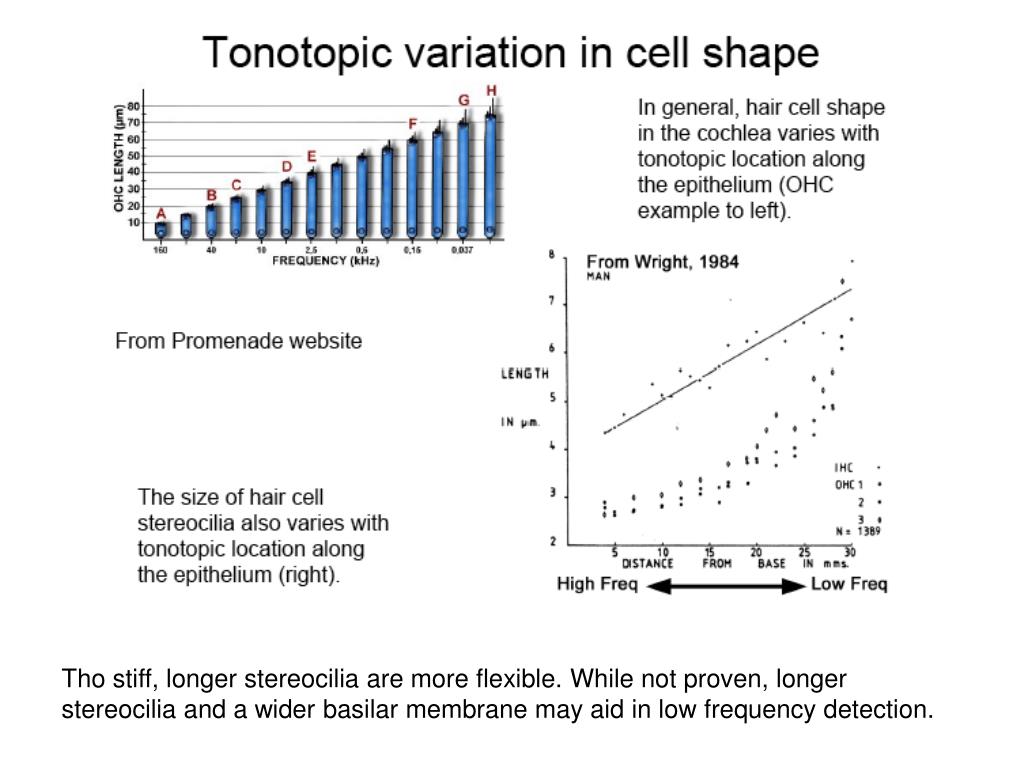
This stiffness is due to, among other things, the thickness and width of the basilar membrane,[5]which along the length of the cochlea is stiffest nearest its beginning at the oval window, where the stapes introduces the vibrations coming from the eardrum. Since its stiffness is high there, it allows only high-frequency vibrations to move ...
What is the cochlea filled with?
The cochlea is filled with a watery liquid, the endolymph, which moves in response to the vibrations coming from the middle ear via the oval window. As the fluid moves, the cochlear partition (basilar membrane and organ of Corti) moves; thousands of hair cells sense the motion via their stereocilia, and convert that motion to electrical signals that are communicated via neurotransmitters to many thousands of nerve cells. These primary auditory neurons transform the signals into electrochemical impulses known as action potentials, which travel along the auditory nerve to structures in the brainstem for further processing.
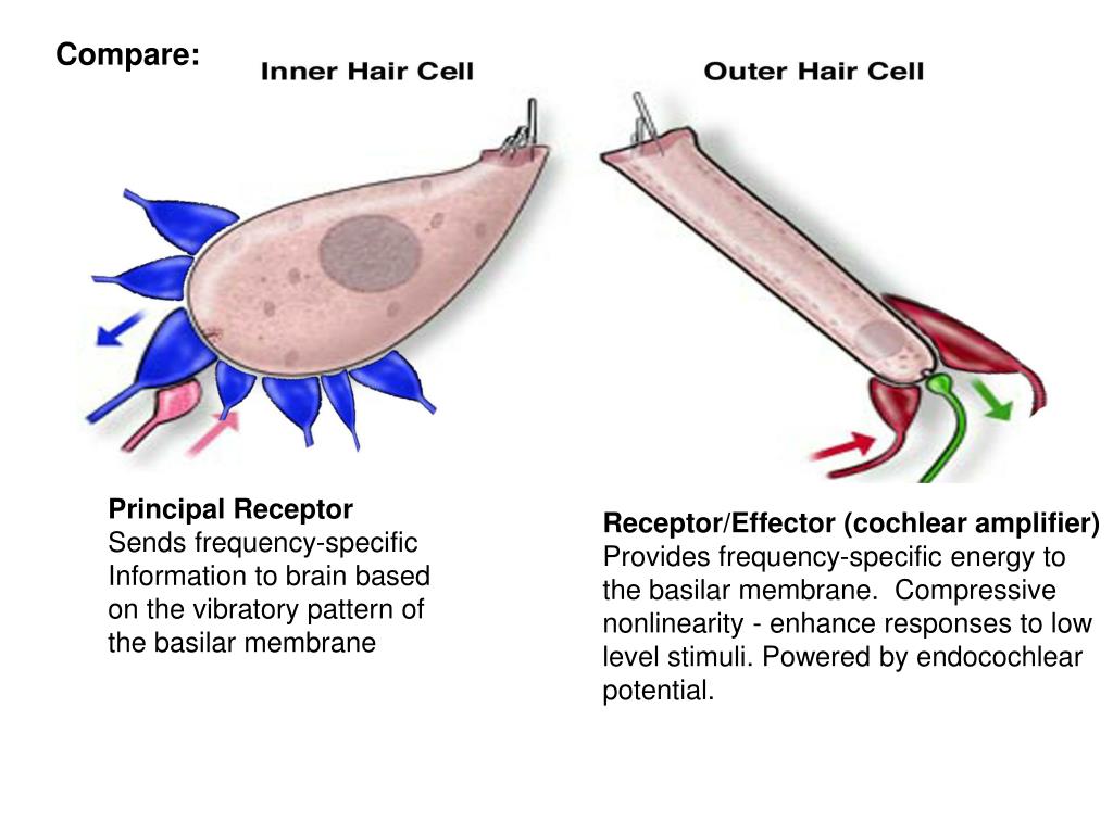
What is the hair cell in the inner ear?
hair cells, sensory cells in the Organ of Corti, topped with hair-like structures called stereocilia. The spiral ligament. The cochlea is a portion of the inner ear that looks like a snail shell (cochlea is Greek for snail.) [3]The cochlea receives sound in the form of vibrations, which cause the stereocilia to move.
Why are otoacoustic emissions important?
Otoacoustic emissions are important in some types of tests for hearing impairment, since they are present when the cochlea is working well, and less so when it is suffering from loss of OHC activity.
What is the cochlea in the human body?
3D model of cochlea and semicircular canals. The cochleais the part of the inner earinvolved in hearing. It is a spiral-shaped cavity in the bony labyrinth, in humans making 2.75 turns around its axis, the modiolus. [1][2]A core component of the cochlea is the Organ of Corti, the sensory organ of hearing, which is distributed along ...
Which part of the cochlea contains electrolytes?
The lengthwise partition that divides most of the cochlea is itself a fluid-filled tube, the third 'duct'. This central column is called the cochlear duct. Its fluid, endolymph, also contains electrolytes and proteins, but is chemically quite different from perilymph. Whereas the perilymph is rich in sodium ions, the endolymph is rich in potassium ions, which produces an ionic, electrical potential.
Which structure separates the vestibular duct from the cochlear duct?
The helicotrema, the location where the tympanic duct and the vestibular duct merge, at the apex of the cochlea. Reissner's membrane, which separates the vestibular duct from the cochlear duct. The osseous spiral lamina, a main structural element that separates the cochlear duct from the tympanic duct.
Who developed the cochlea?
Most of the early morphology and physiology of the cochlea was developed by one man during the 1930'sand 1940's, Georg von Bekesy . The intellectual intensity of his work won him a Nobel Prize in 1961,although his work only provided a conceptual explanation of how the cochlea worked. The problem wasthe tools available to him; he was forced to try and drive a pin into gelatin using a sledge hammer. Paraphrasing another theory-oriented researcher, this work does not refute Bekesy’s, rather this workextends Bekesy’s into realms that Bekesy did not deal with.
Why is the acoustic frequency response of human hearing difficult to quantify?
The acoustic frequency response of human hearing is difficult to quantify because the ultimate measure ofperformance is a psychological phenomenon. A unit of perceptual loudness has been identified anddefined as the phon. A condition of equal loudness must be determined by using a two-frequency test,where the more distant the two frequencies, the more difficult the perceptual comparison. This situationresults in an equal loudness contour that is precise for adjacent frequencies but suffers from a potentialrun-out error as the frequencies become farther apart. The determination of the equal-loudness-levelcontour as a function of frequency is found to also be subject to the state of adaptation of the hearingmodality (and the performance of the Eustachian tube. Thus, tests have been used to determine thestatistical mean sensitivity (energy required to achieve a constant loudness across the audio frequencyband) of the human auditory modality under various states of adaptation. The statistical variation in theseprotocols is quite high, resulting in high standard deviations in the reported data.
What is the Marcatili effect?
Application of the Marcatili Effect to the theoretical understanding of the hearing modality has asignificant impact on the past literature and future research. The Effect, combined with the slow surfaceacoustic wave (SAW) filter of the tectorial membrane coiled into a modified Hankel Function shows thecochlea employs a dispersive filter with the properties associated with a multi-channel spectrometer. As aresult, the cochlea;
Which organs detect head tilt?
the vestibular system (apparatus) - utricle/saccule (the otolith organs; Detect head tilt/linear acceleration or deceleration) and the semicircular canals (Detect head rotation in all three planes of space; near the end of each canal is an enlargement called the ampulla)
Which nerve fibers are responsible for hair cells?
via the sensory ganglion and the VIII cranial nerve (vestibulocochlear nerve); these nerve fibers contact the hair cells at their peripheral end and at their central ends form synapses with neurons in the vestibular nucleus (aka cochlear nucleus) in the medulla

Overview
Function
The cochlea is filled with a watery liquid, the endolymph, which moves in response to the vibrations coming from the middle ear via the oval window. As the fluid moves, the cochlear partition (basilar membrane and organ of Corti) moves; thousands of hair cells sense the motion via their stereocilia, and convert that motion to electrical signals that are communicated via neurotransmitters to many thousands of nerve cells. These primary auditory neurons transform …
Structure
The cochlea (plural is cochleae) is a spiraled, hollow, conical chamber of bone, in which waves propagate from the base (near the middle ear and the oval window) to the apex (the top or center of the spiral). The spiral canal of the cochlea is a section of the bony labyrinth of the inner ear that is approximately 30 mm long and makes 23⁄4 turns about the modiolus. The cochlear structure…
Clinical significance
In 2009, engineers at the Massachusetts Institute of Technology created an electronic chip that can quickly analyze a very large range of radio frequencies while using only a fraction of the power needed for existing technologies; its design specifically mimics a cochlea.
Other animals
The coiled form of cochlea is unique to mammals. In birds and in other non-mammalian vertebrates, the compartment containing the sensory cells for hearing is occasionally also called "cochlea," despite not being coiled up. Instead, it forms a blind-ended tube, also called the cochlear duct. This difference apparently evolved in parallel with the differences in frequency range of hearing between mammals and non-mammalian vertebrates. The superior frequency range i…
History
The name cochlea is derived from the Latin word for snail shell, which in turn is from the Greek κοχλίας kokhlias ("snail, screw"), from κόχλος kokhlos ("spiral shell") in reference to its coiled shape; the cochlea is coiled in mammals with the exception of monotremes.
Additional images
• Right osseous labyrinth. Lateral view.
• Interior of right osseous labyrinth.
• The cochlea and vestibule, viewed from above.
• Cross-section of the cochlea.
See also
• Bony labyrinth
• Membranous labyrinth
• Cochlear implant
• Cochlear nerve
• Cochlear nuclei