Are ultrasound waves longitudinal or transverse waves?
Interestingly ultrasound waves are both longitudinal and transverse waves. They behave as longitudinal waves when they propagate through fluids. When they move through solids they can either be both longitudinal and transverse waves. What type of wave is a ultrasound? sound waves Ultrasound uses sound waves.
What type of waves are used in ultrasonic waves?
Ultrasound waves can propagate in two different waves. One type is longitudinal, In the case of longitudinal waves, the particles are oscillating along the wave propagation axis. Usually, longitudinal waves are used in solid, liquid and gases.
What is a beam of ultrasound like?
A beam of ultrasound is similar to a beam of light; both are waves. The velocity of light waves and ultrasonic wave depends on the properties of medium they travel, not on the properties of the wave. Ultrasonic waves like light waves, can reflect, refract at the interface between two mediums, and diffracts at the edges or around obstacles.
Why does an ultrasound machine produce longitudinal waves?
This is because sound is a longitudinal wave. The molecules move back and forth along the direction of the wave in order to propagate a pressure wave. Because the ultrasound probe is made in such a way that it produces longitudinal waves. It’s the easiest way to do it and works fine.

Is an ultrasound wave longitudinal or transverse?
longitudinal wavesIn longitudinal waves , the vibrations are parallel to the direction of wave travel. Examples of longitudinal waves include: sound waves. ultrasound waves.
What type of wave is an ultrasound?
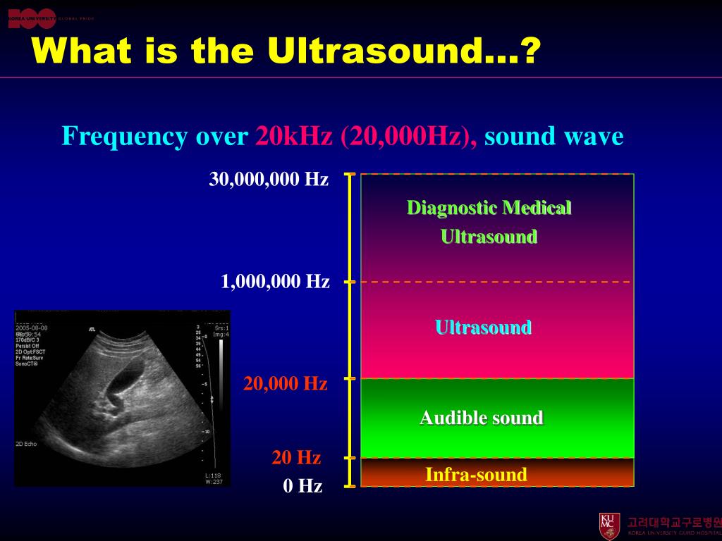
sound wavesUltrasound is the name given to sound waves that have frequencies greater than 20,000Hz (20 kHz). This is above the normal hearing range for humans, so we cannot hear ultrasound.
Why ultrasound waves are longitudinal waves?
Ultrasound tomography Longitudinal or compression waves are defined as waves where the particle motion is in the same direction in which the wave is propagating. The oscillations in pressure are sinusoidal in nature and are characterised by their frequency, amplitude and wavelength (Figure 9.1).
Does ultrasound use mainly transverse or longitudinal vibrations?
7.1, 7.2). The characteristic features of ultrasonography are sound waves that are bound to matter and propagate in a longitudinal direction. The movement of the material particles results in compression and decompression in the tissue.
What are the examples of longitudinal waves?
Some examples of longitudinal waves are sound waves, seismic P-waves, and ultrasound waves. Transverse waves examples include electromagnetic waves and ocean waves.
Are ultrasound waves long or short?
In air at atmospheric pressure, ultrasonic waves have wavelengths of 1.9 cm or less.
How do you know if a wave is transverse or longitudinal?
1:232:56Transverse & Longitudinal Waves | Waves | Physics | FuseSchoolYouTubeStart of suggested clipEnd of suggested clipSo the particles vibrate at 90 degrees to the direction that the energy is moving we can simplyMoreSo the particles vibrate at 90 degrees to the direction that the energy is moving we can simply think of it as in longitudinal waves the hand pulses horizontally pushing and pulling.
What is the transverse wave and ultrasound?
Out-of-plane alignment (transverse or short axis): This refers to when the transducer and the needle are perpendicular to each other.
What are transverse ultrasonic waves?
Transverse ultrasonic waves - waves propagating in a direction perpendicular to the plane in which the directions of displacements and velocities of body particles lie, the same as shear waves [2].
What is the difference between ultrasound and ultrasonic?
ultrasound, also called ultrasonography, in medicine, the use of high-frequency sound (ultrasonic) waves to produce images of structures within the human body. Ultrasonic waves are sound waves that are above the range of sound audible to humans.
Is ultrasound an electromagnetic wave?
Since all sound waves aren't electromagnetic waves, ultrasound waves are NOT electromagnetic waves. The waves are ultra because of the frequency and not the speed and the speed of ultrasound waves is the same as the speed of sound waves (340 m/s) which carry according to the medium.
Is ultrasound a mechanical wave?
Ultrasounds are mechanical waves that necessitate an elastic medium to spread over and differ from sounds by the wave frequency (Figure 4). Sounds are at human hearing frequencies (from 16 Hz to 16–20 kHz), while ultrasounds have frequencies above human hearing but below microwave frequencies (from 20 kHz to 10 MHz).
Is ultrasound an acoustic wave?
Two decades ago, in the field of medical imaging, the terms “acoustic imaging” and “ultrasonic imaging” were synonymous. The only acoustic waves used for imaging biological structures were ultrasonic compressional (or longitudinal) waves.
Does ultrasound use radio waves?
Ultrasound offers many benefits. For example, it does not use electromagnetic radio waves and therefore avoids issues with radio interference, crowding, and privacy.
What is longitudinal wave?
In the case of longitudinal waves, the particles are oscillating along the wave propagation axis. Usually, longitudinal waves are used in solid, liquid and gases. The second type of waves is transverse (shear) when particles oscillate perpendicular to wave propagation direction. Those are used in solid object analysis and not possible on liquids ...
What are the two types of waves that ultrasound waves can propagate?
If we take biological tissue, those are blood vessels, fibres, lesions, organs. Those irregularities are detected due to different density areas. Ultrasound waves can propagate in two different waves. One type is longitudinal, In the case of longitudinal waves, the particles are oscillating along the wave propagation axis.
Why is ultrasound penetration depth minimal?
Higher frequency ultrasound is strongly attenuated; this is why penetration depth is minimal. There is always a compromise between resolution and scanning depth. Ultrasound visualization is mainly based on its ability to reflect from any irregularities in media.
What is ultrasound used for?
In medical diagnostics, ultrasound is mostly used for visualization of tissues and inner organs.
Why is ultrasound important?
Ultrasound became popular and widespread due to the number of benefits. Probably one of the essential features is that it is non-destructive . The other is simplicity and precision. Of course when we talk about price and simplicity we have X-ray or tomography in mind. Ultrasound can be used in many areas and environments. You can measure geometrical properties of internal structures, physical properties like density, tension. In medical diagnostics, ultrasound is mostly used for visualization of tissues and inner organs.
Can you detect a half wavelength object?
Usually, it is possible to detect λ/2 – half-wavelength size objects. It may seem logical to increase ultrasound frequency to get better resolution and sensitivity, but in practice, the other limiting factors appear. Higher frequency ultrasound is strongly attenuated; this is why penetration depth is minimal.
What is longitudinal wave?
Longitudinal waves, also known as Compression waves is one of the most widely used in the inspection of the materials. In the longitudinal wave, the particles are displaced parallel to the direction of wave propagation.
What is the difference between a mechanical wave and an ultrasound wave?
Ultrasonic waves are mechanical waves (compared to X-Ray, Gamma Rays, Lightwaves, radio waves etc. which are all electromagnetic waves ). The mechanical waves need a medium such as solid, liquid, gas, etc., to propagate, They cannot propagate through a vacuum (Just space, no solid/liquid/gas). A beam of ultrasound is similar to a beam ...
How fast do ultrasonic waves travel?
The velocity of longitudinal ultrasonic waves is about 6000 m/s. (20,000 ft/s) in steel, 1500 m/s (5000 ft/s) in water, ...
What type of wave is used to inspect the surface of a material?
Rayleigh waves are another type of ultrasonic wave used for inspecting the surface of the material. Rayleigh waves travel along the free boundary (surface) of a relatively thick solid. These are also referred to as surface waves.
Why do ultrasonic waves differ in amplitude and velocity?
The amplitude and velocity of the ultrasonic waves differ in solids, liquids, and gases because of the large differences in the distance between particles in these forms of matter. These differences influence the forces of attraction between particles and the elastic behaviour of the materials.
How is the speed of a wave determined?
In ultrasonic testing, the speed is measured in Meter per second (m/sec). The speed of a wave is determined by the type of wave and the nature of the medium. As a wave enters a different medium/material, the wave’s speed changes. Waves travel at different speeds in different media.
What is shear wave?
Shear Waves. Shear waves, also known as Transverse waves are extensively used in the inspection of the materials. In the shear wave, the particles are displaced perpendicular to the direction of wave propagation. Air and water will not support transverse waves.
What is the amplitude of an ultrasound?
Amplitude is an important parameter and is concerned with the strength of the ultrasound beam. It is defined as the difference between the peak value and the average value of the waveform. It is expressed in decibels or dB, which is a logarithmic scale. It can be changed by a sonographer. Amplitude decreases as the ultrasound moves through tissue, this is called attenuation. Amplitude decreases usually by 1 dB per 1 MHz per 1 centimeter traveled. For example, if we have a 5 MHz probe and the target is located at 12 cm (24 cm total distance), then the amplitude attenuation will be 1 dB x 5 MHz x 24 cm = 120 dB which nearly 6000 fold decrease.
How does ultrasound work in clinical imaging?
In clinical imaging, the ultrasound beam is electronically focused as well as it is steered. This became possible after phased array technology was invented. By applying electrical current in a differential manner and adjusting the timing of individual PZT excitation, the beam can travel in an arch producing a two-dimensional image. If one applies electricity in a differential manner from outside inward to the center of the transducer, differential focusing can be produced resulting in a dynamic transmit focusing process.
How does ultrasound affect tissue?
As we discussed in the section of amplitude, the energy of ultrasound decreases (attenuation) as it travels through tissue. The stronger the initial intensity or amplitude of the beam, the faster it attenuates. Standard instrument output is ~ 65 dB. So for a 10 MHz transducer, the maximum penetration would be as follows: 1 dB/cm/MHz x 10 MHz x (2 x max depth) = 65 dB. Max depth = 65/20 = 3.25 cm. If we use a 3.5 MHz transducer and apply the same formula for max depth, will get Max depth = 65/7 = 9.3 cm. Attenuation of ultrasound in soft tissue depends on the initial frequency of the ultrasound and the distance it has to travel. As we saw in the example above, in soft tissue the greater the frequency the higher is the attenuation. So we can image deeper with lower frequency transducer. The further into the tissue the ultrasound travels, the higher the attenuation is, so it is ultimately the limiting factor as to how deep we can image clinically relevant structures.
What is the term for the time required to capture one cycle of an ultrasound wave?
This is called attenuation and is more pronounced in tissue with less density (like lung). There are seven parameters that describe ultrasound waves. Period of an ultrasound wave is the time that is required to capture one cycle, i.e., the time from the beginning of one cycle till the beginning of the next cycle.
Why is second harmonic imaging preferential?
There are several parameters that make second harmonic imaging preferential. Since it is produced by the tissue, the deeper the target the more second harmonic frequency is returned. As the ultrasound beam travels through tissue, new frequencies appear that can be interrogated. Second harmonic data gets less distortion, thus it produces better picture. Also, the second harmonic is strongest in the center of the beam, thus it has less side lobe artifacts. At the chest wall the fundamental frequency gets the worst hit due to issues that we have discussed (reflection, attenuation) – if one can eliminate the fundamental frequency data then these artifacts will not be processed. One concept of eliminating fundamental frequency data is called pulse inversion technology. The transducer sends out 2 fundamental frequency pulses of the same amplitude but of different phase. As these pulses are reflected back to the transducer, because of the different phase they cancel each other out (destructive interference) and what is left is the second harmonic frequency data which is selectively amplified and used to generate an image.
What frequency is ultrasound transmitted?
When used in diagnostic echocardiography, the frequency is usually above 20,000 Hz (20 kHz), and it is not audible to a human ear. Fig. 1.
How is density related to ultrasound?
Density of the medium is related to its weight and the stiffness of the medium is related to its “squishability”. As the medium becomes more dense, the slower is speed of ultrasound in that medium (inverse relationship). The stiffer the tissue, the faster will the ultrasound travel in that medium (direct relationship).
