Initially test the sensory branches by lightly touching the face with a piece of cotton wool followed by a blunt pin in three places on each side of the face:
- Around the jawline.
- On the cheek and.
- On the forehead.
Do I have a facial nerve problem?
Various medical conditions can cause facial nerve damage, resulting in a range of symptoms developing. Facial nerve damage leads to muscles in the face becoming weak; speech problems are also common. Other signs of facial nerve damage include headaches, dizziness, nausea, loss of some senses and dry eyes. What Does the Facial Nerve do?
What are the five branches of the facial nerve?
- The cranial nerves
- The facial nerve Components and branches Origin and course
- The muscles of facial expression Orbital group Nasal group Oral group Other muscles
- The facial nerve as part of the corticobulbar tract
- Clinical notes Surgical removal of the parotid gland Facial nerve palsy Inferior medial pontine syndrome Clinical case
How to deal with exam nerves?
To deal with exam anxiety, stay physically active in the days before the exam by going for a walk or playing sports, which will distract you and give your brain a chance to relax. On the day of the exam, avoid negative thoughts and try to say positive things out loud, such as “I can do this,” or, “Everything will be ok.” ...
How to deal with test nerves?
To deal with exam anxiety, stay physically active in the days before the exam by going for a walk or playing sports, which will distract you and give your brain a chance to relax. On the day of the exam, avoid negative thoughts and try to say positive things out loud, such as “I can do this,” or, “Everything will be ok.”

How do you test the branches of the trigeminal nerve?
Trigeminal motor function is tested by palpating the masseter muscles while the patient clenches the teeth and by asking the patient to open the mouth against resistance. If a pterygoid muscle is weak, the jaw deviates to that side when the mouth is opened.
How do you test for facial nerve damage?
Diagnostic imaging using magnetic resonance imaging (MRI) or a computed tomography (CT) scan can rule out other structural causes of pressure on the facial nerve (such as an artery compressing the nerve) and also check the other nerves.
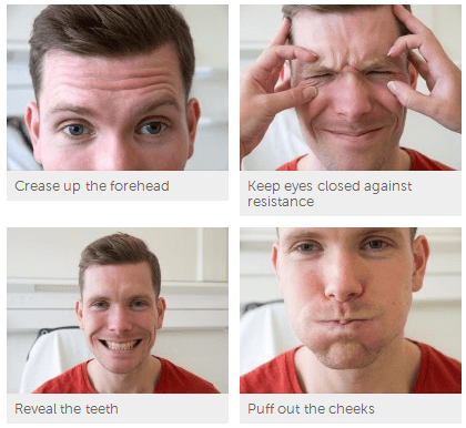
How do you test facial muscles?
Motor Function: Facial MusclesInspect the face. ... Ask the patient to wrinkle his forehead or raise his eyebrows, enabling you to test the upper face (frontalis).Next, have the patient tightly close his eyes. ... Instruct him to puff out both cheeks. ... Have the patient smile broadly and show his teeth, testing the lower face.
What is facial nerve conduction test?
A nerve conduction velocity (NCV) test — also called a nerve conduction study (NCS) — measures how fast an electrical impulse moves through your nerve. NCV can identify nerve damage. During the test, your nerve is stimulated, usually with electrode patches attached to your skin.
What are the branches of facial nerve?
The facial nerve has five main branches, although the anatomy can vary somewhat between individuals. The branches are, from top to bottom: frontal (or temporal), zygomatic, buccal, marginal mandibular, and cervical. Each of these branches provides input to a group of muscles of facial expression.
How do you test facial nerve 7?
1:162:51Cranial Nerve 7 | Facial Nerve Assessment for PhysiotherapistsYouTubeStart of suggested clipEnd of suggested clipAsk the patient to close the eyes tightly. While you try to force them. Open muscles in the lowerMoreAsk the patient to close the eyes tightly. While you try to force them. Open muscles in the lower half of the face can be tested by asking the patient to show their teeth smile or puff out the cheeks.
How do you test for the 11th cranial nerves?
The 11th (spinal accessory) cranial nerve is evaluated by testing the muscles it supplies: For the sternocleidomastoid, the patient is asked to turn the head against resistance supplied by the examiner's hand while the examiner palpates the active muscle (opposite the turned head).
What are the symptoms of facial nerve damage?
Facial paralysisRapid onset of mild weakness to total paralysis on one side of your face — occurring within hours to days.Facial droop and difficulty making facial expressions, such as closing your eye or smiling.Drooling.Pain around the jaw or in or behind your ear on the affected side.More items...•
How do you test for cranial nerve 10?
Cranial Nerves 9 & 10 - Motor The motor division of CN 9 & 10 is tested by having the patient say "ah" or "kah". The palate should rise symmetrically and there should be little nasal air escape. With unilateral weakness the uvula will deviate toward the normal side because that side of the palate is pulled up higher.
Can MRI show facial nerve damage?
An MRI may be able help identify structural lesions that may be pressing against the nerve so the problem can be corrected before permanent nerve damage occurs. Nerve damage can usually be diagnosed based on a neurological examination and can be correlated by MRI scan findings.
Can you see facial nerve on MRI?
MRI can be used to image the facial nerve from the brainstem to the fundus of the internal auditory canal and to determine the presence of perineural spread from parotid malignancies (Figure 9).
Can an EMG be done on the face?
Where is facial EMG commonly used? Facial EMG has a wide variety of uses in the medical field, particularly in movement and rehabilitation research pertaining to neuromuscular diseases like ALS, Parkinson's disease and stroke. Facial EMG has also been used in emotion studies with individuals with Autism.
What are the symptoms of facial nerve damage?
Facial paralysisRapid onset of mild weakness to total paralysis on one side of your face — occurring within hours to days.Facial droop and difficulty making facial expressions, such as closing your eye or smiling.Drooling.Pain around the jaw or in or behind your ear on the affected side.More items...•
How do you treat facial nerve damage?
There are a few different treatments for facial nerve damage that does not repair itself. These can include physical therapy, facial exercise, and surgery. Surgery can be performed by a reconstructive surgeon or our ENT specialist, Dr. Michael Barakate.
How long does it take for facial nerve damage to heal?
Sensory nerves can be accessed by various routes, all of which leave minimal scarring. Peripheral nerves have potential for self-repair, but it is a slow process that may take 3-4 months or longer. Minor and superficial nerve injuries will often heal themselves.
What causes nerve damage in face?
The most common cause of facial paralysis is Bell's palsy, which is thought to be a viral infection of the facial nerve, although the exact cause is not well known. Other causes of facial nerve paralysis include head trauma, parotid tumors, head or neck cancers, infections, brain tumors or stroke.
How to test for sour taste?
Place sweet, salt, bitter and sour taste solutions on one side of tongue one by one using cotton-bud. Patient should be asked to rinse his/her mouth after test for each taste. Test the opposite side also. Other tests (usually not done): Corneal reflex (Afferent is by trigeminal nerve while efferent is by facial nerve)
What nerve supplies stapedius?
Ask if there has been any hearing changes? – facial nerve supplies stapedius – paralysis results in hyperacusis
Overview
The facial nerve is a pathway from your brain to certain muscles in your face. It controls muscles that help you make expressions like raising an eyebrow, smiling or frowning. This nerve is also responsible for most of your tongue’s taste sensations.
Function
The facial nerve performs these motor (movement) and sensory functions:
Anatomy
The facial nerve is the seventh of 12 cranial nerves in your nervous system. You have two facial nerves, one on each side of your head.
Conditions and Disorders
Several conditions can cause weakness or paralysis of the facial nerve, including:
What is the facial nerve?
The facial nerve is one of a group of nerves called the cranial nerves (CN), twelve pairs of nerves that , with the exception of the spinal accessory nerve (CN XI), originate in the brain and contribute to the peripheral nervous system (PNS).
What are the components of the facial nerve?
The facial nerve contains many different types of fibers, including general sensory (afferent) fibers, special sensory fibers, visceral/autonomic motor (efferent) fibers, and somatic motor fibers. General sensory fibers in the facial nerve are responsible for transmitting signals to the brain from ...
Where do facial nerve fibers travel?
The fibers travel towards the floor of IV ventricle and go around the abducens nucleus and descend. The facial nerve emerges from the lateral surface of brainstem at the pontine-medullary junction between the VI and VIII nerves.
What is the vascular damage of the facial nerve?
Vascular damage to the facial nerve usually occurs at the supranuclear, pontine, and (rarely) cerebellopontine angle. Upper motor neuron (UMN) lesions occur in strokes and can easily be differentiated with lower motor neuron (LMN) lesions by their presentation. A LMN lesion causes paralysis of the whole side of face, ...
What is the function of the facial nerve?
While it is indeed responsible for innervating the muscles of facial expression, the facial nerve is a complex structure containing many fiber types with a variety of functions, including motor, sensory, and autonomic. The following article will discuss the importance and versatility facial nerve.
Which nerve is responsible for innervating the lacrimal gland, submandibular gland, sublingual?
Visceral/autonomic motor fibers in the facial nerve are responsible for innervating the lacrimal gland, submandibular gland, sublingual gland, and the mucous membranes of the nasal cavity and hard and soft palates, allowing for production of tears, saliva, etc., from these locations.
Which nerve contains motor fibers?
the hypoglossal nerve (CN XII) Some of these contain motor fibers, some contain autonomic fibers, some contain somatic sensory fibers, some contain special sensory fibers, and some contain combinations of a number of these aforementioned fiber types.
Where does the facial nerve enter the middle ear?
Before the facial nerve exits the cranium via the stylomastoid foramen, it gives off the chorda tympani. The chorda tympani travels superiolaterally and enters the middle ear, arches across pars flaccida medial to the superior portion of the handle of malleus, and traverses above the insertion of tensor tympani.
Which muscle is the motor fiber of the facial nerve?
Nerve to the stylohyoid muscle (raises hyoid bone) The remaining motor fibers of the facial nerve continue anteriorly/inferiorly and enter the parotid gland. Within the parotid gland, the facial nerve divides into five branches: Temporal. Zygomatic.
Which nerve fibers innervate the submandibular and sublingual salivary glands?
The chorda tympani provides taste sensation from the anterior 2/3 tongue and also carries preganglionic parasympathetic nerve fibers that innervate the submandibular and sublingual salivary glands. The nerve fibers of the chorda tympani arise from 2 distinct nuclei in the pons: Superior salivary nucleus:
Which nerve fibers synapse in the pterygopalatine ganglion?
Here, the preganglionic parasympathetic nerve fibers synapse in the aptly named pterygopalatine ganglion.
Where do parasympathetic preganglionic fibers travel?
The parasympathetic preganglionic fibers that join the greater petrosal nerve travel anteromedialy and enter the middle cranial fossa, passing over the foramen lacerum. These fibers then enter the pterygoid canal of the temporal bone (along with sympathetic fibers of the deep petrosal nerve) and form the pterygoid nerve.
Which parasympathetic fibers innervate the submandibular, sublingual, and la?
Preganglionic parasympathetic fibers innervating the submandibular, sublingual, lacrimal glands as well as the mucous membranes of the nose, palate, and oropharynx
Where does the lingual nerve exit the cranium?
The lingual nerve travels anteroinferiorly through the floor of the mouth, where the preganglionic fibers synapse in the submandibular ganglion.