
Lateral humeral condyle fractures also referred to simply as lateral condyle fractures (in the appropriate context), are relatively common elbow fractures that predominantly occur in children.
How to recover from lateral epicondylitis?
- Wrist Extension and Flexion: Straighten your arm, bend your wrist back with your fingers pointing upwards, and pull it with your other hand until you feel a stretch in your ...
- Wrist Turn: Bend the elbow at a right angle and extend the palm outwards facing up. ...
- Elbow Bend: Stand up straight and hold your arm down to one side. ...
How long does it take to heal a broken humerus?
This will obviously vary from case to case, depending on the severity of the injury. In most cases, however, it will take around three months for a broken humerus bone to fully heal. This will be achieved by engaging in regular exercises, and potentially physiotherapy – with these timelines potentially being reduced with the aid of a professional.
How painful is a broken humerus?
Humerus fractures cause severe pain and swelling. On a scale of 10, pain in patients following a humerus fracture is eight or more. Shortening of the arm is apparent with significant deformity of the bones. Humerus fractures are a very painful injury, and patients may need to take pain relief medications regularly as prescribed by the doctor.
Can a lucency indicate a fracture?
Healio goes on to point out that fractures also cause bone lucencies. Dr. Joseph Accurso on HealthTap describes lucencies as areas on an X-ray where the bone is less dense, which can be a sign of bone lesions. Many other factors can cause bone lucencies.

What is a condyle fracture?
condyle neck fracture, which occurs at the inferior attach area of the joint capsule, refers to an area that becomes narrow from the condyle head. It is an extracapsular fracture as it is not included in the joint capsule, and exists at the inferior attach area of the lateral pterygoid.
What is lateral condyle?
Medical Definition of lateral condyle : a condyle on the outer side of the lower extremity of the femur also : a corresponding eminence on the upper part of the tibia that articulates with the lateral condyle of the femur — compare medial condyle.
What is lateral condyle fracture with humerus?
Lateral condyle fractures are the second most common elbow fracture after the supracondylar humerus fracture in children. This fracture pattern is typically through the lateral metaphysis extending into the epiphysis and often extends into the articular surface.
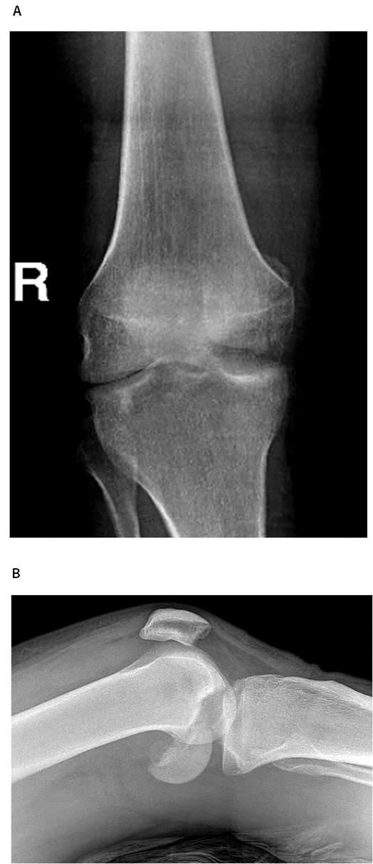
What is a lateral femoral condyle fracture?
Osteochondral fracture of the lateral femoral condyle is a rare injury of the knee joint, which mostly occurs in adolescence1. In adolescence, the cartilage‐bone interface is the weakest transitional area in the knee joint, and there is no obvious boundary between calcified and uncalcified cartilage2.
How long does it take to heal a lateral condyle fracture?
The most common timeline for this injury is four to six weeks in a long arm cast. Your child will then come back to get the cast removed and have an x-ray. Pins from surgery are removed once the bone has healed and is stable. If screws were used, they may not need to be removed.
How long does a condyle fracture take to heal?
Bony healing typically takes 6 weeks to complete; therefore, a soft diet may be necessary for 1 to 2 months in the absence of other fixation methods to prevent jaw displacement while the fracture is healing.
Where is the lateral condyle located?
The lateral condyle is the lateral portion of the upper extremity of tibia. Upper surface of right tibia. (Anterior is at top.) It serves as the insertion for the biceps femoris muscle (small slip).
Where is the lateral condyle of the humerus?
elbow jointThe lateral epicondyle of the humerus is a large, tuberculated eminence, curved a little forward, and giving attachment to the radial collateral ligament of the elbow joint, and to a tendon common to the origin of the supinator and some of the extensor muscles.
What is the lateral condyle of the humerus called?
lateral epicondyle. what is the name of the lateral condyle on the humerus? medial epicondyle.
Does a lateral femoral condyle fracture require surgery?
Isolated coronal fracture of medial femoral condyle with intact lateral femoral condyle is extremely rare [,,], caused by a direct impact on the flexed knee during weight bearing [3]. Surgery is the gold standard for displaced fractures or to enable rapid return of knee function.
How is femoral condyle fracture treated?
For young patients with good compliance, simple medial or lateral condylar fractures can be treated via a medial or lateral parapatellar approach. After fracture exposure, headless compression screws can be inserted perpendicularly to the fracture line from posterior to anterior.
What is the lateral condyle of the knee?
The lateral condyle is one of the two projections on the lower extremity of the femur. The other one is the medial condyle. The lateral condyle is the more prominent and is broader both in its front-to-back and transverse diameters.
What is the function of the lateral condyle?
The lateral condyle is the lateral portion of the upper extremity of tibia. Upper surface of right tibia. (Anterior is at top.) It serves as the insertion for the biceps femoris muscle (small slip).
What is the function of the condyle?
Condyle - Refers to a large prominence, which often provides structural support to the overlying hyaline cartilage. It bears the brunt of the force exerted from the joint. Examples include the knee joint (hinge joint), formed by the femoral lateral and medial condyles, and the tibial lateral and medial condyles.
What does condyle mean in medical terms?
Medical Definition of condyle : an articular prominence of a bone —used chiefly of such as occur in pairs resembling a pair of knuckles (as those of the occipital bone for articulation with the atlas, those at the distal end of the humerus and femur, and those of the lower jaw) — see lateral condyle, medial condyle.
What is the function of the lateral condyle of the femur?
The larger lateral femoral condyle provides a bony buttress that helps provide lateral patellar stability. The trochlear groove is shallower proximally than distally, indicating that bony stability is compromised as the patella moves superiorly during terminal knee extension.
What is the second most common fracture in the pediatric elbow?
Lateral Condyle Fractures are the second most common fracture in the pediatric elbow and are characterized by a higher risk of nonunion, malunion, and AVN than other pediatric elbow fractures.
Where do fractures originate?
fractures originate proximally at the posterior aspect of the distal humerus metaphysis and extend distally and anteriorly across the physis and epiphysis into the elbow joint. fracture may extend medially into the trochlear groove, making the elbow unstable and prone to dislocation. posteromedial elbow dislocation.
How to see a lateral condyle fracture?
The best view to see the lateral condyle fracture is an internal oblique and this should always be performed when a lateral condyle fracture has been diagnosed. This is done by pronating the arm, however, it is important to be aware that by placing the arm in pronation the fracture may be further displaced 14 .
What is lateral condyle?
Lateral humeral condyle fractures also referred to simply as lateral condyle fractures (in the appropriate context), are relatively common elbow fractures that predominantly occur in children. They may be subtle but are hugely important to diagnose in a timely manner because if they are missed, they have a tendency to migrate dorsally ...
What is supracondylar fracture?
supracondylar fracture (a transverse fracture usually through the supracondylar fossa) lateral epicondyle avulsion fracture (avulsion of the ossification center, not a transverse fracture through the condyle)
What classification is used for fractures?
The Milch and Weiss classifications have been used for these fractures. The Weiss classification uses the degree of displacement of the fracture and is a more relevant measure of severity.
How long does it take to cast a fractured arm?
It involves long-arm casting for approximately 4-6 weeks 2, 14.
What is a lateral condyle fracture?
Lateral condyle fractures involve a fracture line entering into the elbow joint. The Milch classification system (Table1) classifies the fracture according to the location of the fracture line in relation to the capitellar ossification centre.
Why do you need prompt orthopaedic consultation for lateral condyle fracture?
Due to the potential poor outcomes , all lateral condyle fractures require prompt orthopaedic consultation. Prompt recognition and treatment are essential to avoid complications and prevent functional impairment.
How common are condyle fractures in children?
They account for 12-20% of elbow fractures in children. The peak age of incidence is six years.
How long does it take to follow up on an undisplaced fracture?
Follow-up in fracture clinic should occur within 7 days with x-ray.
Can a lateral condyle fracture be seen on an x-ray?
If a lateral condyle fracture is suspected or minimally displaced on x-rays, then oblique views are often useful. These fractures do not require splinting prior to imaging as there is usually less pain and swelling. This allows subtle fracture lines to be more visible on x-ray. 6.
Can a condyle fracture be benign?
Lateral condyle fractures can frequently look benign with minimal swelling and minimal deformity, which can lead to delays in presentation and recognition of the fracture. 5.
Can an undisplaced fracture be immobilised?
Undisplaced fractures can be immobilised in an above-elbow backslab with the elbow flexed to 90 degrees. No reduction is required.
What is the diagnosis of a lateral condylar fracture?
A child with an elbow fracture invariably presents with a swollen, tender elbow. The lateral condylar fracture patients do not have as much deformity as the supracondylar fracture or elbow dislocation patients. Tenderness and ecchymosis on the lateral elbow are very helpful in proper clinical diagnosis, especially in the stable and/or minimally displaced fractures. 4 There may be crepitus with range of motion, though most children will not tolerate movement of the elbow in the acute setting. The diagnosis is really made by radiographs, with specific anteroposterior (AP), lateral, and internal oblique views of the distal humerus being necessary for accurate diagnosis of fracture displacement and risk of further displacement with cast treatment. 5
What percentage of lateral condylar fractures occur in children?
Lateral condylar fractures of the distal humerus occur less frequently than supracondylar fractures, accounting for 15% to 20% of all pediatric elbow fractures. Like most children’s upper limb injuries, they occur secondary to a fall or traumatic collision with another person or immobile (ground, building, tree) or mobile (car, bike) solid object. There is some debate as to whether during the injury the radial head “pushes off” the lateral condyle with a compression force or the forearm and extensor-supinator muscle origins “pull off” the lateral condyle with valgus-extension stress. The controversy makes for interesting academic discussion and paper publications 1 – 3 but really matters little in the simple “bone be broke, bone needs to be fixed” clinical scenario that we, the surgeon, the affected child, and his or her family confront.
How to reduce a type II condylar fracture?
Closed reduction of almost all type II and some rare, malrotated type III lateral condylar fractures is feasible. 5 Varus stress with the forearm supinated is utilized for reduction. Elbow extension with the varus stress and supination position is most reliable for reduction of most fractures. After successful closed reduction , percutaneous pin fixation is performed to stabilize the fracture. The results of CRPP are better than closed reduction and immobilization in terms of malunion and nonunion. 18 Two divergent, lateral entry pins that penetrate the far cortex (similar to supracondylar fractures [see Chapter 27 ]) are used (Figure 28-7 ). 19 A third pin indentllel to the joint from the lateral to the medial condyle helps with articular reduction and stability. In situations when there is uncertainty about the articular reduction and/or stability, an arthrogram for confirmation and stress testing is indicated. 20
How long does it take for a condylar fracture to heal?
Bivalved long-arm cast, collar and cuff, splint and sling have all been used successfully 14–16 The fracture will heal in 3 to 6 weeks. Immobilization in forearm supination and wrist extension has been used to lessen muscle tension on the lateral condyle and therefore the risk of fracture displacement. These nondisplaced fractures should be stable, and supination and wrist extension positioning may be overkill. However, there will be a rare fracture that you never expected to displace, yet it does. 17 Vigilant use of close, follow-up radiographs is necessary to catch the “rule breaker.” The false assumptions in these rare situations are that: (1) the articular surface is always intact and stable and (2) the soft tissue injury is minor. Protective immobilization should continue until there are clinical and radiographic signs of bony healing. If the fracture displaces or fails to heal in a timely manner, then CRPP is required (Figure 28-6 ).
What is the distal humeral articular anatomy?
Clearly understanding the distal humeral articular anatomy and soft tissue attachments about the elbow is key to proper diagnosis and management of this problem. The extensor-supinator muscle mass of the forearm, wrist, and hand originates from the metaphysis of the lateral condyle of the humerus. The lateral collateral ligament complex consists of the radial collateral ligament (from the isometric point on the lateral epicondyle to the annular ligament), lateral ulnar collateral ligament (from the lateral epicondyle to the crista supinatoris), and annular ligament (Figure 28-2 ).
Can a capitellar shear fracture be missed on a CT scan?
Finally, the capitellar shear fracture is a seindentte entity that can be missed on plain radiographs (Figure 28-5 ). This fracture is high risk for long-term complications, even with early accurate diagnosis and appropriate ORIF. Analysis with CT scan is very helpful to define the fracture location, pattern, and size of the fracture fragment in preindenttion for surgery.
What is the condylar fracture on a radiograph?
Panoramic radiograph of left condylar fracture shows enlargement of the condyle due to overlap of the fractured fragment on the condylar neck stump.
How to heal condylar fracture?
A condylar fracture that has healed following closed reduction with a minimal early posterior molar contact and a contralateral anterior openbite can be managed by occlusal grinding of the ipsilateral posterior maxillary teeth. A dental splint would be of no use in the secondary rehabilitation of this injury. If the condylar motion is good, the best procedure for restoring good occlusion would be a sagittal splint osteotomy lengthening the ramus while avoiding any secondary injury to the area of the joint. If the joint motion is not good or an ankylosis is present, a costochondral graft can be used to restore the ramus height and improve limited motion of the joint.
What is a closed mouth fracture?
Closed-mouth fractures tend to distribute some of the energy to the occlusal surface of the teeth, and cuspal fractures are common. Under the influence of the masticatory muscles, the mandibular ramus may shorten vertically and produce premature occlusal contacts distally.
Why are condylar fractures so common in children?
First, a relatively significant number of these injuries are never diagnosed, and second, regardless of whether they are diagnosed, condylar fractures can cause significant lower facial asymmetry and masticatory dysfunction, particularly in growing patients. In fact, Proffit and co-authors reported that up to 10% of patients with dentofacial deformities have evidence of previously undiagnosed condylar fractures.17 The mandible is the last bone in the face to reach skeletal maturity, and consequently, it is vulnerable to injury-related disturbances in growth. This is magnified by the fact that mandibular trauma has a greater incidence in late childhood and adolescence. Classically caused by a fall and commonly heralded by a laceration in the submental region, condylar fractures are characterized by shortening of the ramus on the affected side, which causes deviation of the chin to the affected side. On the unaffected side, open bite and flattening of the body of the mandible are noted. In bilateral fractures of the condyle, posterior displacement of the mandible is seen with an anterior open bite. Occasionally, despite a condylar fracture the child will be able to hold projection, symmetry, and occlusion in the mandible without difficulty. In such cases, observation with diet modification is usually sufficient for treatment. The immediate sequelae of condylar fractures are the same in children and adults. However, the manner in which the fractures propagate can be substantially different. Because the immature mandible has a relatively thick and short condylar neck, children have a propensity for fracture through the condylar head rather than the low neck pattern seen more commonly in adults. In addition, compression injuries of the fossa and condylar head, as well as medial pole fractures, also occur more commonly in children.
What bone is the left condyle on a radiograph?
The trabecular bone of the lesion is continuous with the trabecular bone of the condyle.
What is a low Jupiter fracture?
Low Jupiter fractures are equivalent to Milch type I fractures and high Jupiter fractures to Milch type II fractures. Preferred treatment in adults is ORIF with early ROM exercises.
Where is the bone plate used to stabilize a mandibular fracture?
CBCT surface rendering shows bone plates used to stabilize a mandibular fracture are noted on midline of the mandible. A fracture in this location is associated with compressive forces to the TMJs.
What is lateral condyle fracture?
A lateral condyle fracture is type of broken elbow that kids can get after falling. Lets quickly review some anatomy of the elbow to better understand the injury. Our elbow is a joint where the arm bone (humerus) meets up with your two forearm bones (the radius and the ulna).
Which bone is on the lateral side of the elbow?
The radius bone is on the lateral side (outside) of the elbow, while the ulna bone is on the medial side (inside). The lateral condyle is outer part of the humerus bone that makes contact with the radius bone at the elbow joint. The lateral condyle can break after falling onto the elbow.
Can a condyle break?
The lateral condyle can break after falling onto the elbow. It is often difficult to diagnose (more on this in the diagnosis section), and the break enters into the elbow joint, which means that this type of injury often needs surgery (also more on this later).
Where is the lateral humeral condylar fracture in dogs?
What are humeral condylar fractures? Fractures of the humerus (upper arm bone) are common in dogs and cats with approximately half of all humeral fractures occurring in the distal (lower) portion of the bone, at or near the elbow joint. The vast majority of distal humeral fractures involve ...
How long does it take for a dog to bear weight after a condylar fracture?
However, with appropriate surgical techniques from a veterinarian with extensive humeral condylar fracture repair and sugery experience, most dogs are very comfortable after surgery and can start to bear weight on the limb in as little as one or two days.
What is the best treatment for humeral condylar fracture?
Surgery is the best treatment for humeral condylar fractures. Because these fractures run through the joint surface, perfect alignment of the fractured fragment with inter-fragmentary compression is necessary in order to restore elbow function.
What is the most common fracture in dogs?
Distal humeral condylar fractures are much more common in dogs than they are in cats. Lateral condylar fractures occur when forces concentrated on the lateral portion of the elbow joint cause breakage along this vertical growth plate and through the lateral portion of the bone above it. This type of fracture is most common in young dogs ...
What is the end of the humerus called?
The end of the humerus that forms the upper part of the elbow joint is composed of two halves, called the medial and lateral condyles, which are separated down the middle in young dogs by a growth plate that normally fuses upon reaching adulthood. Distal humeral condylar fractures are much more common in dogs than they are in cats.
How to diagnose humeral condyles?
Fractures of the humeral condyles are diagnosed by taking x-rays of the elbows. Because of the discomfort caused by this injury and the need for very precise positioning, sedation and pain medication are necessary to take diagnostic x-rays. Occasionally, more advanced forms of diagnostic imaging of both elbow joints, such as a CT scan, can provide additional information that will be necessary for treatment.
Is humeral condyle surgery good?
The prognosis following surgery for fractures of the outside (lateral aspect) of the humeral condyle is generally good, although because of joint surface involvement, some degree of osteoarthritis will eventually develop in the elbow joint.
What is a fracture of the femoral condyle?
A femoral condyle is the ball-shape located at the end of the femur (thigh bone). There are two condyles on each leg known as the medial and lateral femoral condyles. If there is a fracture (break) in part of the condyle, this is known as a fracture of the femoral condyle. Physiotherapy is very important during the rehabilitation following a femoral condyle fracture.
What to do if you think you have a femoral condyle fracture?
You should visit your nearest accident and emergency department if you think you may have a femoral condyle fracture. You will need to have an X-ray to locate the exact area of damage. Other tests may need to be carried out including MRI scans, if trauma to other non-bony structures are suspected.
What is the procedure called when a fracture is realigned?
However, if other structures are involved or an open fracture is shown, the bone will need to be realigned by a procedure referred to as ‘open reduction internal fixation ’ which is a surgical procedure.
Could there be any long-term effects from a fracture of the femoral condyle?
However, if a thorough physiotherapy programme is followed, long-term complications should be minimal and you will be able to return to your sport or normal activities of living.
Summary
How Are They Classified?
- Lateral condyle fractures involve a fracture line entering into the elbow joint. The Milch classification system (Table1) classifies the fracture according to the location of the fracture line in relation to the capitellar ossification centre. Table 1: Milch classification of lateral condyle fractures.
How Common Are They and How Do They occur?
- Lateral condyle fractures of the elbow are the second most common paediatric elbow fracture after supracondylar fractures. They account for 12-20% of elbow fractures in children. The peak age of incidence is six years. They usually occur as a result of indirect forces being applied to the elbow following a fall on an outstretched hand. Angular and rotational forces are thought to cont…
What Do They Look Like - clinically?
- The child will present with pain, limited elbow range of motion (ROM) and swelling at the lateral aspect of the elbow.
What Radiological Investigations Should Be ordered?
- Anteroposterior (AP) and lateral x-rays of the elbow should be obtained without splinting. If there is clinical suspicion of injury in the forearm or wrist then separate films of these areas should be ordered. If a lateral condyle fracture is suspected or minimally displaced on x-rays, then oblique views are often useful.
When Is Reduction (Non-Operative and Operative) Required?
- Undisplaced fractures can be immobilised in an above-elbow backslab with the elbow flexed to 90 degrees. No reduction is required. Minimally displaced ( <2 mm gap) can either be managed with immobilisation alone or with closed reduction and percutaneous pinning. All displaced fractures (>2 mm gap and/or angulation of the lateral condyle) will need to go to theatre either for closed r…
Do I Need to Refer to Orthopaedics Now?
- Patients should be kept nil orally until a decision about surgery is made. For open fractures, tetanus immunisation status should be assessed.
What Is The Usual Ed Management For This Fracture?
- All lateral condyle fractures require an orthopaedic consult in the ED (see above). Undisplaced fractures can be immobilised in an above-elbow backslab with the elbow flexed to 90 degrees and supported in a sling.
What Follow-Up Is Required?
- For undisplaced fractures, follow-up in the fracture clinic within 7 days with repeat x-ray is required. It is usually necessary to remove the backslab to obtain adequate views of the fracture at review. For fractures that go to theatre, follow-up should be arranged by the consulting orthopaedic team.
What Advice Should I Give to Parents?
- The backslab and sling should be worn under clothing (e.g. loose fitting shirt) and not through the sleeve. Close follow-up is required to ensure that the fracture remains in the correct position. In the majority of minimally displaced fractures, union will occur. Fractures managed with operative intervention have a high union rate.