What's the function of the axon hillock?
The axon hillock acts as an administrator, sums up the total signals received, both inhibitory and excitatory signals. If this sum exceeds the limiting threshold, the action potential is triggered. This results in the transmission of the generated electrical signal through the axon away from the neuronal cell body.
What is axon hillock in psychology?
a cone-shaped part of the cell body of a neuron from which the axon originates. Depolarization must reach a critical threshold at the axon hillock for the axon to propagate a nerve impulse.
What is the difference between axon and axon hillock?
The axon arises from the cell body at a small elevation called the axon hillock. The proximal part of the axon, adjacent to the axon hillock, is the initial segment. The cytoplasm of the axon (axoplasm) contains dense bundles of microtubules and neurofilaments (Figs.
Where is the axon hillock of a neuron?
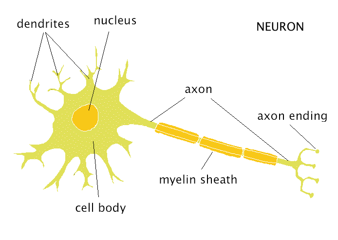
A typical neuron. Dendrites branch out from the cell body, where the nucleus is located. The axon hillock is located where the cell body transitions into the axon. The axon begins at the axon hillock and ends at the presynaptic terminal, which can branch into multiple terminals.
Why is axon hillock called trigger zone?
It is located between the axon and the cell body. The axon hillock normally contains the trigger zone of the neurone. This is the area that must be depolarised to above threshold potential before it initiates the all-or-nothing response of an action potential.
What is the function of the axon hillock quizlet?
> Axon Hillock: Located at the end of the Soma and controls the firing of the neuron. If total strength of signal exceeds threshold, an action potential will occur.
What happens at the axon hillock action potential?
Action Potentials. The action potential is an all-or-nothing electrical wave that is initiated at the axon hillock and propagates toward the axon terminal via highly coordinated sequential activation of various ion channels that have differential selectivities with respect to ion permeation (Figure 1).
Why does action potential start in axon hillock?
If several such events occur in a short time, the axon hillock may become sufficiently depolarized for the voltage-gated sodium channels to open. This initiates an action potential that then propagates down the axon.
What happens at the axon hillock action potential?
Action Potentials. The action potential is an all-or-nothing electrical wave that is initiated at the axon hillock and propagates toward the axon terminal via highly coordinated sequential activation of various ion channels that have differential selectivities with respect to ion permeation (Figure 1).
What is the function of Neurofibrils?
These are similar to intermediate filaments and microtubules found in other types of cells. The main component of the cytoskeleton is formed by neurofilaments....The cell body.StructuresFunctionsNeurofibrilsDetermine shape of neuronMicrofilamentsHelp to form and retract cell processes; assist in cellular transport11 more rows
Why do action potentials start at the axon hillock?
A long-standing hypothesis is that action potentials initiate first in the axon hillock/initial segment (AH–IS) region because of a locally high density of Na+ channels.
Why is action potential initiated at axon hillock?
If several such events occur in a short time, the axon hillock may become sufficiently depolarized for the voltage-gated sodium channels to open. This initiates an action potential that then propagates down the axon.
What is an Axon Hillock?
Imagine for a moment that you are one of many departmental managers at a large business. Each department deals with something different, such as payroll or legal issues. The employees under your supervision are responsible for receiving information from other departments and even other businesses and then sending this input to you. Your function is to arrive at a consensus, then decide whether or not to send your decision throughout the business's hierarchy.
Why is the axon hillock unique?
The axon hillock is a unique area within neurons because of its ability to process the incoming signals from other cells. To understand how this works, let's look at what a chemical impulse really is. A neuron's outer membrane is semi-permeable, which means some materials are allowed into the cell and some are not.
What is the control area that is in charge of initiating neuron chemical impulses after receiving them from other neurons?
An axon hillock is the control area that is in charge of initiating neuron chemical impulses after receiving them from other neurons. Identify the various functions of neurons and the specifics of the axon hillock. Updated: 11/18/2021
How many axons do neurons have?
Neurons only have one axon, which is the extension that allows nerve impulses to move away from the cell body and toward other nerve cells. The axon then communicates with the dendrites or the cell body of the next neuron. However, axons do not make direct contact with these structures, and instead just get real close.
When does the Axon Hillock turn on?
Like the thermostat in an air conditioner, the axon hillock will only turn on if its threshold is reached. For example, when the surrounding temperature exceeds the setting at which the air conditioner turns on, that is its threshold.
What is the gap between two structures called?
The resulting gap between structures is called the synapse. Synapses are often named by their location. For example, those that only communicate with cell bodies are called axosomatic, meaning 'axo' for axon and 'somatic' for cell body.
What are the elements in the axon hillock?
The axon hillock may contain fragments of Nissl substance, including abundant ribosomes, which diminish as the hillock continues into the initial segment. Here, the various axoplasmic components begin to align longitudinally. A few ribosomes and the smooth ER persist, and some axoaxonic synapses occur. The axolemma of the initial segment where the action potential originates exhibits a dense granular layer similar to that seen at the nodes of Ranvier, consistent with a specialized membrane cytoskeleton. Also present in this region are microtubules, neurofilaments and mitochondria. The arrangement of the microtubules in the initial segment is distinctive in forming fascicles interconnected by side arms. Beyond the initial segment, the axon maintains a relatively uniform caliber even after branching with little or no diminution until the very terminal arbors (Fig. 1-7 ). One exception is a reduction of caliber for myelinated axons at the peripheral node of Ranvier ( Hsieh et al., 1994) (see Fig. 1-2 and below). Myelinated axons show granular densities on the axolemma at nodes of Ranvier ( Raine, 1982) that correspond to adhesion molecules and high densities of sodium channels. In myelinated fibers, there is a concentration of sodium channels at the nodal axon, a feature underlying the rapid, saltatory conduction of such fibers (Ch. 4).
Where does the axon come from?
The axon arises from the cell body at a small elevation called the axon hillock. The proximal part of the axon, adjacent to the axon hillock, is the initial segment. The cytoplasm of the axon (axoplasm) contains dense bundles of microtubules and neurofilaments ( Figs. 2.1 and 2.5A, B ). These function as structural elements, and the microtubules also play key roles in the transport of metabolites and organelles along the axon. Axons are typically devoid of ribosomes, a feature that distinguishes them from dendrites at the ultrastructural level.
What are microtubules in axons?
6). Microtubules are present in loose groupings rather than bundles and vary in their spacing ( Fig. 1-7 A). Vesicles and mitochondria are typically seen in association with these microtubule domains, consistent with their movement in fast axonal transport (Ch. 8). In axons less than a micron in diameter, which are usually unmyelinated, microtubules are the primary structural cytoskeletal elements, with sparse neurofilaments and gaps in the neurofilament cytoskeleton. As axons get larger, the number of neurofilaments increases dramatically, becoming the primary determinant of axonal caliber. For large, myelinated axons, neurofilaments occupy the bulk of an axon cross-section (Ch. 6) with microtubules found in small groups along with membrane profiles.
What type of interneuron has a single axon?
Whereas most thalamic interneurons have several characteristic axoniform dendrites, they have only a single axon that is recognizable on the basis of an axon hillock and a slim initial segment and, at least in some instances, myelin, and this axon in turn leads into a long branched process that does not taper significantly and that generally branches in the neighborhood of the cell body. On the basis of the axonal ramification, Tömböl (1969) has described two sorts of interneuron, one with a locally ramifying axon and one with an axon that goes beyond the dendritic arbor but generally stays in the same nucleus or nuclear group. In the lateral geniculate nucleus this type of axon may go into an adjacent lamina, in the medial geniculate nucleus, Tömböl describes that it can go from one nuclear subdivision to another. She described such cells in the ventral posterior nucleus and the mediodorsal nucleus as well, but comparable descriptions have not appeared subsequently, apart from the interneurons described by Winer and Morest (1983 and 1984) in the medial geniculate nucleus of the cat, where they distinguished large and small interneurons and suggested that some of the interneuronal axons passed from one subdivision of the medial geniculate nucleus to another.
Why are axons swollen?
With such stains damaged axons appear swollen because of the interruption in their fast transport system and the proximal accumulation of organelles and fluid.
Where does action potential originate?
Action potentials can originate not only at the axon hillock, but also in the axon initial segment, 30–40 μm from the soma and close to the first myelinated segment. In some neurons the action potential even originates at the first node of Ranvier, where sodium channels are highly concentrated ( Figure 1 ). For both myelinated and unmyelinated axons, once the action potential begins in the axon, it not only propagates orthodromically toward the nerve terminals but also propagates antidromically, back into the soma and dendrites.
Does F response mean proximal or distal?
It is important to emphasize that although F responses usually are thought of as assessing the proximal nerve segments, they actually check the entire nerve. For example, any nerve with a prolonged distal motor latency on routine nerve conduction studies will also have prolonged F responses, because the F response must travel through the distal segment of nerve as well as the proximal segment. This situation is commonly seen in patients with median neuropathy at the wrist, wherein the median minimal F latency is often prolonged; in this situation, the depolarization travels antidromically from the stimulation site at the wrist up the nerve to the anterior horn cell, and then back down the nerve to the point of stimulation. However, once the depolarization proceeds past the point of stimulation, through the area of slowing at the wrist, this results in prolongation of the F response. Likewise, if there is generalized conduction velocity slowing from a polyneuropathy, the F response will also be slowed, reflecting the slowed conduction velocity of the entire nerve. The F response latency is shorter in the arms than in the legs, reflecting the shorter length of nerve traveled. Therefore, it should be no surprise that taller patients have longer F responses than do shorter patients. Thus, the distal motor latency, the conduction velocity, and the height of the patient must all be taken into account before a prolonged F response is interpreted as indicating a proximal nerve lesion.