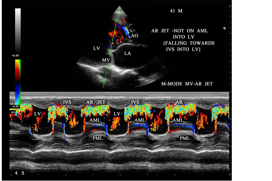
The normal area of the mitral valve orifice is about 4 to 6 cm2. In normal cardiac physiology, the mitral valve opens during left ventricular diastole, to allow blood to flow from the left atrium to the left ventricle.
How do you calculate the area of a mitral valve?
- Implications of the New Cardiac Catheterization Laboratory in the 21st Century. ...
- Principles of Cardiac Catheterization. ...
- Valve Stenosis. ...
- Evaluation of Unexplained Dyspnea. ...
- Pulmonary Hypertension. ...
- Constrictive Pericarditis Versus Restrictive Cardiomyopathy. ...
- Hypertrophic Cardiomyopathy. ...
- Conclusion. ...
- Disclosures
- Footnotes. ...
What is a normal aortic valve area?
Keep reading. What Is Normal Aortic Valve Area? A normal aortic valve area is greater than or equal to 2.0 cm2. In people with normal aortic valves, the normal aortic valve area range is 2.0 cm2 and greater.
Can doctors replace the mitral valve?
Treatment for mitral valve disease depends on the severity of your condition. Doctors may recommend surgery to repair or replace mitral valves for some people with mitral valve disease. Several surgical procedures exist to repair or replace mitral valves, including open-heart surgery or minimally invasive heart surgery.
Is mitral valve disease a serious condition?
Mitral valve prolapse rarely becomes a serious condition. However, in the most serious cases it can cause abnormal heartbeats (arrhythmias) that eventually may become life-threatening and lead to a heart attack or stroke. In some instances, patients may need to have a valve repair or even replacement.

What is normal mitral valve stenosis?
Mitral valve stenosis, shown in the heart on the right, is a condition in which the heart's mitral valve is narrowed. The valve doesn't open properly, blocking blood flow coming into the left ventricle, the main pumping chamber of the heart. A typical heart is shown on the left.
What is moderate to severe mitral stenosis?
Mitral valve stenosis (sometimes called mitral stenosis) is a disease caused by narrowing or blockage of the mitral valve inside your heart. While it doesn't cause symptoms in many people, in some it can cause heart rhythm problems and an increased risk of stroke.
What is normal mitral valve area by pressure half time?
Mitral valve area by pressure half-time had a range of 1.6–4.5 cm2, whereas mitral valve area by the continuity equation had a range of 1.0–3.6 cm2.
What is abnormal mitral valve?
An abnormal mitral valve may become “floppy” and not close well (prolapse), it may allow blood to leak back from the left ventricle into the left atrium (regurgitation) or it may become narrow or tight (stenosis)..
How do you determine the severity of mitral stenosis?
Two major factors determine the severity of mitral stenosis:the size of the mitral orifice during diastole (mitral valve area) and the magnitude of the gradients across the valve. The mitral vale area (MVA) can be determined with 2D echo (planimetry and by Doppler techniques - the pressure half time method).
What are the first symptoms of mitral stenosis?
Mitral Stenosis SymptomsShortness of breath: You may have a hard time breathing, especially after being active or when you lie down.Fatigue: You may tire easily during increased physical activity.Swollen ankles and feet: Swelling may occur when blood flow is disturbed.More items...
What is considered severe mitral stenosis?
A pressure gradient across the mitral valve of 20 mmHg due to severe mitral stenosis will cause a left atrial pressure of about 25 mmHg. This left atrial pressure is transmitted to the pulmonary vasculature resulting in pulmonary hypertension.
What is MVA in cardiology?
We sometimes encounter patients with microvascular angina (MVA), a disease characterized by anginal pain without abnormal coronary arteriographic findings or coronary spasm. More than 40 years have passed since MVA was first confirmed.
What gradient is severe mitral stenosis?
The gradient can be measured by tracing the dense outline of mitral diastolic inflow and the mean pressure gradient is automatically calculated. The severity can be assessed as mild (<5), moderate (5–10) and severe (>10).
What is mild mitral insufficiency?
Mitral insufficiency, the most common form of valvular heart disease, occurs when the mitral valve does not close properly, allowing blood to flow backwards into the heart. As a result, the heart cannot pump efficiently, causing symptoms like fatigue and shortness of breath.
What are the symptoms of mitral valve disease?
Symptoms of Mitral Valve ProlapseFluttering or rapid heartbeat called palpitations.Shortness of breath, especially with exercise.Dizziness.Passing out or fainting , known as syncope.Panic and anxiety.Numbness or tingling in the hands and feet.
What is mild mitral regurgitation?
Overview. Mitral valve regurgitation is a type of heart valve disease in which the valve between the left heart chambers doesn't close completely, allowing blood to leak backward across the valve. It is the most common type of heart valve disease (valvular heart disease).
Where is the mitral valve located?
Where is the mitral valve? The mitral valve is located in the left side of the heart, between the left atrium and left ventricle. Oxygen-rich blood flows into the left atrium from the pulmonary veins. When the left atrium fills with blood, the mitral valve opens to allow blood to flow to the left ventricle. It then closes to prevent blood ...
What is the mitral valve?
This is one of the heart’s four valves that help prevent blood from flowing backward as it moves through the heart. Read on to learn more about the mitral valve, including its location and anatomy.
What valve opens to allow blood to flow to the left ventricle?
When the left atrium fills with blood, the mitral valve opens to allow blood to flow to the left ventricle. It then closes to prevent blood from flowing back into the left atrium. All of this happens in a matter of seconds as the heart beats.
How many leaflets does the mitral valve have?
The mitral valve has two leaflets. These are projections that open and close. One of the leaflets is called the anterior leaflet. This is a semicircular structure that attaches to two-fifths of the mitral valve’s area. The other is called the posterior leaflet. It attaches to the remaining three-fifths of the valve.
Why does my mitral valve prolapse?
Mitral valve prolapse is the most common reason for mitral valve repair in the United States. This condition occurs when the valve doesn’t completely close because it’s loose. Mitral valve prolapse doesn’t always cause symptoms. But in some people, it can cause mitral valve regurgitation, which can cause some symptoms.
What is the name of the process where blood flows backwards through the mitral valve?
Mitral valve regurgitation refers to extra blood flowing backward through the mitral valve and into the left atrium. This makes the heart work harder to move blood, causing an enlarged heart.
What is the zone of coaptation?
Zone of coaptation. The zone of coaptation is a rough area on the top side of the valve’s surface. It’s where the chordae tendinae attach the mitral valve to the papillary muscles. This area makes up a small portion of the mitral valve, but any irregularities in it can prevent the valve from functioning properly.
What is the area of the mitral valve?
Normal mitral valve orifice has an area of about 4.0 – 6.0 cm 2. Mitral stenosis defines the mechanical obstruction in this blood flow due to different causes, such as thickening and immobility of the leaflets, ...
What happens when the mitral valve area becomes less than 1 cm2?
When the mitral valve area becomes less than 1 cm 2, the subsequent increase in the left atrial pressure is then transmitted to the pulmonary vasculature, which will in turn result in pulmonary hypertension and eventually pulmonary congestion and edema.
What is the mitral valve?
Indeed, the mitral valve apparatus is a dynamic three-dimensional (3D) structure composed of the saddle-shaped annulus; two asymmetric leaflets; multiple chordae tendineae of various lengths, thicknesses, and points of attachment; the left ventricular wall and the attached papillary muscles; and parts of the left atrium ( Figure 5-1 ). During normal function, this array of parts constantly shifts in a complex but defined pattern. Normal alignment of all aspects of this biologic machine is required to avoid dysfunction. Therefore, full understanding of the mitral valve anatomy is not ascertained without 3D imaging. Real-time 3D echocardiography (RT3DE) gives echocardiographers a rapid and easily accessible method of identifying the entire mitral valve apparatus in most patients. As a prominent structure in the posterior left heart, the mitral valve often can be displayed in 3D using either a transthoracic approach or a transesophageal approach. Furthermore, detailed volumetric and positional analysis of the various mitral valve components often is possible ( Table 5-1 ). RT3DE, however, is a novel modality, and its use in mastering the analysis of the intricate mitral valve requires a substantial time commitment. This chapter highlights the use of RT3DE in imaging the normal mitral valve, demonstrating usual transthoracic and transesophageal views for each of its components. It also provides an overview of typical 3D quantitative analyses of the size and position of the mitral valve structures.
What is the annulus of the mitral valve?
3D reconstructions identified the mitral valve annulus as a hyperbolic paraboloid or a saddle-shaped structure with curved planes parallel to the anteroposterior axis opening upward and orthogonal curved planes that open downward ( Figure 5-2 ). 1 This complex shape is not appreciated on 2D imaging, which tends to simplify the annulus as a planar ring. In fact, the anterior and posterior portions of the mitral valve annulus are about 5 mm higher than the medial and lateral commissural points. 1 This height, along with the commissural diameter (the distance between the two low points) and the anteroposterior diameter (the distance between the two high points), can be easily measured with 3D imaging. The nonplanar shape of the mitral valve annulus is very important in reducing leaflet stress, which is increased when the annulus flattens. 2 As such, the hyperbolic paraboloid annular shape with a height/commissural width ratio of 15% to 20% is preserved in many mammalian species, including humans. 2–4
Why is the annulus of the mitral valve important?
The nonplanar shape of the mitral valve annulus is very important in reducing leaflet stress, which is increased when the annulus flattens. 2 As such, the hyperbolic paraboloid annular shape with a height/commissural width ratio of 15% to 20% is preserved in many mammalian species, including humans. 2–4.
Where is the anterior leaflet located?
The anterior leaflet covers two thirds of the orifice and is attached to the part of anterior annulus adjacent to the aortic valve near the right fibrous trigone, aortic-mitral fibrosa, and left fibrous trigone ( Figure 5-5 ).
