Clear layer (stratum lucidum): Cells are tightly compressed, flattened and indistinguishable from one another. Capa clara (estrato lúcido): Las células están densamente comprimidas, aplanadas y no pueden distinguirse unas de otras. ParaCrawl Corpus
Why is stratum lucidum called the clear layer?
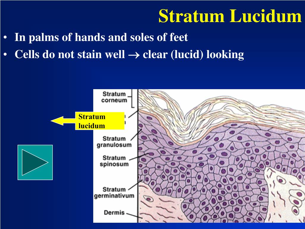
The stratum lucidum (Latin, 'clear layer') is a thin, clear layer of dead skin cells in the epidermis named for its translucent appearance under a microscope. It is readily visible by light microscopy only in areas of thick skin, which are found on the palms of the hands and the soles of the feet.
Is stratum lucidum clear?
Keratinocytes have granules within them, and in this layer they're visible under a microscope. Between the stratum granulosum and the stratum corneum. The stratum lucidum is a thin, transparent layer of keratinocytes that are becoming less round and have a flatter shape.
What is unique about stratum lucidum?
The presence of the stratum lucidum makes the palmar and plantar skin characteristically thick. The skin in other body parts lacks stratum lucidum. Its presence indicates the importance of having thicker skin to protect against frequent exposure to mechanical stress.
What is the clear transparent layer just under the skin surface?
The stratum lucidum (STRAT-um LOO-sih-dum) is the clear, transparent layer under the stratum corneum; it consists of small cells through which light can pass.
What part of the epidermis is known as the clear cells?
What part of the epidermis is known as the clear cells? The stratum lucidum is a thin, clear layer of dead skin cells that allow light to pass through.
Which epidermis is transparent?
Upper EpidermisUpper Epidermis - Single layer of tightly packed cells with a thin waxy coating called a cuticle. The cuticle prevents water loss and creates a physical barrier to protect against insects and microorganisms. These cells do not contain chloroplasts, so the layer is transparent and allows light to pass through.
What is stratum lucidum and its function?
It regulates the temperature and the amount of water released in the environment. Stratum lucidum is a thin, clear layer of dead skin cells in the epidermis of the skin. It is composed of five layers of dead flattened keratinocytes.
Why is stratum lucidum absent in thin skin?
Thick skin has more epidermal layers present, and in fact, has a stratum lucidum layer present. This particular layer consists of dead cells and it is not found in thin skin at all. In fact, thick skin is lacking many of the structures that are present in thin skin.
Is lucidum only in thick skin?
Only thick skin contains the stratum lucidum layer. The stratum lucidum is a thin, transparent layer consisting of two to three layers of cells. It contains a protein called eleidin.
Which layer is seen only in the skin?
Stratum Lucidum This thin layer of cells is found only in the thick skin of the palms, soles, and digits. The keratinocytes that compose the stratum lucidum are dead and flattened (see [link]).
What layer of the skin is just below the surface?
Skin has three layers: The epidermis, the outermost layer of skin, provides a waterproof barrier and creates our skin tone. The dermis, beneath the epidermis, contains tough connective tissue, hair follicles, and sweat glands. The deeper subcutaneous tissue (hypodermis) is made of fat and connective tissue.
What epidermal layer is found in thick skin but not in thin skin?
The stratum lucidum is a smooth, seemingly translucent layer of the epidermis located just above the stratum granulosum and below the stratum corneum. This thin layer of cells is found only in the thick skin of the palms, soles, and digits.
What layer of the epidermis is clear and only found in thick skin?
stratum lucidum layerOnly thick skin contains the stratum lucidum layer. The stratum lucidum is a thin, transparent layer consisting of two to three layers of cells. It contains a protein called eleidin.
Is a clear intracellular protein present in the stratum lucidum?
Eleidin is clear intracellular protein which is present in the stratum lucidum of the skin. Eleidin is a transformation product of the amino acid complex keratohyalin, the lifeless matter deposited in the form of minute granules within the protoplasm of living cells.
Is plant epidermis transparent?
Epidermis – transparent, physical defence layer that does not contain chloroplasts. It allows light into the leaf.
Which stratum is responsible for skin color?
stratum basaleThe stratum basale also contains melanocytes, cells that produce melanin, the pigment primarily responsible for giving skin its color.
What is the stratum lucidum?
The stratum lucidum is responsible for the capability of the skin to stretch. It also contains a protein that is responsible for the degeneration of skin cells. This thick layer also lowers the effects of friction in skin, especially in regions like the soles of feet and palms of hands. It is responsible for making the skin waterproof.
How many layers are there in the stratum lucidum?
The stratum lucidum layer is composed of three to five layers of dead flattened keratinocytes.
Where is the epidermis located?
Explanation: It is a thin transitional layer of epidermis found between the horny and granular layers of the skin. It is readily visible by light microscopy in areas of thick skin, which are formed on the palms of the hands and soles of the feet.
What is the role of GluR5 in MFT?
Research into the subunit (s) responsible for the presynaptic KAR actions have produced conflicting data; pharmacological studies using compounds that selectively activate and inhibit (ATPA and LY382884, respectively) GluR5-containing receptors have implicated GluR5 as mediating the KAR-dependent actions in MFT transmission ( Bortolotto et al., 1999 ), though studies from Heinemann and colleagues using GluR5 and GluR6 transgenic mouse models have found that GluR6 knockout mice fail to exhibit many actions of kainate-induced MFT plasticity ( Contractor et al., 2000, 2001 ), suggesting that it is the GluR6 subunit, and not the GluR5, that plays a presynaptic autoreceptor role in facilitating MFT plasticity. A more recent study has also evaluated the role of presynaptic GluR7-containing receptors using transgenic mice, and reported that disruption of the GluR7 gene reduced paired-pulse facilitation, low-frequency facilitation, and MFT LTP (see below); thus, GluR7-containing presynaptic autoreceptors also facilitate plasticity at the MFT–CA3 synapse ( Pinheiro et al., 2007 ). Considering this study also demonstrated a physical interaction between GluR6 and GluR7 proteins, reasonable hypothesis is that a GluR6/7 KAR complex in MFTs may be critical in these plasticity modes. Generation of KA2 knockout mice has demonstrated altered heterosynaptic facilitation in the presynaptic MFT, likely as a result of a decreased affinity for endogenously released glutamate; furthermore, these mice also demonstrate altered postsynaptic excitatory postsynaptic currents ( Contractor et al., 2003 ).
What is the role of KARs in glutamatergic transmission?
While most studies have focused on AMPAR and NMDAR-dependent postsynaptic mechanisms of synaptic plasticity on CA3–CA1 synapses, synaptic plasticity such as long-term potentiation (LTP) at DG-CA3 MFTs may involve NMDAR-independent, KAR-dependent pre synaptic modulation of glutamate release. In hippocampal synapses, KARs play a role in glutamatergic transmission through multiple mechanisms, including bidirectionally modulating MFT glutamate release ( Schmitz et al., 2001a ), increasing excitability on commissural/perforant path inputs to CA3 neurons ( Contractor et al., 2000 ), and exerting direct actions on postsynaptic neurons via KARs on CA3 neurons ( Castillo et al., 1997 ). In addition, KARs are postulated to have indirect effects on CA1 neurons through the modulation of GABA release via KAR-containing CA1 interneurons, a mechanism that is presumed to play a role in KA-induced epileptogenesis ( Min et al., 1999 ).
What are the synaptic connections between mossy fibers?
As noted above, the mossy fiber axons make contact onto both pyramidal neurons and stratum lucidum interneurons in the CA3 region. These two synaptic connections demonstrate a remarkable segregation in their functional properties. High frequency stimulation of mossy fibers, which causes LTP at pyramidal cell synapses, actually depresses mossy fiber interneuron synapses through activation of presynaptic mGluR7 and PKC signaling. Naïve interneuron synapses are not potentiated by elevating cAMP by application of forskolin, suggesting that these synapses are under regulation by fundamentally different mechanisms, despite being formed by the same axons and being closely spatially arranged (filopodial synapses onto interneurons emanate from the main mossy fiber boutons that form the synapses onto the pyramidal neuron). However, one interesting report demonstrated that activation of mGluR7, and depression of the mossy fiber interneuron synapse, uncovers a subsequent sensitivity to cAMP-dependent potentiation. This state-dependent responsiveness to cAMP suggests that this signaling pathway is more ubiquitous in mossy fibers than was originally thought to be the case.
What is the downstream target of mGluR7?
Recently one of the downstream targets of mGluR7 activation at these synapse s was elucidated. Pelkey et al. (2006) observed that transmission at MF filopodia is supported primarily by P/Q-type voltage-gated calcium channel activity, with only a minor role for N-type channels. This presynaptic calcium channel arrangement is identical to that observed at the large MF bouton inputs into pyramidal cells and ruled out a role for differential voltage-gated calcium channel distribution as a mechanism to explain the functional divergence of the two presynaptic release sites. Of particular importance, they showed that LTD induced by either exogenous application of L-AP4 or HFS resulted in a persistent depression of the presynaptic voltage-gated calcium channel signal monitored by two-photon microscopy. Importantly, this depression of the calcium transient arose through a preferential depression of P/Q calcium channels: Blockade of P/Q channels prevented both the depression of synaptic events and calcium transients by either L-AP4 or HFS. These data indicate that synaptic depression at MF–CP-AMPAR interneuron synapses arises by an mGluR7-dependent persistent reduction in P/Q calcium channel function.
What are the interneurons of the stratum lucidum?
Interneurons of the stratum lucidum are primarily targeted by the filopodial extensions and en passant MF synapses, but rarely by the larger MF boutons that typically contact principal cells (Acsady et al., 1998) ( Figure 1). Given that the filopodial extensions originate from the large MF terminal, it might be assumed that any change in presynaptic release probability arising from long-term potentiation (LTP) in the large MF bouton would distribute evenly to all synapses, resulting in long-term changes at both MF–pyramidal cell and interneuron synapses. However, quite the opposite is the case. At MF–CA3 st. lucidum interneuron synapses, the same high-frequency nonassociative protocol that induces LTP at principal cell synapses induces two forms of long-term depression (LTD) at interneuron synapses (Figure 1; Lei and McBain, 2004; Pelkey et al., 2005 ). Postsynaptically, AMPARs at MF-SLIN synapses comprise a continuum ranging from GluR2-lacking, Ca 2+ -permeable channels (CP-AMPARs) to GluR2-containing, Ca 2+ -impermeable channels (CI-AMPARs) ( Toth et al., 2000; Lei and McBain, 2002 ). Both CI-AMPAR- and CP-AMPAR-containing MF-SLIN synapses exhibit LTD in response to high-frequency stimulation (HFS) that arises from two distinct mechanisms ( Figure 2; Maccaferri et al., 1998; Toth et al., 2000; Lei and McBain, 2002, 2004; Pelkey et al., 2005 ). Induction of each is blocked by inclusion of the Ca 2+ chelator BAPTA in the postsynaptic compartment, indicating a postsynaptic induction locus for each (cf. MF CA3 pyramidal cell LTP described following). Activity-induced LTD of MF-SLIN transmission at CI-AMPAR synapses is blocked by inclusion of the N -methyl- d-aspartate receptor (NMDAR) antagonist, D-AP5, indicating that this form of plasticity is mediated in part by postsynaptic NMDAR activation. Expression of this form of LTD proceeds via subsequent AMPAR internalization by a mechanism similar, but not identical, to postsynaptically expressed LTD observed at excitatory synapses throughout the central nervous system (Lei and McBain, 2002, 2004). Consistent with a postsynaptic expression locus, this form of LTD is not accompanied by changes in the paired pulse ratio, the CV (coefficient of variation), or the synaptic failure rate – parameters typically used to monitor presynaptic mechanisms. Although none of these parameters on their own rigorously supports presynaptic changes (e.g., postsynaptic conversion of silent to active synapses will also change these parameters), an independent test of whether this NMDA receptor–dependent form of mossy fiber plasticity was postsynaptic made use of the low-affinity α-amino - 3-hydroxy-5-methyl-isoxazole-4-propionic acid (AMPA) receptor antagonist γ-D-glutamyl glycine (γ-DGG) (Lei and McBain, 2004 ). In these experiments, Lei and McBain (2004) demonstrated that the magnitude of block of the evoked synaptic event by γ-DGG was unaltered following induction of NMDA-dependent MF-interneuron long-term depression. These data argue that the transmitter-release concentration profile is unchanged following LTD expression. Taken together, these data support the conclusion that this form of plasticity has both its induction and expression locus within the postsynaptic compartment.
Where can you see the most complex synapse?
The greatest complexity of synapse—spine shape and interactions can be seen in CA3 of the hippocampus where the mossy fibers from the dentate granule cells contact the thorny excrescences of stratum lucidum of the giant pyramidal neurons (Figure 6 (B)). The contacts take the form of giant boutons (GBs) (also named detonator synapses because of their size) and there is an abundance of spheroid vesicles (glutamate containing) in the GBs which make contact on the thorns that are unique to CA3 in the hippocampus.
Where are the synapse and spine similar?
Spine and synapse forms are similar throughout the hippocampus except in CA3 in stratum lucidum where the dendrites are covered with thorny excrescences which are contacted by the giant boutons of the mossy fibers as seen in (B) and shown in the 3-D reconstructions in (F).
What is the epidermis made of?
The epidermis is composed of keratinized, stratified squamous epithelium. It is made of four or five layers of epithelial cells, depending on its location in the body. It does not have any blood vessels within it (i.e., it is avascular). Skin that has four layers of cells is referred to as “thin skin.” From deep to superficial, these layers are the stratum basale, stratum spinosum, stratum granulosum, and stratum corneum. Most of the skin can be classified as thin skin. “Thick skin” is found only on the palms of the hands and the soles of the feet. It has a fifth layer, called the stratum lucidum, located between the stratum corneum and the stratum granulosum ( Figure 5.1.2 ).
Why is the stratum spinosum spiny?
As the name suggests, the stratum spinosum is spiny in appearance due to the protruding cell processes that join the cells via a structure called a desmosome. The desmosomes interlock with each other and strengthen the bond between the cells. It is interesting to note that the “spiny” nature of this layer is an artifact of the staining process. Unstained epidermis samples do not exhibit this characteristic appearance. The stratum spinosum is composed of eight to 10 layers of keratinocytes, formed as a result of cell division in the stratum basale ( Figure 5.1.5 ). Interspersed among the keratinocytes of this layer is a type of dendritic cell called the Langerhans cell, which functions as a macrophage by engulfing bacteria, foreign particles, and damaged cells that occur in this layer.
Which layer of the epidermis is the deepest?
The stratum basale (also called the stratum germinativum) is the deepest epidermal layer and attaches the epidermis to the basal lamina, below which lie the layers of the dermis. The cells in the stratum basale bond to the dermis via intertwining collagen fibers, referred to as the basement membrane.
How many layers of cells are there in the stratum corneum?
There are usually 15 to 30 layers of cells in the stratum corneum. This dry, dead layer helps prevent the penetration of microbes and the dehydration of underlying tissues, and provides a mechanical protection against abrasion for the more delicate, underlying layers.
Why is the dermis reticulated?
deeper layer of the dermis; it has a reticulated appearance due to the presence of abundant collagen and elastin fibers
What are the layers of the skin?
Figure 5.1.1 – Layers of Skin: The skin is composed of two main layers: the epidermis, made of closely packed epithelial cells, and the dermis, made of dense, irregular connective tissue that houses blood vessels, hair follicles, sweat glands, and other structures. Beneath the dermis lies the hypodermis, which is composed mainly ...
What is the integumentary system?
The skin and its accessory structures make up the integumentary system, which provides the body with overall protection. The skin is made of multiple layers of cells and tissues, which are held to underlying structures by connective tissue ( Figure 5.1.1 ). The most superficial layer of the skin is the epidermis which is attached to ...
